. 2013 Sep;33(18):3540-8.
doi: 10.1128/MCB.00771-13. Epub 2013 Jul 8.

Interaction between 25S rRNA A loop and eukaryotic translation initiation factor 5B promotes subunit joining and ensures stringent AUG selection

Affiliations

Interaction between 25S rRNA A loop and eukaryotic translation initiation factor 5B promotes subunit joining and ensures stringent AUG selection

Hiroyuki Hiraishi et al. Mol Cell Biol. 2013 Sep.

Abstract

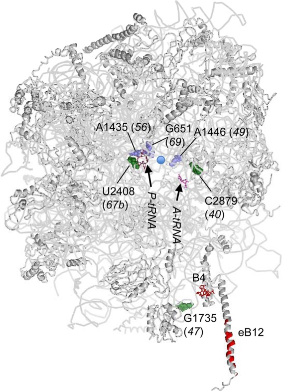
In yeast, 25S rRNA makes up the major mass and shape of the 60S ribosomal subunit. During the last step of translation initiation, eukaryotic initiation factor 5B (eIF5B) promotes the 60S subunit joining with the 40S initiation complex (IC). Malfunctional 60S subunits produced by misfolding or mutation may disrupt the 40S IC stalling on the start codon, thereby altering the stringency of initiation. Using several point mutations isolated by random mutagenesis, here we studied the role of 25S rRNA in start codon selection. Three mutations changing bases near the ribosome surface had strong effects, allowing the initiating ribosomes to skip both AUG and non-AUG codons: C2879U and U2408C, altering the A loop and P loop, respectively, of the peptidyl transferase center, and G1735A, mapping near a Eukarya-specific bridge to the 40S subunit. Overexpression of eIF5B specifically suppressed the phenotype caused by C2879U, suggesting functional interaction between eIF5B and the A loop. In vitro reconstitution assays showed that C2879U decreased eIF5B-catalyzed 60S subunit joining with a 40S IC. Thus, eIF5B interaction with the peptidyl transferase center A loop increases the accuracy of initiation by stabilizing the overall conformation of the 80S initiation complex. This study provides an insight into the effect of ribosomal mutations on translation profiles in eukaryotes.


Figures

Fig 1
Fig 1
Locations of 25S rRNA point mutations in the yeast 60S subunit structure. The yeast 60S subunit (Protein Data Bank [PDB] 3O58) is shown with RNA and protein backbones in gray and viewed from the 40S interface side. 25S rRNA residues altered by rdn mutations (rdn allele numbers are in parentheses) are highlighted as green or slate spheres. Nucleotides in gray are located near the surface, whereas those in slate are located deep inside the ribosome structure. The blue circle indicates the location of the PTC (A2820 and C2821). Functional rRNA bases relevant to this study are shown as colored sticks; G2619 and G2620 are in raspberry (P loop), G2922 is in magenta (A loop), and A846 and A847 are in red (a 40S interface termed B4). Amino acids of Rpl19e involved in eB12, another 40S interface, are also in red. PYMOL was used to draw this figure.
Fig 2
Fig 2
C2879U (rdn40), G1735A (rdn47), and U2408C (rdn67b) allow bypass of AUG codons. (A) Gcn phenotype test. Fixed amounts (5 μl of culture diluted to an A600 of 0.15) of NOY908 (WT), KAY968 (rdn40), KAY971 (rdn47), KAY964 (rdn49), KAY777 (rdn56), KAY1016 (rdn67b), and KAY963 (rdn69) (Table 1) and their 10-fold serial dilutions were spotted onto synthetic complete (SC) medium lacking histidine but supplemented with 40 mM leucine and with (panel 2) or without (panel 1) 50 mM 3AT and incubated for 3 days at 36°C. The schematic at the top depicts the structure of the GCN4-lacZ leader region. (B) Transformants of designated strains in panel A carrying p180 (GCN4-lacZ) were grown in the absence (−) or presence (+) of 30 mM 3AT for 6 h following preculturing for 2 h and assayed for β-galactosidase (26) at 36°C (panel 1) or 34°C (panel 2). The graphs show the averages using 2 or more independent transformants, with bars indicating standard errors (SE) (n = 4 to 10). (C) Leaky scanning of the uORF1 start codon. The graph presents β-galactosidase activities expressed from pM226 in transformants of rdn strains listed to the left in panel A that were grown at 30°C and subjected to the assay, as described previously (26). The schematic at the top depicts the structure of the modified GCN4-lacZ leader carried on the plasmid used. Bars indicate SE from 4 reactions using two independent transformants. Each of the rdn mutations tested increased expression from pM226 compared to the wild type (P < 0.002).
Fig 3
Fig 3
C2879U (rdn40), G1735A (rdn47), and U2408C (rdn67b) increase the initiation accuracy by bypassing UUG codons (Ssu phenotype test). Fixed amounts of transformants of KAY165 (his4-306 RDN+) and its 25S rdn derivatives (Table 1) carrying YDpU-SUI3 (SUI3) or YDpU-SUI3-2 (SUI3-2) (44) and their 10-fold serial dilutions were spotted onto synthetic complete (SC) medium lacking uracil (+His) or lacking uracil and histidine (−His) and incubated for 4 or 6 days, respectively. The column to the right summarizes the Ssu phenotypes of the rdn mutants.
Fig 4
Fig 4
C2879U (rdn40) is suppressed by overexpression of eIF5B. (A) Effect on Gcn phenotype. Cultures of transformants of NOY908 (RDN+; WT) and KAY968 (C2879U) bearing pC565-3 (hc eIF5B) (24) or a vector control (Vec) and their 10-fold serial dilutions were spotted onto SC medium plates lacking histidine and uracil with (panel 2) or without (panel 1) 50 mM 3AT. The plates were incubated for 3 (panel 1) or 7 (panel 2) days at 34°C. (B and C) Effect on GCN4 expression and leaky scanning of the uORF1 start codon. Transformants of NOY908 (WT) or KAY968 (C2879U) carrying p180 (B) or pM226 (C) and pD401-4 (hc eIF5B TRP1) (5B) or a vector control (Vec) were assayed for β-galactosidase as for Fig. 2B and C. (B) Transformants were grown at 34°C in SC medium lacking His, Trp, and uracil (SC-His-ura) supplemented (+) or not supplemented (−) with 30 mM 3AT. The β-galactosidase units presented are the averages from 10 reactions using 5 independent transformants. *, P = 0.02; **, P = 0.0003; ***, P = 0.03. (C) Transformants were grown at 30°C in SC-Trp-ura. Shown are the averages from 16 reactions using 4 independent transformants. *, P = 0.04. (D) Effect on Ssu phenotype. Transformants of KAY165 (WT) and KAY427 (C2879U) carrying pD401-4 (hc eIF5B) or a vector control (Vec) and YDpU-SUI3 (SUI3) or YDpU-SUI3-2 (SUI3-2) were assayed except as follows, and the results are presented as for panel A. We used SC medium lacking uracil and tryptophan (+His) or lacking uracil, tryptophan, and histidine supplemented with 0.3 μM histidine (−His), and the plates were incubated for 4 and 7 days, respectively.
Fig 5
Fig 5
Primer extension analysis of purified 40S subunits. The homogeneity of the C2879U mutant 40S subunit was confirmed by primer extension using 25S rRNA isolated from the purified sample (right lane of the gel). Wild-type (WT) 40S subunit was used as a control (left lane). The mutation site for C2879U, the primer, and expected cDNAs obtained from primer extensions are shown below the gel.
Fig 6
Fig 6
The C2879U (rdn40) mutation in 25S rRNA impairs 80S complex formation. (A) Scheme of 80S complex formation assay. A final concentration of 1 nM [35S]Met-tRNAiMet was used as a limiting component for the assay. (B) 80S complex formation assay. 48S complexes were formed using a limited amount of 35S-labeled Met-tRNAiMet, and, with GTP chase, subunit joining was performed according to the scheme described in panel A. The reactions were quenched at the indicated time by loading onto a 4% acrylamide native gel, and subunit joining was monitored by electrophoresis. The results of experiments done in the absence of eIF5B or in the presence of 0.5 or 2 μM eIF5B are presented. *, smearing bands below the 80S complex. (C) 80S complex formation assay in the presence of two different concentrations of eIF5B with GDPNP chase, as shown in the scheme in panel A. (D) Kinetics of subunit joining with 0.5 μM eIF5B. Amounts of [35S]Met-tRNAiMet that were free or bound to 80S complexes at the indicated times were quantified, and the fraction of [35S]Met-tRNAiMet in the 80S complexes was calculated. The fitting curve shown here is from one of two independent experiments.
Fig 7
Fig 7
Effect of the 25S rRNA mutations on translation initiation and quality control coupled to ribosome bypass and recycling. The left column describes translation initiation steps until the subunit joining. The gray oval represents the 40S subunit with the AUG codon (box AUG) of the mRNA (line), loaded on its P site (P) and base-paired to the anticodon of Met (M)-tRNAiMet (a plug). eIF1A (circle 1A) is bound to the 40S A site (indicated by A), while eIF5B (rectangle 5B) forms a bridge between the 40S subunit and Met-tRNAiMet. When a wild-type 60S subunit (a large gray circle with A and P denoting the A site and P site, respectively) joins, eIF5B and eIF1A are released to produce the 80S IC, which binds aa-tRNA and undergoes elongation (right column, step 1). However, when a mutant 60S subunit (a gray circle labeled 60S*) joins, the 40S IC components (eIF5B, Met-tRNAiMet, or 40S subunit) sense the misalignment (dashed arrows for each rdn mutation indicated) and reject the mutant 60S subunit (right column, steps 2 and 3). The 40S subunit may remain bound to the mRNA for ribosome bypass and reinitiation (step 2) or become dissociated from it for recycling (step 3). In this way, the 25S rdn mutations increase the frequency of reinitiation or recycling, bypassing AUG or UUG codons as observed in this study. Mutant 60S subunits released from misaligned complexes may be targeted for degradation (? in steps 2 and 3). Alternatively, they are degraded after stalling the elongation complex (dashed arrows).

Similar articles

Cited by

References

    1. Jackson RJ, Hellen CUT, Pestova TV. 2010. The mechanism of eukaryotic translation initiation and principles of its regulation. Nat. Rev. Mol. Cell Biol. 10:113–127 - PMC - PubMed
    1. Sonenberg N, Hinnebusch AG. 2009. Regulation of translation initiation in eukaryotes: mechanisms and biological targets. Cell 136:731–745 - PMC - PubMed
    1. Narla A, Ebert BL. 2010. Ribosomopathies: human disorders of ribosome dysfunction. Blood 115:3196–3205 - PMC - PubMed
    1. Nemoto N, Udagawa T, Singh CR, Wang S, Thorson E, Winter ZA, Ohira T, Brown SJ, Asano K. 2010. Yeast 18S rRNA is directly involved in the ribosomal response to stringent AUG selection during translation initiation. J. Biol. Chem. 285:32200–32212 - PMC - PubMed
    1. Algire MA, Maag D, Lorsch JR. 2005. Pi release from eIF2, not GTP hydrolysis, is the step controlled by start-site selection during eukaryotic translation initiation. Mol. Cell 20:251–262 - PubMed

Publication types

MeSH terms

LinkOut - more resources