. 2013 Jul 11;4(1):66-75.
doi: 10.1016/j.celrep.2013.06.012. Epub 2013 Jul 3.

FGF14 regulates presynaptic Ca2+ channels and synaptic transmission

Affiliations

FGF14 regulates presynaptic Ca2+ channels and synaptic transmission

Haidun Yan et al. Cell Rep. .

Abstract

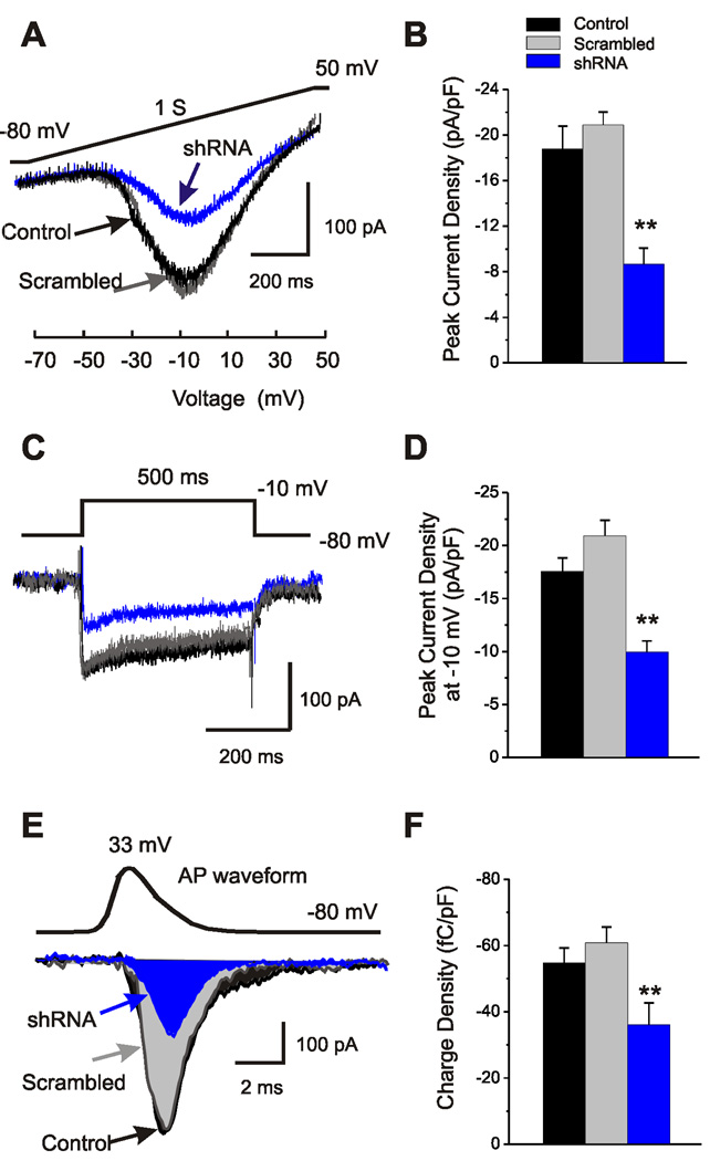
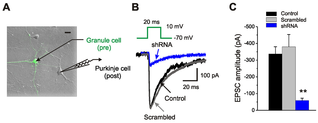
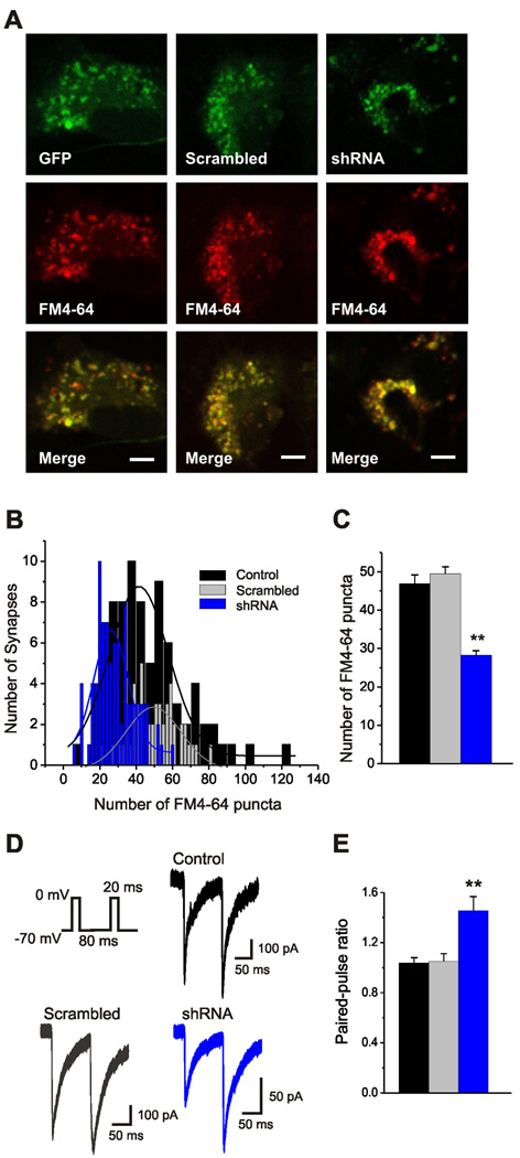
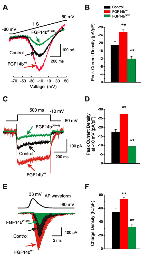
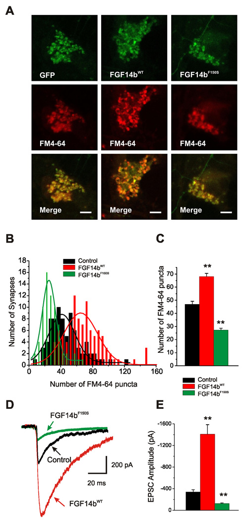
Fibroblast growth factor homologous factors (FHFs) are not growth factors, but instead bind to voltage-gated Na+ channels (NaV) and regulate their function. Mutations in FGF14, an FHF that is the locus for spinocerebellar ataxia 27 (SCA27), are believed to be pathogenic because of a dominant-negative reduction of NaV currents in cerebellar granule cells. Here, we demonstrate that FGF14 also regulates members of the presynaptic CaV2 Ca2+ channel family. Knockdown of FGF14 in granule cells reduced Ca2+ currents and diminished vesicular recycling, a marker for presynaptic Ca2+ influx. As a consequence, excitatory postsynaptic currents (EPSCs) at the granule cell to Purkinje cell synapse were markedly diminished. Expression of the SCA27-causing FGF14 mutant in granule cells exerted a dominant-negative reduction in Ca2+ currents, vesicular recycling, and the resultant EPSCs in Purkinje cells. Thus, FHFs are multimodal, regulating several discrete neuronal signaling events. SCA27 most likely results at least in part from dysregulation of Ca2+ channel function.


Figures

Figure 1
Figure 1. Endogenous FGF14 regulates Ca2+ channel currents in granule cells
A. Example Ca2+ channel current traces (using Ba2+ as the charge carrier) recorded from a cerebellar granule cell transfected with GFP-control (black), scrambled control shRNA (gray), or FGF14 shRNA (blue). The currents were evoked by a ramp protocol from a holding potential of −80 mV to 50 mV in 1 sec. B. Summary data from granule cells expressing GFP control (n=17), scrambled control shRNA (n=10), or FGF14 shRNA (n=12). C. Example Ca2+ channel current traces recorded from a cerebellar granule cell transfected with GFP-control (black), scrambled control shRNA (gray), or FGF14 shRNA (blue). The currents were evoked by a step protocol from a holding potential of −80 mV to −10 mV in 500 ms. D. Summary data from granule cells expressing GFP control (n=22), scrambled control shRNA (n=11), or FGF14 shRNA (n=10). E. Representative Cd2+-sensitive Ba2+ currents evoked by a single action potential waveform (APW, top) command recorded from granule cells transfected with GFP control (black), scrambled control shRNA (gray), or FGF14 shRNA (blue). The integrated current (Q) is colored with black, gray, or blue. F. Summary data of the integrated current (Q) normalized to each cell capacitance. Summary results were obtained from granule cells expressing GFP control (n=25), scrambled control shRNA (n=11), or FGF14 shRNA (n=10). **p<0.01 versus Control.
Figure 2
Figure 2. FGF14 modulates CaV2.1 and CaV2.2 channels
A. Example Ca2+ channel current traces (using Ba2+ as the charge carrier) recorded from HEK293T cells in which CaV2.1 channels were co-expressed with GFP control (black) or FGF14WT (red). The currents were elicited by depolarizing pulses of 300 ms from −80 mV to +60 mV (in 10-mV increments). B. Current-voltage relationships (normalized to cell capacitance) for cells in which CaV2.1 was co-transfected with GFP control (black) or FGF14WT (red). C. Representative Cd2+-sensitive Ba2+ currents evoked by a single action potential waveform (APW, top) command from HEK293T cells in which CaV2.1 channels were co-transfected with GFP control (black) or FGF14WT (red). The integrated current (Q) is colored with black or red. D. Summary data of the integrated current (Q) normalized to cell capacitance for each cell expressing GFP (n=9) or FGF14WT (n=9). E-G. Current-voltage relationships (normalized to cell capacitance) for cells in which CaV2.2 (E), CaV1.2 (F) or CaV2.3 (G) were co-transfected with GFP (black) or FGF14WT (red). The current amplitude values were divided by the capacitance of each cell to obtain current density (pA/pF). *p<0.05, **p<0.01 versus Control.
Figure 3
Figure 3. FGF14 increases Ca2+ gating charge in CaV2.1 channels
A. Example Ca2+ channel current traces (using Ba2+ as the charge carrier) recorded from HEK293T cells in which CaV2.1 channels were co-expressed with GFP control (black) or FGF14WT (red). The example traces were elicited by a 20 ms step from the holding potential (− 80 mV) to the reversal potential. Magnification of the integrated gating currents is shown below. B. Summary data of the integrated current (Q) normalized to each cell capacitance for cells expressing CaV2.1 or GFP control (n=14) or FGF14WT (n=13). **p<0.01 versus Control.
Figure 4
Figure 4. Endogenous FGF14 regulates synaptic transmission at the granule cell to Purkinje cell synapse
A. Evoked EPSCs in an untransfected Purkinje cell were elicited by a 20 ms depolarization of a transfected granule cell from a holding potential of −70 mV to −10 mV. B. Representative EPSC traces recorded from Purkinje cells in which the presynaptic granule cell was transfected with GFP control (black), scrambled control shRNA (gray), or FGF14 shRNA (blue). C. Averaged amplitude of unitary Purkinje cell EPSCs when the presynaptic granule cells expressed GFP control (n=14), scrambled control shRNA (n=25), or FGF14 shRNA (n=20). **p<0.01 versus Control.
Figure 5
Figure 5. Endogenous FGF14 regulates vesicular recycling and short-term synaptic plasticity
A. Confocal images from cultured cerebellar neurons expressing GFP (left), scrambled control shRNA (middle), or FGF14 shRNA (right) that were loaded with FM4-64 by a 90 sec depolarization using 90 mM KCl. Scale bar, 5 µm. B. The distribution of FM4-64 puncta per synapse within a 45×45 µm2 region of interest (ROI) in neurons transfected with control GFP (black, n=81), scrambled control shRNA (gray, n=49), or FGF14 shRNA (blue, n=87). The data for each group were fit to a Gaussian distribution. C. Average puncta per 45×45 µm2 ROI for neurons over-expressing control GFP, scrambled control shRNA, or FGF14 shRNA. D. Representative EPSCs evoked by a paired-pulse protocol (indicated in inset; 80 ms interstimulus interval) when the presynaptic granule cells expressed GFP control (black), scrambled control shRNA (gray), or FGF14 shRNA (blue). E. Averaged paired-pulse ratio (amplitude of the second EPSC divided by the amplitude of the first EPSC). **p<0.01 versus Control.
Figure 6
Figure 6. The SCA27 FGF14 mutant reduces granule Ca2+ currents
A. Example Ca2+ current traces (using Ba2+ as the charge carrier) recorded from a cerebellar granule cell transfected with GFP-control (black), FGF14WT (red), or FGF14bF150S (green). The currents were evoked by a ramp protocol from a holding potential of −80 mV to 50 mV in 1 sec. B. Summary data from granule cells expressing GFP control (n=17), FGF14WT (n=25), or FGF14bF150S (n=16). C. Example Ca2+ channel current traces recorded from a cerebellar granule cell transfected with GFP-control (black), FGF14WT (red), or FGF14bF150S (green). The currents were evoked by a step protocol from a holding potential of −80 mV to −10 mV in 500 ms. D. Summary data from granule cells expressing GFP control (n=22), FGF14WT (n=37), or FGF14bF150S (n=13). E. Representative Cd2+-sensitive Ba2+ currents evoked by a single action potential waveform (APW, top) command recorded from granule cells transfected with GFP control (black), scrambled control shRNA (gray), or FGF14 shRNA (blue). The integrated current (Q) is colored with black, red, or green. F. Summary data of the integrated current (Q) normalized to each cell capacitance. Summary results were obtained from granule cells expressing GFP control (n=25), FGF14WT (n=28), or FGF14bF150S (n=16). **p<0.01 versus Control.
Figure 7
Figure 7. The SCA27 FGF14 mutant in granule cells reduces presynaptic Ca2+ influx and EPSCs at a granule cell to Purkinje cell synapse
A. Confocal images from cultured cerebellar neurons expressing GFP (left), FGF14WT (middle), or FGF14bF150S (right) that were loaded with FM4-64 by a 90 sec depolarization using 90 mM KCl. Scale bar, 5 µm. B. The distribution of FM4-64 puncta per synapse within a 45×45 µm2 ROI in neurons transfected with GFP (black, n=81), FGF14WT (red, n=98), or FGF14bF150S (green, n=69). The data for each group were fit to a Gaussian distribution. C. Averaged puncta number per 45×45 µm2 ROI for neurons over-expressing control GFP (black, n=81), FGF14WT (red, n=98), or FGF14bF150S (green, n=69). D. Representative EPSC traces recorded from Purkinje cells evoked by a 20 ms depolarization of the granule cell from a holding potential of −70 mV to −10 mV in which the presynaptic granule cell was transfected with GFP (black), FGF14WT (red), or FGF14bF150S (green). E. Averaged amplitude of Purkinje cell EPSCs when the presynaptic granule cell expressed GFP control (n=14), FGF14WT (n=19), or FGF14bF150S (n=10). **p<0.01 versus Control.

Similar articles

Cited by

References

    1. Bannister RA, Melliti K, Adams BA. Differential modulation of CaV2.3 Ca2+ channels by Galphaq/11-coupled muscarinic receptors. Mol Pharmacol. 2004;65:381–388. - PubMed
    1. Borst JG, Helmchen F, Sakmann B. Pre- and postsynaptic whole-cell recordings in the medial nucleus of the trapezoid body of the rat. J Physiology. 1995;489(Pt 3):825–840. - PMC - PubMed
    1. Dalski A, Atici J, Kreuz FR, Hellenbroich Y, Schwinger E, Zuhlke C. Mutation analysis in the fibroblast growth factor 14 gene: frameshift mutation and polymorphisms in patients with inherited ataxias. Eur J Hum Genet. 2005;13:118–120. - PubMed
    1. Evans GJ, Cousin MA. Simultaneous monitoring of three key neuronal functions in primary neuronal cultures. J Neurosci Methods. 2007;160:197–205. - PMC - PubMed
    1. Fletcher CF, Lutz CM, O'Sullivan TN, Shaughnessy JD, Jr., Hawkes R, Frankel WN, Copeland NG, Jenkins NA. Absence epilepsy in tottering mutant mice is associated with calcium channel defects. Cell. 1996;87:607–617. - PubMed

Publication types

Substances