. 2013;9(6):e1003480.
doi: 10.1371/journal.ppat.1003480. Epub 2013 Jun 27.

Negative regulation of TLR inflammatory signaling by the SUMO-deconjugating enzyme SENP6

Affiliations

Negative regulation of TLR inflammatory signaling by the SUMO-deconjugating enzyme SENP6

Xing Liu et al. PLoS Pathog. 2013.

Abstract

The signaling of Toll-like receptors (TLRs) induces host defense against microbial invasion. Protein posttranslational modifications dynamically shape the strength and duration of the signaling pathways. It is intriguing to explore whether de-SUMOylation could modulate the TLR signaling. Here we identified SUMO-specific protease 6 (SENP6) as an intrinsic attenuator of the TLR-triggered inflammation. Depletion of SENP6 significantly potentiated the NF-κB-mediated induction of the proinflammatory genes. Consistently, SENP6-knockdown mice were more susceptible to endotoxin-induced sepsis. Mechanistically, the small ubiquitin-like modifier 2/3 (SUMO-2/3) is conjugated onto the Lysine residue 277 of NF-κB essential modifier (NEMO/IKKγ), and this impairs the deubiquitinase CYLD to bind NEMO, thus strengthening the inhibitor of κB kinase (IKK) activation. SENP6 reverses this process by catalyzing the de-SUMOylation of NEMO. Our study highlights the essential function of the SENP family in dampening TLR signaling and inflammation.


Conflict of interest statement

The authors have declared that no competing interests exist.

Figures
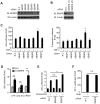
Figure 1
Figure 1. Identification of SENP6 as a new regulator of TLR signaling.
A, Immunoblot analysis of the lysates from 293T cells transfected with the indicated siRNA. B, Immunoblot analysis of the lysates from MEF cells transfected with the indicated siRNA. C, SENP6 potentiates TNF-α-induced NF-κB activation. 5×κB-luciferase (left panel) or E-selectin-luciferase (right panel) and pTK-Renilla reporters were transfected into HEK293T cells together with the indicated siRNA. Forty-eight hours after transfection, cells were stimulated with TNF-α (10 ng/ml) for eight hours before luciferase reporter assays were performed. D, SENP6 potentiates TLR-triggered NF-κB activation. The nonspecific control (N.C.) or SENP6 siRNA were transfected into RAW264.7 cells with 5×κB-luciferase and pTK-Renilla reporters. Forty-eight hours after transfection, cells were stimulated with LPS (1 µg/mL), poly (I:C) (50 µg/ml), R837 (10 µg/ml) for 8 h before luciferase assays were performed. E, SENP6 has no effect on the NF-κB activation in response to DNA damage. HEK293T cells were transfected with 5×κB-luciferase and pTK-Renilla reporters together with the nonspecific control (N.C.), SENP6 siRNA or SENP2 siRNA. Forty-eight hours after transfection, cells were stimulated with doxorubicin (500 ng/ml) for eight hours before luciferase reporter assays were performed. F, SENP6 has no influence on the activation of AP-1. HEK293T cells were transfected with AP-1-luc and pTK-Renilla reporters together with the nonspecific control (N.C.) or SENP6 siRNA. Forty-eight hours after transfection, cells were stimulated with PMA (20 ng/ml) for eight hours before luciferase reporter assays were performed. Data from C-F are presented as means ±S.D. from three independent experiments. n.s., not significant; *, P<0.05; **, P<0.01.
Figure 2
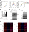
Figure 2. SENP6 negatively regulates TLR4-induced NF-κB signaling.
A, Loss of SENP6 promotes LPS-induced NF-κB-responsive genes. MEF cells transfected with the indicated siRNAs were incubated with LPS (1 µg/mL) for the indicated time periods. Induction of IL-6, TNF-α, and ICAM-1 mRNA was measured by quantitative PCR. B, Catalytic activity of SENP6 is required for its action in TLR signaling. MEF cells were transfected with the nonspecific control (N.C.) or SENP6 siRNA and then rescued with the indicated siRNA-resistant SENP6 constructs. After LPS (1 µg/mL) stimulation, induction of IL-6 mRNA was measured by quantitative PCR. C, SENP6 has no effects on LPS-induced auto-ubiquitination of TRAF6. Immunoprecipitation of endogenous TRAF6 from lysates of LPS-treated MEF cells transfected with the nonspecific control (N.C.) or SENP6 siRNA, followed by immunoblot analysis with the indicated antibodies. D, SENP6 knockdown enhances the phosphorylation of IκBα and NF-κB p65. Immunoblot analysis of phosphorylated (p-) IκBα, NF-κB p65 and JNK in WT and SENP6 knockdown MEFs stimulated with LPS (1 µg/mL). E, RNAi of SENP6 accelerates the nuclear translocation of p65. Immunofluorescence microscopy of NF-κB p65 (red) in WT and SENP6 knockdown MEF cells stimulated with LPS (1 µg/mL); nuclei were counterstained with DAPI (blue). Scale bar, 50 µm. Original magnification, ×63. Data in A–B are presented as means ± S.D. from three independent experiments. *, P<0.05; **, P<0.01.
Figure 3
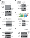
Figure 3. NEMO is modified on K277 by SUMO-2/3.
A, NEMO, rather than IKKα or IKKβ, is modified by SUMO-3 in an overexpression system. Flag-IKKα, IKKβ or NEMO were individually transfected into HEK293T cells along with HA-SUMO-3. Cell lysates were subjected to denaturing immunoprecipitation with Flag antibody and then immunoblotted with the indicated antibodies. B, Either SUMO-2 or SUMO-3 could be attached to NEMO. Flag-NEMO was co-transfected into HEK293T along with HA-SUMO-2 or HA-SUMO-3. Cell lysates were subjected to denaturing immunoprecipitation with Flag antibody and then immunoblotted with the indicated antibodies. C, The SUMOylation of NEMO is independent of its IKK-binding domain. Expression vectors for Flag-NEMO and its mutants as shown in upper panel were transfected into 293T cells along with HA-SUMO-3 (lower panel). Cell lysates were subjected to immunoprecipitation with Flag antibody and then immunoblotted with the indicated antibodies. KBD, IKK-binding domain; CC1 and CC2, coiled-coil domain 1 and 2; LZ, leucine-zipper motif; ZF, zinc-finger domain. D, Endogenous NEMO is covalently modified by endogenous SUMO-2/3. After mock or LPS (1 µg/ml) stimulation, lysates from RAW264.7 cells were immunoprecipitated with NEMO antibody or control IgG and then immunoblotted with the indicated antibodies. The intensity of the SUMOylated NEMO was quantified and normalized to that of the corresponding immunoprecipitated NEMO. The relative levels of SUMOylated NEMO are shown as fold change compared with the control. E, K277 was the major acceptor site on NEMO for SUMO-3. HEK293T cells were transfected with Flag-NEMO or its mutants along with HA-SUMO-3. Cell lysates were subjected to immunoprecipitation with Flag antibody and then immunoblotted with the indicated antibodies. F, K277 of NEMO is sufficient for the SUMO-3 modification. HEK293T cells were transfected with the indicated plasmids. Cell lysates were subjected to immunoprecipitation with Flag antibody and then immunoblotted with the indicated antibodies.
Figure 4
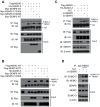
Figure 4. SENP6 catalyzes the de-SUMOylation on NEMO K277.
A, Overexpression of catalytic active SENP6 deconjugates SUMOylated NEMO. Flag-NEMO and HA-SUMO-3 were transfected into HEK293T cells along with Myc tagged SENP6, SENP6 mutants or SENP3, respectively. 36 hours post-transfection, cell lysates were immunoprecipitated with Flag antibody and then immunoblotted with the indicated antibodies. B, SENP6 removes SUMO-3 attached on NEMO K277. HEK293T cells were transfected with the indicated plasmids. 48 hours post-transfection, cell lysates were subjected to immunoprecipitation with Flag antibody and then immunoblotted with the indicated antibodies. C, SENP6 knockdown enhance SUMOylation of exogenous NEMO. HEK293T cells were transfected with the indicated siRNAs. Twenty-four hours later, Flag-NEMO and HA-SUMO-3 were transfected into the knockdown cells. Cell lysates were subjected to immunoprecipitation with Flag antibody and immunoblotted with the indicated antibodies. D, The level of modified endogenous NEMO by SUMO-2/3 is elevated upon SENP6 depletion. HEK293T cells were transfected with the indicated siRNAs. Forty-eight hours later, cell lysates were subjected to immunoprecipitation with NEMO antibody or control IgG and then immunoblotted with the indicated antibodies.
Figure 5
Figure 5. SENP6 selectively interacts with the SUMOylated NEMO.
A, SENP6 associates with the NEMO-SUMO-3 fusion protein. The indicated Flag tagged constructs were individually transfected into HEK293T cells along with Myc-SENP6. Then, cell lysates were subjected to immunoprecipitation with Flag antibody. The immunoprecipitates were immunoblotted with the indicated antibodies. B, The catalytic activity of SENP6 is not required for the association of SENP6 and NEMO-SUMO-3. HEK293T cells were transfected with Flag-NEMO-SUMO-3 along with Myc-SENP6 or SENP6 mutants. Cell lysates were subjected to immunoprecipitation with Flag antibody. The immunoprecipitates were immunoblotted with the indicated antibodies. C, Mapping the binding domain of SENP6 with NEMO-SUMO-3. Myc-SENP6 and its mutants, as shown in upper panel, were individually transfected into HEK293T cells along with Flag-NEMO-SUMO-3. The cell lysates were immunoprecipitated with Flag antibody and then immunoblotted with the indicated antibodies (lower panel). A nonspecific band is indicated by an asterisk. D, SENP6 interacts with the SUMOylated NEMO. The flow chart of the GST-pulldown analysis (right panel). HEK293T cells were transfected with the indicated plasmids. Firstly, the cell extracts were purified by Ni-NTA agarose. Then the Ni-NTA-bound proteins were eluted by imidazole (∼300 mM), and the elute fractions were subjected to GST-PD analysis. The SUMOylated Flag-NEMO was indicated on the right (left panel). E, Flag-NEMO and its mutants were individually transfected into HEK293T cells along with His-SUMO-3, and then the cell lysates were incubated with the extracts from the HEK293T cells transfected with Myc tagged SENP6. The mixture was subjected to immunoprecipitation with Flag antibody and then immunoblotted with the indicated antibodies.
Figure 6
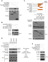
Figure 6. SENP6 attenuates the action of NEMO in TLR-triggered NF-κB signaling.
A, The flow charts of the GST-pulldown analysis. HEK293T cells were transfected with the indicated plasmids. When HA-NEMO (Sample 1) or HA-NEMO-SUMO-3 (Sample 2) was expressed alone, the cell extracts were subjected directly to GST-PD analysis. When HA-NEMO was co-expressed with His-SUMO-3, the cell extracts were firstly purified by Ni-NTA agarose. Then the Ni-NTA-bound His-SUMO-3 modified proteins were eluted by imidazole (∼300 mM), and the elute fractions were subjected to GST-PD analysis (Sample 3). B, HEK293T cells were transfected with HA tagged NEMO or NEMO-SUMO-3, and then equal amounts of cell lysates were subjected to GST-PD as indicated according to the experimental design described in A. C, HEK293T cells were transfected with HA-NEMO together with His-SUMO-3. The His-SUMO-3 modified proteins purified from the cell extracts were analyzed by GST-PD analysis as indicated according to the experimental design described in A. The SUMOylated and unmodified HA-NEMO were indicated on the right. D, SENP6 depletion abrogates the endogenous interaction between NEMO and CYLD. After mock or LPS (1 µg/ml) stimulation, lysates from MEF cells were immunoprecipitated with anti-NEMO antibody or anti-IgG and then immunoblotted with an anti-CYLD antibody. The intensity of the CYLD bound to NEMO was quantified and normalized to that of the corresponding immunoprecipitated NEMO. The relative levels of co-immunoprecipitated CYLD are shown as fold change compared with the control. E, SENP6 inhibits LPS-induced attachment of polyubiquitin chains onto NEMO. MEF cells were transfected with the nonspecific control (N.C.) or SENP6 siRNA and then treated with LPS (1 µg/mL). Cell lysates were immunoprecipitated with anti-NEMO and analyzed by immunoblotting using anti-ubiquitin (Ub). F, Modification of NEMO by SUMO-3 is required for the activation of NF-κB. RAW264.7 cells were transfected with the nonspecific control (N.C.) or NEMO siRNA together with 5×κB-luciferase and pTK-Renilla reporters, as well as the indicated siRNA-resistant NEMO constructs. After LPS (1 µg/ml) stimulation, cell lysates were prepared for luciferase assay. Data from F are presented as means ± S.D. from three independent experiments. *, P<0.05; **, P<0.01.
Figure 7
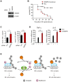
Figure 7. Mice deficient in SENP6 are more susceptible to LPS induced endotoxic shock.
A, Immunoblot analysis of SENP6 in lysates of kupffer cells from mice at forty-eight hours after transfection with SENP6 or nonspecific (N.C.) siRNA. B, Survival of mice (n = 10 per group) transfected with SENP6 or control siRNA and forty-eight hours later injected intraperitoneally with LPS (25 mg/kg). C, ELISA of TNF-α and IL-6 in serum from mice (n = 8 per group) transfected with SENP6 or control siRNA and seventy-two hours later injected intraperitoneally with LPS (25 mg/kg), assessed two hours after LPS injection. D, Quantitative PCR of relative TNF-α and IL-6 mRNA in livers from mice (n = 7 per group) transfected with SENP6 or control siRNA and seventy-two hours later injected intraperitoneally with LPS (25 mg/kg), assessed two hours after LPS injection. Data from C-D are presented as means ±S.D. from three independent experiments. *, P<0.05; **, P<0.01. E, A schematic model of the SENP6 function in TLR signaling. The deubiquitinase CYLD acts as a negative regulator of TLR signaling by interacting with NEMO and cleaving polyubiquitin chains. SUMO-2/3 could be conjugated onto NEMO, which prevents CYLD from binding to NEMO, thus strengthening the IKK activation. SENP6 reverses this process by catalyzing the de-SUMOylation of NEMO.

Similar articles

Cited by

References

    1. Kawai T, Akira S (2010) The role of pattern-recognition receptors in innate immunity: update on Toll-like receptors. Nat Immunol 11: 373–384. - PubMed
    1. Medzhitov R (2007) Recognition of microorganisms and activation of the immune response. Nature 449: 819–826. - PubMed
    1. Cook DN, Pisetsky DS, Schwartz DA (2004) Toll-like receptors in the pathogenesis of human disease. Nat Immunol 5: 975–979. - PubMed
    1. Beutler B (2004) Inferences, questions and possibilities in Toll-like receptor signalling. Nature 430: 257–263. - PubMed
    1. Akira S, Takeda K (2004) Toll-like receptor signalling. Nat Rev Immunol 4: 499–511. - PubMed

Publication types

MeSH terms

Grants and funding

This work was supported by grants from National Natural Science Foundation of China (31030021, 81161120542) and Ministry of Science and Technology of China (2012CB910200, 2011CB910900, 2010CB529703). XL was supported by China Postdoctoral Science Foundation. The funders had no role in study design, data collection and analysis, decision to publish, or preparation of the manuscript.