. 2013;9(6):e1003446.
doi: 10.1371/journal.ppat.1003446. Epub 2013 Jun 27.

Bruton's Tyrosine Kinase (BTK) and Vav1 contribute to Dectin1-dependent phagocytosis of Candida albicans in macrophages

Affiliations

Bruton's Tyrosine Kinase (BTK) and Vav1 contribute to Dectin1-dependent phagocytosis of Candida albicans in macrophages

Karin Strijbis et al. PLoS Pathog. 2013.

Abstract

Phagocytosis of the opportunistic fungal pathogen Candida albicans by cells of the innate immune system is vital to prevent infection. Dectin-1 is the major phagocytic receptor involved in anti-fungal immunity. We identify two new interacting proteins of Dectin-1 in macrophages, Bruton's Tyrosine Kinase (BTK) and Vav1. BTK and Vav1 are recruited to phagocytic cups containing C. albicans yeasts or hyphae but are absent from mature phagosomes. BTK and Vav1 localize to cuff regions surrounding the hyphae, while Dectin-1 lines the full length of the phagosome. BTK and Vav1 colocalize with the lipid PI(3,4,5)P3 and F-actin at the phagocytic cup, but not with diacylglycerol (DAG) which marks more mature phagosomal membranes. Using a selective BTK inhibitor, we show that BTK contributes to DAG synthesis at the phagocytic cup and the subsequent recruitment of PKCε. BTK- or Vav1-deficient peritoneal macrophages display a defect in both zymosan and C. albicans phagocytosis. Bone marrow-derived macrophages that lack BTK or Vav1 show reduced uptake of C. albicans, comparable to Dectin1-deficient cells. BTK- or Vav1-deficient mice are more susceptible to systemic C. albicans infection than wild type mice. This work identifies an important role for BTK and Vav1 in immune responses against C. albicans.


Conflict of interest statement

The authors have declared that no competing interests exist.

Figures

Figure 1
Figure 1. BTK and Vav1 interact with Dectin-1 during C. albicans phagocytosis by macrophages.
(A): Morphology and β-glucan exposure of C. albicans expressing blue fluorescent protein (Candida-BFP) at indicated time points after incubation in DMEM with 10% IFS. Candida-BFP was stained with fluorescent carbohydrate recognition domain of Dectin-1 (Dectin1-CRD-Alexa647) that binds β-glucan. Arrows indicate increases staining at bud scars. (B): Immunoblotting experiment showing expression of different proteins in RAW-Dectin1 cells during co-incubation with live C. albicans for the indicated time points. Phagocytosis of C. albicans occurs throughout the time course, but the morphology of the ingested particles changes over time. (C): Co-incubation of RAW-Dectin1 with C. albicans followed by co-immunoprecipitation with anti-BTK or anti-Vav1 antibody and immunoblotting with anti-HA to detect Dectin-1. BTK/Dectin-1 and Vav1/Dectin-1 complexes were detected at different time points during the co-incubation. (D): Quantification of BTK/Dectin-1 and Vav1/Dectin-1 complexes showing strongest interactions at the 90- and 180-minute time points, respectively. Means +/− SD of three independent experiments are shown.
Figure 2
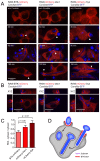
Figure 2. Localization of BTK-mCherry and mCherry-Vav1 to the Candida-containing phagocytic cup.
(A): Confocal images showing localization of BTK-mCherry, mCherry-Vav1 and mCherry-Syk in RAW-Dectin1 macrophages incubated with Candida-BFP. Images were taken without Candida-BFP or after 30 minutes, 90 minutes and 180 minutes of co-incubation with Candida-BFP to study phagocytosis of yeast, hyphae, and very long hyphae, respectively. White arrows indicate areas of mCherry recruitment to the Candida-containing phagocytic cup, visible during ingestion of C. albicans yeast (30 minutes) and C. albicans hyphae (90 and 180 minutes). (B): XYZ images of C. albicans phagocytosis by the BTK-mCherry, mCherry-Vav1 and mCherry-Syk cell lines. Cuff regions of protein recruitment are circular bands around C. albicans hyphae that are being ingested. (C): Quantitation of BTK-mCherry, mCherry-Vav1 and mCherry-Syk recruitment to the phagocytic cup after 90 minutes of coincubation with Candida-BFP. (D): Model showing localization of BTK and Vav1 to the phagocytic cup but not to mature phagosomes during phagocytosis of C. albicans yeast and hyphae by macrophages. Representative micrographs and means +/− SD of 3 independent experiments are shown. For statistical analysis, all data were analyzed by unpaired t test.
Figure 3
Figure 3. Localization of Dectin-1 during phagocytosis of C. albicans hyphae.
Confocal images showing localization of Dectin-1 during phagocytosis of C. albicans hyphae by the BTK-mCherry, mCherry-Vav1 and mCherry-Syk RAW macrophage cell lines. Dectin1-HA was visualized by anti-HA immunofluorescence.
Figure 4
Figure 4. Localization of phospholipids and PKC family proteins during C. albicans phagocytosis.
(A): PH-Akt-RFP and C1-PKCδ-GFP biosensors showing localization of PI(3,4,5)P3/PI(3,4)P2 and DAG, respectively, without challenge or after 30 or 90 minutes of co-incubation with Candida-BFP. White arrows indicate areas of PI(3,4,5)P3/PI(3,4)P2 and DAG co-localization, while red and green arrows indicate areas of speciation. (B): Quantitation of PI(3,4,5)P3/PI(3,4)P2- and DAG-positive phagosomes after 30 minutes of coincubation with Candida-BFP. (C): Model showing localization of PI(3,4,5)P3/PI(3,4)P2 and DAG during engagement and internalization of C. albicans yeast and hyphae by macrophages. (D): Localization of GFP-tagged PKCα, PKCβ, PKCδ, PKCε and PKCζ after 30 or 90 minutes of Candida-BFP phagocytosis. (E): Quantitation of PKCα-GFP, PKCβ-GFP, PKCδ-GFP, PKCε-GFP and PKCζ-GFP recruitment to the phagocytic cup after 90 minutes of coincubation with Candida-BFP. Representative micrographs and means +/− SD of 3 independent experiments are shown. For statistical analysis, all data were analyzed by unpaired t test.
Figure 5
Figure 5. BTK-mCherry and mCherry-Vav1 colocalize with PI(3,4,5)P3 but not with DAG.
(A): Colocalization of BTK-mCherry and PH-BTK-GFP that binds to PI(3,4,5)P3 at 30 and 90 minutes of coincubation with Candida-BFP showing phagocytosis of yeast and hyphae, respectively. (B): Colocalization of mCherry-Vav1 and PH-BTK-GFP at 30 and 90 minutes of coincubation with Candida-BFP. (C): Localization of mCherry-Syk and PH-BTK-GFP at 30 and 90 minutes of coincubation with Candida-BFP. (D): Localization of BTK-mCherry and C1-PKCδ-GFP at 30 and 90 minutes of coincubation with Candida-BFP. (E): Localization of mCherry-Vav1 and C1-PKCδ-GFP at 30 and 90 minutes of coincubation with Candida-BFP. (F): Localization of mCherry-Syk and C1-PKCδ-GFP at 30 and 90 minutes of coincubation with Candida-BFP. White arrows indicate areas of co-localization, while red and green arrows indicate areas of speciation. Experiments were performed at least three times, representative micrographs are shown.
Figure 6
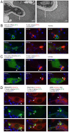
Figure 6. BTK, Vav1 and Syk colocalize with F-actin and PI(3,4,5)P3.
(A): Electron microscopy of C. albicans phagocytosis by RAW-Dectin1 macrophages. Cells were fixed to visualize polymerized actin. (B): Localization of LifeAct-RFP detecting F-actin and C1-PKCδ-GFP that binds to DAG after 30 and 90 minutes of coincubation with Candida-BFP. (C): Colocalization of LifeAct-RFP detecting F-actin and PH-BTK-GFP that binds to PI(3,4,5)P3 after 30 and 90 minutes of coincubation with Candida-BFP. (D): Localization of BTK-mCherry, mCherry-Vav1 and Syk-mCherry with LifeAct-GFP after 90 minutes of coincubation with Candida-BFP. White arrows indicate areas of co-localization, while red and green arrows indicate areas of speciation. Experiments were performed multiple times, representative micrographs are shown.
Figure 7
Figure 7. BTK is involved in DAG production at the phagocytic cup.
(A): RAW-Dectin1 macrophages were pre-incubated with the indicated concentrations of BTK inhibitor PCI-32765 followed by coincubation with Candida-BFP (MOI 10) for 1 hour. Graphs represent number of internalized Candida-BFP per macrophage as determined by microscopy. (B): DAG measurements in RAW-Dectin1 macrophages preicubated with the indicated inhibitors and in the presence or absense of C. albicans. Thin layer chromatography was performed to visualize the phosphatidic acid (PA) product of the DAG kinase assay. (C): Quantification of PA signal from three independent DAG kinase experiments. (D): Confocal images of C1-PKCδ-GFP (DAG) and PKCε-GFP distribution in RAW-Dectin1 macrophages pre-incubated with 0.5 µM BTK inhibitor PCI-32765. (E): Quantification of C1-PKCδ-GFP and PKCε-GFP recruitment to phagosomes in absence and presence of 0.5 µM BTK inhibitor. All graphs display means +/− SD of three independent experiments. (F): Schematic showing localization of PI(3,4,5)P3, BTK, Vav1, DAG and PKC family proteins during engagement and internalization of C. albicans yeast and hyphae by macrophages. (G): Model summarizing this studies findings. BTK, Vav1 and Syk interact with Dectin-1 during phagocytosis of C. albicans (left). Phosphatidylinositol 4,5-biphosphate (PI(4,5)P2) can be converted to phosphatidylinositol 3,4,5-triphosphate (PI(3,4,5)P3) by PI3K or to diacylglycerol (DAG) by phospholipase C γ (PLCγ). Specialized PI(3,4,5)P3- and DAG-rich phagosomal membranes can be distinguished during C. albicans phagocytosis. Bruton's Tyrosine Kinase (BTK) and Vav1 localize to PI(3,4,5)P3-rich membrane regions and colocalize with F-actin. Vav1 might play an active role in actin rearrangements at the phagocytic cup through activation of small GTPases Rac1, Cdc42 and/or Rho1. BTK is involved in the production of DAG at the phagocytic cup, possibly through the activation of PLCγ. Protein Kinase C (PKC) family proteins localize to DAG-rich membranes. For statistical analysis, all data were analyzed by unpaired t test.
Figure 8
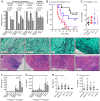
Figure 8. Phenotypic analysis of BTK- and Vav1-deficient macrophages and mice.
(A): Peritoneal macrophages or bone marrow-derived macrophages (BMDM) from wild type, dectin1−/−, btk−/− or vav1−/− mice were incubated with zymosan-Alexa647 or live Candida-BFP for 30 minutes or 1 hour at an MOI of 10 and the number of internalized Candida-BFP was determined by microscopy. Graphs represent means and standard deviations of experiments with three different mice. (B): The contribution of BTK and Vav1 to overall immune responses to C. albicans was determined using the model for systemic candidiasis. Tail vein injection of wild type, dectin1−/−, btk−/− or vav1−/− mice were performed with 0.5×10⁴ colony forming units (CFU) of C. albicans and disease was monitored over time. (C): C. albicans CFU in kidneys of indicated mice at final stage of disease, means +/− SD are indicated. (D): GMS staining of kidney histology slides of wild type, dectin1−/−, btk−/− or vav1−/− mice, at final stage of disease showing extensive fungal invasion of tissues. (E): H&E staining of kidney histology slides of wild type, dectin1−/−, btk−/− or vav1−/− mice, at final stage of disease showing extensive immune cell invasion of tissues. Representative images are shown. TNFα (F) and IL-6 (G) levels in supernatant of mouse peritoneal macrophages 12 hours after incubation without or with C. albicans. Graphs represent means and standard deviations of experiments with three different mice. TNFα (H) and IL-6 (I) levels in kidney lysates of mice infected with 5×10⁴ CFU C. albicans at 11 days after infection. Each dot represents one mouse; means and standard deviations are indicated. Values did not differ significantly. For statistical analysis, all data were analyzed by unpaired t test.

Similar articles

Cited by

References

    1. Cheng SC, Joosten LA, Kullberg BJ, Netea MG (2012) Interplay between Candida albicans and the mammalian innate host defense. Infection and immunity 80: 1304–1313. - PMC - PubMed
    1. Iliev ID, Funari VA, Taylor KD, Nguyen Q, Reyes CN, et al. (2012) Interactions between commensal fungi and the C-type lectin receptor Dectin-1 influence colitis. Science 336: 1314–1317. - PMC - PubMed
    1. Herre J, Marshall AS, Caron E, Edwards AD, Williams DL, et al. (2004) Dectin-1 uses novel mechanisms for yeast phagocytosis in macrophages. Blood 104: 4038–4045. - PubMed
    1. Rogers NC, Slack EC, Edwards AD, Nolte MA, Schulz O, et al. (2005) Syk-dependent cytokine induction by Dectin-1 reveals a novel pattern recognition pathway for C type lectins. Immunity 22: 507–517. - PubMed
    1. Elsori DH, Yakubenko VP, Roome T, Thiagarajan PS, Bhattacharjee A, et al. (2011) Protein kinase Cdelta is a critical component of Dectin-1 signaling in primary human monocytes. Journal of leukocyte biology 90: 599–611. - PMC - PubMed

Publication types

MeSH terms