. 2013 Aug;140(15):3176-87.
doi: 10.1242/dev.092502. Epub 2013 Jul 3.

Tbx20 acts upstream of Wnt signaling to regulate endocardial cushion formation and valve remodeling during mouse cardiogenesis

Affiliations

Tbx20 acts upstream of Wnt signaling to regulate endocardial cushion formation and valve remodeling during mouse cardiogenesis

Xiaoqiang Cai et al. Development. 2013 Aug.

Abstract

Cardiac valves are essential to direct forward blood flow through the cardiac chambers efficiently. Congenital valvular defects are prevalent among newborns and can cause an immediate threat to survival as well as long-term morbidity. Valve leaflet formation is a rigorously programmed process consisting of endocardial epithelial-mesenchymal transformation (EMT), mesenchymal cell proliferation, valve elongation and remodeling. Currently, little is known about the coordination of the diverse signals that regulate endocardial cushion development and valve elongation. Here, we report that the T-box transcription factor Tbx20 is expressed in the developing endocardial cushions and valves throughout heart development. Ablation of Tbx20 in endocardial cells causes severe valve elongation defects and impaired cardiac function in mice. Our study reveals that endocardial Tbx20 is crucial for valve endocardial cell proliferation and extracellular matrix development, but is not required for initiation of EMT. Elimination of Tbx20 also causes aberrant Wnt/β-catenin signaling in the endocardial cushions. In addition, Tbx20 regulates Lef1, a key transcriptional mediator for Wnt/β-catenin signaling, in this developmental process. Our study suggests a model in which Tbx20 regulates the Wnt pathway to direct endocardial cushion maturation and valve elongation, and provides new insights into the etiology of valve defects in humans.

Keywords: Cardiac valve; Heart development; Mouse; Tbx20.


Figures
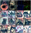
Fig. 1.
Fig. 1.
Dynamic Tbx20 expression in endocardial cushions and valves during mouse heart development. (A) Whole-mount RNA in situ hybridization to assess Tbx20 expression in E10.0 mouse embryos. (B) X-Gal staining of a Tbx20nlacZ embryo faithfully recapitulates endogenous Tbx20 expression in the heart (left arrow), neural tube (right arrow) and optic vesicle (arrowhead). (C-F) GFP from Tbx20H2BGFP and Nfatc1 immunostaining in the endocardium at multiple stages (arrows in D,F). Tbx20 expression in cushion mesenchymal cells (arrowheads in D,F). The boxed regions in C and E are shown at higher magnification in D and F. (G-N) X-Gal staining of E12.5 and E14.5 cryosectioned Tbx20nlacZ hearts. Tbx20nlacZ is present in the endocardial cushions (H, arrows in I,J) and valve leaflets (arrows in L-N). H and L are higher magnification images of the atrioventricular cushion region from G and K. (O-R) Tbx20nlacZ is present in the remodeled valve leaflets from neonatal stages through adulthood (arrows). P1, postnatal day 1. Ao, aorta; AVCu, atrioventricular cushion; PV, pulmonary valve; AoV, aortic valve; RA/LA, right/left atrium; RV/LV, right/left ventricle; TV, tricuspid valve; MV, mitral valve.
Fig. 2.
Fig. 2.
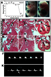
Tbx20 endocardial deletion causes embryonic lethality with cushion and valve developmental defects. (A) Tbx20 CKO embryos die by E16.5. Incidence of Tbx20 CKO embryos at each embryonic stage and the expected survival percentage (25%, dashed line). Numbers indicate total embryos genotyped. (B,C) Tbx20 CKO embryos exhibit peripheral hemorrhage over the body at E15.5. (D-I) Transverse sections of Tbx20 CKO mutant (E,G,I) and control (D,F,H) hearts at E14.5. The mutant valves, including the pulmonary valve (arrows in D,E), aortic valve (arrows in F,G), tricuspid valve and mitral valve (arrows in H,I), are shorter and blunt relative to those of controls. (J,K) Doppler in utero echocardiography. In the E14.5 control embryos (J), the waves above the baseline represent normal systolic transvalvular flow from left ventricle to aorta during systole. Tbx20 CKO mutant hearts (K) show regurgitant flow (arrow) across the aortic valve, which is indicative of valve incompetence.
Fig. 3.
Fig. 3.
Tbx20 endocardial deletion disrupts valve elongation and endocardial cell proliferation. (A-F) Tbx20 CKO mutant and control valves at E14.5. Arrows indicate pulmonary valve (A,B), aortic valve (C,D), tricuspid valve and mitral valves (E,F). Valve length is indicated by doubleheaded arrows. Dashed lines mark the junctions between valve base and cusp. (G) Relative valve length. (H,I) Proliferating cells in mitral valves detected by EdU staining (green) at E13.5. CD31 co-staining (red) marks valve endocardial cells. Arrowheads indicate proliferating endocardial cells. (J,K) Apoptotic cells (green) detected by TUNEL in control and mutant mitral valves at E13.5. Dashed lines in H-K indicate the junction between valve base and cusp. (L) Statistical comparison of valve endocardial and mesenchymal cell proliferation at E13.5. Decreased cell proliferation is observed in the valve endocardium but not the mesenchyme. *P<0.05, **P<0.01, versus control. n.s., not significant. Error bars indicate s.d.
Fig. 4.
Fig. 4.
Defective atrioventricular cushion formation in Tbx20 CKO embryos. (A-H) Transverse sections of Tbx20 CKO mutant and control hearts at E12.5. Arrows indicate endocardial cushions in the pulmonary valve (A,B) and aortic valve (C,D). G and H are higher magnification images of the atrioventricular cushion region (boxed) from E and F. (I) Relative cushion size was determined for pulmonary valve, aortic valve and atrioventricular cushion. The mutant atrioventricular cushions are smaller than in the control, whereas the pulmonary valve and aortic valves are normal in size at E12.5. (J-M) Proliferative cells are labeled by EdU staining (green) in the atrioventricular cushions at E11.5. CD31 co-staining (red) labels cushion endocardial cells. Arrowheads indicate proliferating endocardial cells. L and M are higher magnification images of the atrioventricular cushion region (boxed) from J and K. (N,O) Few apoptotic cells (green) are found in the atrioventricular cushion region of mutant and control embryos. MF20 antibody (red) labels myocardial cells. (P) At E11.5, mutant atrioventricular cushion endocardial cell proliferation is significantly slower than in the control, whereas mesenchymal cell proliferation is relatively normal. *P<0.05, versus control. n.s., not significant. Error bars indicate s.d.
Fig. 5.
Fig. 5.
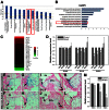
Tbx20 loss of function in endocardium disrupts Wnt/β-catenin signaling in developing valves. (A) Significant canonical pathways identified from genes differentially expressed in Tbx20 CKO versus control by ingenuity pathway analysis. The y-axis is the -log10 of the P-value for the enrichment of a pathway in the input gene list compared with genes in the whole genome. Red boxes highlight Wnt/β-catenin signaling pathway. The zigzag orange line indicates the enrichment ratio of the percentage of genes in a particular pathway over the percentage of pathway genes in the genome. (B) Significant Gene Ontology (GO) terms identified in the differentially expressed genes. x-axis is the -log10 of the P-value for the enrichment of a GO term in the input gene list compared with genes in the whole genome. (C) Heat map of Wnt signaling pathway genes. Red and green denotes upregulated and downregulated genes, respectively, in the Tbx20 CKO. (D) Relative expression of Wnt pathway genes at E10.5-12.5 determined by RT-qPCR. (E-L) X-Gal staining of TOPGAL indicator mice to detect active Wnt/β-catenin signaling at E11.5 (E-H) and E12.5 (I-L). The mutant tricuspid and mitral valves display fewer X-Gal-stained cells and fainter staining in the positive cells in both endocardium and adjacent mesenchyme (arrows in F,H,J,L versus E,G,I,K). G,H and K,L are higher magnification images of the tricuspid valve region (boxed) from E,F and I,J. (M) Relative expression of lacZ at different stages as determined by RT-qPCR. *P<0.05, versus control. Error bars indicate s.d.
Fig. 6.
Fig. 6.
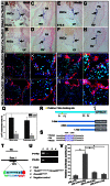
Misexpression of Wnt/β-catenin pathway genes in the valve endocardium of Tbx20 CKO hearts. (A-H) RNA in situ hybridization of Wnt4 and Wnt9b in the mitral (A,B,E,F) and pulmonary (C,D,G,H) valve endocardial cells in control (A,C,E,G) and Tbx20 CKO mutant (B,D,F,H). Arrows indicate valve endocardial cells. (I-P) Downregulated Lef1 (green) expression in mitral (I-L) and pulmonary (M-P) valve endocardial cells in the mutants (arrowheads in K,L,O,P). CD31 (red) co-staining marks endocardium. K,L and O,P are higher magnification images of the boxed regions from I,J and M,N. Nuclei are labeled by DAPI (blue). (Q) Comparison of the number of Lef1+ endocardial cells in mitral and pulmonary valves in control and mutant hearts at E12.5. *P<0.05, versus control. (R) The putative T-box binding site in the Lef1 promoter and luciferase reporter constructs containing 3.9 kb, 2.9 kb and 1.7 kb Lef1 promoter fragments (in the pGL3-TK vector). P1-P4 refer to the ChIP-PCR primers. (S) Alignment of Lef1 genomic sequences in human, rat and mouse. The putative T-box binding site is highlighted (red circle). (T) In Tbx20V5-Avi biotin tag knock-in mice, full-length Tbx20 cDNA is fused with V5 and Avi (V5-Tbx20-Avi-polyA) and the cassette is inserted into the start codon of the Tbx20 genomic locus. (U) ChIP-PCR on E12.5 heart tissues (from Tbx20V5-Avi/V5-Avi;Rosa26BirA/BirA doubly homozygous mice) using the primers shown in R demonstrates recruitment of Tbx20 to regions containing the T-box consensus site within the Lef1 promoter (P1+P2); ChIP-PCR with primers against a different region reveals no Tbx20 recruitment (P3+P4). No recruitment was found with heart tissues from Rosa26BirA/BirA mice (lane 3, control). Negative control, H2O as PCR template. (V) Luciferase reporter assays of the Tbx20 expression vector with various Lef1 promoter fragments. The 3.9 kb fragment containing the putative T-box binding site activates the luciferase reporter significantly, whereas the 2.9 kb and 1.7 kb fragments without the binding site do not. *P<0.01, paired t-test. Error bars indicate s.d. PA, pulmonary artery.
Fig. 7.
Fig. 7.
Altered expression of ECM genes in Tbx20 CKO heart. (A) Relative expression of ECM genes as determined by RT-qPCR on E12.5 hearts. Postn, Mmp13 and Acan are significantly downregulated in the Tbx20 CKO heart. *P<0.05, versus control. Error bars indicate s.d. (B-E) RNA in situ hybridization of Postn expression in atrioventricular (B,C) and pulmonary (D,E) cushion endocardial and mesenchymal cells at E12.5. Arrows indicate cushion endocardial cells. (F-I) Immunostaining of Mmp13 (F,G) and Acan (H,I) in atrioventricular cushion at E12.5. Arrows indicate cushion endocardial cells. (J-M) Alcian Blue staining of hyaluronic acid in atrioventricular and pulmonary valves at E14.5.

Similar articles

Cited by

References

    1. Akiyama H., Chaboissier M. C., Behringer R. R., Rowitch D. H., Schedl A., Epstein J. A., de Crombrugghe B. (2004). Essential role of Sox9 in the pathway that controls formation of cardiac valves and septa. Proc. Natl. Acad. Sci. USA 101, 6502–6507 - PMC - PubMed
    1. Alfieri C. M., Cheek J., Chakraborty S., Yutzey K. E. (2010). Wnt signaling in heart valve development and osteogenic gene induction. Dev. Biol. 338, 127–135 - PMC - PubMed
    1. Armstrong E. J., Bischoff J. (2004). Heart valve development: endothelial cell signaling and differentiation. Circ. Res. 95, 459–470 - PMC - PubMed
    1. Bancroft J. D., Gamble M. (2008). Theory and Practice of Histological Techniques. Philadelphia, PA: Churchill Livingstone Elsevier;
    1. Basson C. T., Bachinsky D. R., Lin R. C., Levi T., Elkins J. A., Soults J., Grayzel D., Kroumpouzou E., Traill T. A., Leblanc-Straceski J., et al. (1997). Mutations in human TBX5 cause limb and cardiac malformation in Holt-Oram syndrome. Nat. Genet. 15, 30–35 - PubMed

Publication types

MeSH terms

LinkOut - more resources