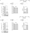
Dyskerin depletion increases VEGF mRNA internal ribosome entry site-mediated translation
- PMID: 23821664
- PMCID: PMC3783170
- DOI: 10.1093/nar/gkt587
Dyskerin depletion increases VEGF mRNA internal ribosome entry site-mediated translation
Abstract
Dyskerin is a nucleolar protein encoded by the DKC1 gene that (i) stabilizes the RNA component of the telomerase complex, and (ii) drives the site-specific pseudouridilation of rRNA. It is known that the partial lack of dyskerin function causes a defect in the translation of a subgroup of mRNAs containing internal ribosome entry site (IRES) elements such as those encoding for the tumor suppressors p27 and p53. In this study, we aimed to analyze what is the effect of the lack of dyskerin on the IRES-mediated translation of mRNAs encoding for vascular endothelial growth factor (VEGF). We transiently reduced dyskerin expression and measured the levels of the IRES-mediated translation of the mRNA encoding for VEGF in vitro in transformed and primary cells. We demonstrated a significant increase in the VEGF IRES-mediated translation after dyskerin knock-down. This translational modulation induces an increase in VEGF production in the absence of a significant upregulation in VEGF mRNA levels. The analysis of a list of viral and cellular IRESs indicated that dyskerin depletion can differentially affect IRES-mediated translation. These results indicate for the first time that dyskerin inhibition can upregulate the IRES translation initiation of specific mRNAs.
Figures


Similar articles
-
Novel dyskerin-mediated mechanism of p53 inactivation through defective mRNA translation.Cancer Res. 2010 Jun 1;70(11):4767-77. doi: 10.1158/0008-5472.CAN-09-4024. Epub 2010 May 25. Cancer Res. 2010. PMID: 20501855
-
The translation initiation factor DAP5 promotes IRES-driven translation of p53 mRNA.Oncogene. 2014 Jan 30;33(5):611-8. doi: 10.1038/onc.2012.626. Epub 2013 Jan 14. Oncogene. 2014. PMID: 23318444
-
Control of the vascular endothelial growth factor internal ribosome entry site (IRES) activity and translation initiation by alternatively spliced coding sequences.J Biol Chem. 2004 Apr 30;279(18):18717-26. doi: 10.1074/jbc.M308410200. Epub 2004 Feb 5. J Biol Chem. 2004. PMID: 14764596
-
An atypical IRES within the 5' UTR of a dicistrovirus genome.Virus Res. 2009 Feb;139(2):157-65. doi: 10.1016/j.virusres.2008.07.017. Epub 2008 Sep 11. Virus Res. 2009. PMID: 18755228 Review.
-
Role of RNA structure motifs in IRES-dependent translation initiation of the coxsackievirus B3: new insights for developing live-attenuated strains for vaccines and gene therapy.Mol Biotechnol. 2013 Oct;55(2):179-202. doi: 10.1007/s12033-013-9674-4. Mol Biotechnol. 2013. PMID: 23881360 Review.
Cited by
-
RNA methylation-related inhibitors: Biological basis and therapeutic potential for cancer therapy.Clin Transl Med. 2024 Apr;14(4):e1644. doi: 10.1002/ctm2.1644. Clin Transl Med. 2024. PMID: 38572667 Free PMC article. Review.
-
Implications of telomeres and telomerase in endometrial pathology.Hum Reprod Update. 2017 Mar 1;23(2):166-187. doi: 10.1093/humupd/dmw044. Hum Reprod Update. 2017. PMID: 27979878 Free PMC article. Review.
-
RNA Pseudouridylation in Physiology and Medicine: For Better and for Worse.Genes (Basel). 2017 Nov 1;8(11):301. doi: 10.3390/genes8110301. Genes (Basel). 2017. PMID: 29104216 Free PMC article. Review.
-
Cap-Independent Translational Control of Carcinogenesis.Front Oncol. 2016 May 25;6:128. doi: 10.3389/fonc.2016.00128. eCollection 2016. Front Oncol. 2016. PMID: 27252909 Free PMC article. Review.
-
Translation Stress Regulates Ribosome Synthesis and Cell Proliferation.Int J Mol Sci. 2018 Nov 27;19(12):3757. doi: 10.3390/ijms19123757. Int J Mol Sci. 2018. PMID: 30486342 Free PMC article. Review.
References
-
- Heiss NS, Knight SW, Vulliamy TJ, Klauck SM, Wiemann S, Mason PJ, Poustka A, Dokal I. X-linked dyskeratosis congenita is caused by mutations in a highly conserved gene with putative nucleolar functions. Nat. Genet. 1998;19:32–38. - PubMed
-
- Watkins NJ, Bohnsack MT. The box C/D and H/ACA snoRNPs: key players in the modification, processing and the dynamic folding of ribosomal RNA. Wiley Interdisci.p Rev. RNA. 2012;3:397–414. - PubMed
-
- Mitchell JR, Wood E, Collins K. A telomerase component is defective in the human disease dyskeratosis congenital. Nature. 1999;402:551–555. - PubMed
Publication types
MeSH terms
Substances
LinkOut - more resources
Full Text Sources
Other Literature Sources
Research Materials
Miscellaneous

