. 2013 Aug 6;52(31):5195-205.
doi: 10.1021/bi400335g. Epub 2013 Jul 24.

A novel role for coenzyme A during hydride transfer in 3-hydroxy-3-methylglutaryl-coenzyme A reductase

Affiliations

A novel role for coenzyme A during hydride transfer in 3-hydroxy-3-methylglutaryl-coenzyme A reductase

C Nicklaus Steussy et al. Biochemistry. .

Abstract

In this study, we take advantage of the ability of HMG-CoA reductase (HMGR) from Pseudomonas mevalonii to remain active while in its crystallized form to study the changing interactions between the ligands and protein as the first reaction intermediate is created. HMG-CoA reductase catalyzes one of the few double oxidation-reduction reactions in intermediary metabolism that take place in a single active site. Our laboratory has undertaken an exploration of this reaction space using structures of HMG-CoA reductase complexed with various substrate, nucleotide, product, and inhibitor combinations. With a focus in this publication on the first hydride transfer, our structures follow this reduction reaction as the enzyme converts the HMG-CoA thioester from a flat sp(2)-like geometry to a pyramidal thiohemiacetal configuration consistent with a transition to an sp(3) orbital. This change in the geometry propagates through the coenzyme A (CoA) ligand whose first amide bond is rotated 180° where it anchors a web of hydrogen bonds that weave together the nucleotide, the reaction intermediate, the enzyme, and the catalytic residues. This creates a stable intermediate structure prepared for nucleotide exchange and the second reduction reaction within the HMG-CoA reductase active site. Identification of this reaction intermediate provides a template for the development of an inhibitor that would act as an antibiotic effective against the HMG-CoA reductase of methicillin-resistant Staphylococcus aureus.


Conflict of interest statement

Notes

The authors declare no competing financial interests.

Figures

Figure 1
Figure 1
Chemistry of hydride transfer and the structure of HMG-CoA. (A) Schematic of the first hydride transfer of HMG-CoA reductase. On the left is HMG-CoA schematically arranged with some of the residue side chains known to be important in catalysis. Here the thioester is in a flat, sp2 orbital configuration with the carbonyl carbon colored red. This cartoon represents the RPMS structure. NADH is then bound and the hydride transferred from the C4 position of the nicotinamide ring to the sp2 carbon. The previously published structure of the nonproductive complex of NAD and HMG-CoA (Protein Data Bank entry 1QAX) serves as the best representative of this dynamic state. The hydride transfer then converts the thioester to the hemiacetal with a tetrahedral sp3 orbital configuration (RPMU). Lys267 has been suggested as the proton donor for the reaction based on the earlier 1QAX structure. Our hemiacetal structure suggests that Glu83 is a more likely candidate, and this has been supported with computational experiments. The mechanism of the first hydride transfer of the reaction mechanism only is illustrated; the entire mechanism is outlined in ref . (B) 3-Hydroxy-3-methylglutaryl-coenzyme A (HMG-CoA). Structure of the ligand for HMG-CoA reductase with emphasis on the portions discussed in the text. The first amide bond that forms the basis of the hydrogen bonding network with hemiacetal formation is colored red. Sulfur is substituted for the carbonyl oxygen of the thioester bond in the dithio-HMG-CoA slow substrate used to trap the hemiacetal intermediate (in blue). This figure was prepared using ChemDraw.
Figure 2
Figure 2
Domain structure of HMGR from P. mevalonii. (A) A dimer of HMGR (structure RPMU) with the bound substrate and cofactor. The two monomers intertwine with the active site at their interface. The 53 residues of the C-terminal flap domain have been removed to expose the configuration of the ligands in the active site. (B) One monomer, rotated around a complex axis to best demonstrate the domain structure, bringing the green NAD binding domain to the bottom of the figure and the central helix of the triangular large domain to a 45° position in the plane of the figure. In this panel, the large domain (red) is composed of residues 1–108 and 220–349 and binds the dithio-HMG-CoA. The NAD binding domain (green) contains residues 109–219 and binds the NAD(H) for the opposite active site. The C-terminal flap domain (blue) contains three helices of residues 375–421 and is seen only when both ligands are bound. This figure was prepared using Pymol.
Figure 3
Figure 3
Electrostatic surface of HMGR with HMG-CoA and NAD(H). (A) HMG-CoA (data set RPMU) is shown binding to the enzyme in a shallow groove along the surface. The adenine of the CoA, on the left of the figure, is obscured by the side chain of Arg11 that covers the nucleotide. The dithioester between CoA and HMG is shown here in the hemiacetal configuration, with the sulfurs (yellow) toward the middle of the figure. NAD(H) is bound in a surface groove to the right. Its adenine is partially obscured by the side chains of the conserved DAMG sequence associated with the non-Rossmann nucleotide-binding domain of the enzyme. The nicotinamide ring of NAD(H) and the thioester bond of HMG-CoA come into catalytic contact in the center of the figure, shown in more detail in panel B. (C) Flap domain in a ribbon cartoon form, positioned over the active site by the hydrogen bonding interactions associated with hemiacetal formation. This figure was prepared using Pymol.
Figure 4
Figure 4
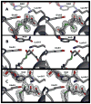
Evolution of the thioester bond during the first hydride transfer. (A) Initial binding (data set RPMS) of HMG-CoA to HMG-CoA reductase from P. mevalonii. Depicted is the structure with the slow substrate, dithio-HMG-CoA, where the carbonyl carbon is replaced by a (green) sulfur atom. The electron density shown is a simulated annealing omit FoFc map (CNS version 1.3) contoured at 3.0σ. The NAD+ in the panel is not actually present in the structure but is there for the purpose of orientation, shown at the location of the cofactor in RPMU. Note the flat geometry of the dithioester bond consistent with an unreduced sp2 orbital and its orientation away from the nicotinamide ring (HMGCoA C1–NAD C4 distance of 5.1 Å). (B) Nonproductive complex containing NAD+ and HMG-CoA (PDB entry 1QAX). This models the movement of the thioester as the nucleotide binds with the thioester bond rotated up toward the NAD+ while retaining its flat sp2 orbital-like geometry (HMGCoA C1–NAD C4 distance of 4.0 Å). (C) Dithioester (data set RPMU) of the slow substrate reduced to the hemiacetal form, and therefore the end result of the first hydride transfer. The dithioester now has a pyramidal geometry consistent with a reduced, sp3 orbital configuration oriented roughly parallel to the nicotinamide ring, with the former “carbonyl” atom directed upward toward active site residues Glu83 and Lys267 (HMGCoA C1–NAD C4 distance of 3.2 Å). The electron density shown is a simulated annealing omit FoFc map (CNS version 1.3) contoured at 3.0σ. This figure was prepared using Pymol.
Figure 5
Figure 5
Hydrogen bonding between HMG-CoA and Arg261 through the reaction cycle. (A) Structure of RPMS, with only the dithio-HMG-CoA bound. The ester bond is in a flat, sp2 orbital, and there are two hydrogen bonds between the glutaryl portion of HMG-CoA and Arg261. (B) Structure developed from the nonproductive complex of NAD+ and HMG-CoA (PDB entry 1QAX) that serves here as a model for the intermediate in the reaction in which HMG-CoA and NADH are both bound but the hydride transfer has not yet taken place. The thioester thus remains in the flat sp2 orbital configuration but has reoriented toward the active site residues. Adjusting to this motion, the glutaryl group changes its orientation relative to Arg261, and the side chain of the amino acid flips, such that only one hydrogen bond is formed between the two. (C) RPMU in which the thioester of dithio-HMG-CoA has been reduced to the hemiacetal. The tetrahedral configuration of the bond is evident. This conversion allows the glutaryl moiety and Arg261 to regain their original orientation, forming two hydrogen bonds. The electron density shown in panels A and C is 1.4σ, 2FoFc density from the refined model. The red and green β-strands toward the back of the figure are from monomers A and B of the dimer, respectively.
Figure 6
Figure 6
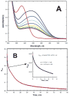
DithioHMG-CoA is a slow substrate. (A) Plots of absorbance vs wavelength at different times during the reaction of dithio-HMG-CoA and NADH in the presence of HMGR. The scan with a maximum near 312 nm (red line) is the spectrum of the HMGR–dithio-HMG-CoA complex before the reaction was initiated. The reaction was started by adding NADH to this mixture and restarting the spectrophotometer immediately at a scan rate of 60 nm/min from 450 to 240 nm. Scans were collected manually at 4 min intervals with the exception of the last scan, which was at an 8 min interval. The reaction mixture contained 20 μM S. aureus HMGR, 30 μM dithio-HMG-CoA, 80 μM NADH, and 50 mM HEPES (pH 7.0). The decrease in the NADH absorbance at 340 nm can be clearly followed (first scan, black line; final scan, purple line). (B) Plot of absorbance at 340 nm vs time for the reaction of dithio-HMG-CoA and NADH in the presence of S. aureus HMGR. In this study, the reaction was started by adding enzyme to the reaction mixture. The other conditions were the same as those described for Figure 6A, except that the reaction was blanked with a completed reaction mixture. The reaction is clearly triphasic. The initial burst phase and part of the intermediate zero-order phase were fit by nonlinear least squares to the equation, which contains both exponential and linear terms, shown in the inset. The half-life of the burst phase is ~0.5 min, and the magnitude of the burst corresponds to the oxidation of one enzyme equivalent of NADH.
Figure 7
Figure 7
Adaptation of serine 85 to the formation of the hemiacetal. (A) Configuration of dithio-HMG-CoA and the protein loop containing serine 85 in the single-substrate complex (RPMS). The nucleotide is not present in this structure but is imported from the ternary complex structure (RPMU) for the purpose of reference. The hydroxyl of the serine is oriented away from the ligand, and the first amide bond of the HMG-CoA is turned away and distant from the serine. The electron density shown is the final refined 2FoFc density contoured at 1.2σ. The Real Space correlation coefficient for the dithio-HMG-CoA is 0.884. (B) These same structures, but after the formation of the hemiacetal (RPMU). Note the thioester of the dithio-HMG-CoA has taken on the tetrahedral configuration of the hemiacetal and has been reoriented close to the nicotinamide ring. The first pantothenic amide bond has rotated such that its nitrogen can make a 2.6 Å hydrogen bond to the hydroxyl of the rotated Ser85. The electron density shown is again the refined 2FoFc density contoured at 1.2σ. The Real Space correlation coefficient for the NAD ligand is 0.939 and that of the dithio-HMG-CoA 0.926. The red and green ribbons show the β-strands of monomers A and B, respectively. This figure was prepared using Pymol.
Figure 8
Figure 8
Hydrogen bonding network formed with creation of the hemiacetal intermediate. The ligands modeled here are from the structure (RPMU) in which the NADH has reduced the dithio-HMG-CoA to its hemiacetal form. Serine 85 has rotated to interact with the first amide bond of CoA and His381. The hydrogen bonding network continues with connections through Asn188, and from there through the NAD phosphate to His385. The first helix of the flap domain, which contains His381 and His385, is colored light blue extending down the center of the figure. Red and green β-strands are from monomers A and B, respectively. This figure was prepared using Pymol.

Similar articles

Cited by

References

    1. Goldstein JL, Brown MS. Regulation of the mevalonate pathway. Nature. 1990;343:425–430. - PubMed
    1. Johnson EA, Schroeder WA. Microbial carotenoids. Adv Biochem Eng Biotechnol. 1996;53:119–178. - PubMed
    1. Reusch VM., Jr Lipopolymers, isoprenoids, and the assembly of the Gram-positive cell wall. Crit Rev Microbiol. 1984;11:129–155. - PubMed
    1. Bochar DA, Stauffacher CV, Rodwell VW. Sequence comparisons reveal two classes of 3-hydroxy-3-methylglutaryl coenzyme A reductase. Mol Genet Metab. 1999;66:122–127. - PubMed
    1. Istvan ES. Bacterial and mammalian HMG-CoA reductases: Related enzymes with distinct architectures. Curr Opin Struct Biol. 2001;11:746–751. - PubMed

Publication types

LinkOut - more resources