. 2013 Sep;87(17):9590-603.
doi: 10.1128/JVI.00994-13. Epub 2013 Jun 19.

The Us2 gene product of herpes simplex virus 2 is a membrane-associated ubiquitin-interacting protein

Affiliations

The Us2 gene product of herpes simplex virus 2 is a membrane-associated ubiquitin-interacting protein

Ming-Hsi Kang et al. J Virol. 2013 Sep.

Abstract

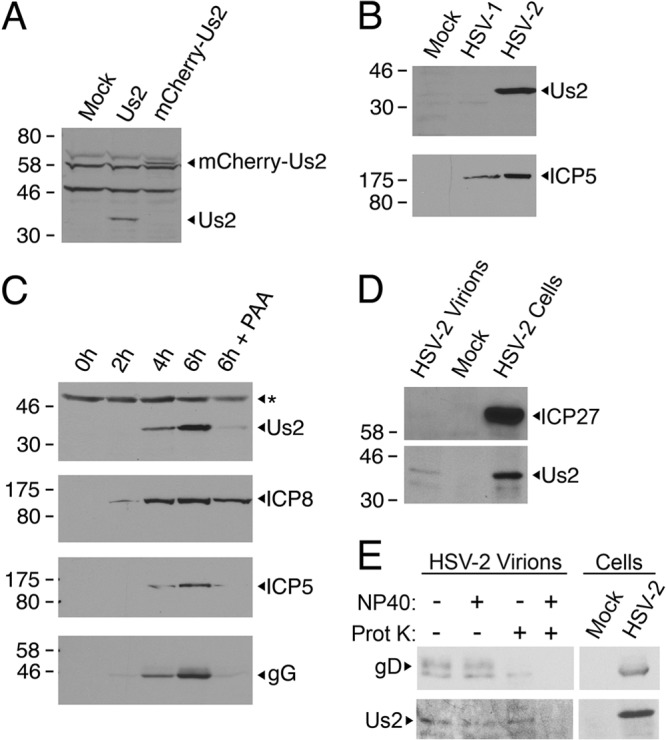
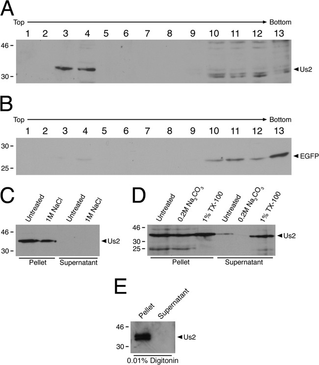
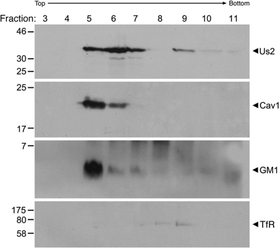
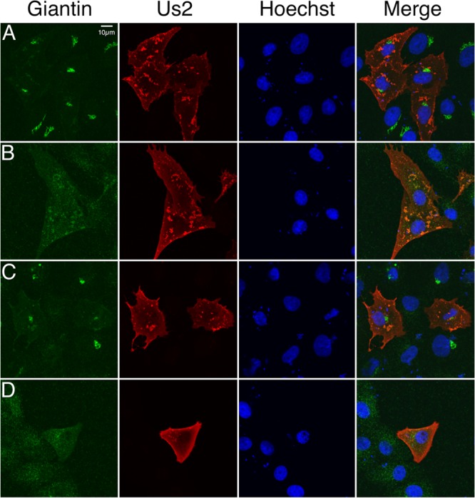
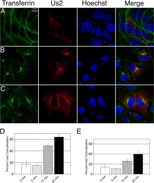
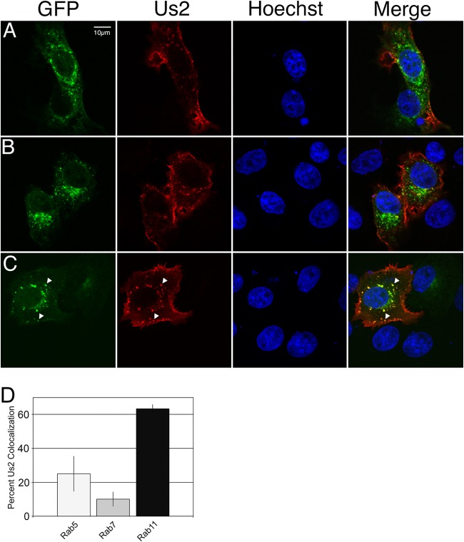
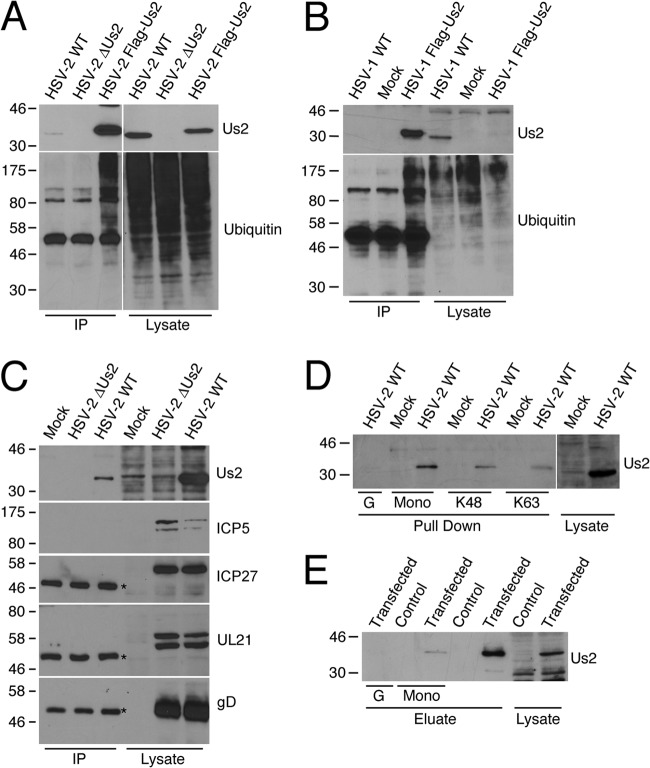
The Us2 gene encodes a tegument protein that is conserved in most members of the Alphaherpesvirinae. Previous studies on the pseudorabies virus (PRV) Us2 ortholog indicated that it is prenylated, associates with membranes, and spatially regulates the enzymatic activity of the MAP (mitogen-activated protein) kinase ERK (extracellular signal-related kinase) through direct binding and sequestration of ERK at the cytoplasmic face of the plasma membrane. Here we present an analysis of the herpes simplex virus 2 (HSV-2) Us2 ortholog and demonstrate that, like PRV Us2, HSV-2 Us2 is a virion component and that, unlike PRV Us2, it does not interact with ERK in yeast two-hybrid assays. HSV-2 Us2 lacks prenylation signals and other canonical membrane-targeting motifs yet is tightly associated with detergent-insoluble membranes and localizes predominantly to recycling endosomes. Experiments to identify cellular proteins that facilitate HSV-2 Us2 membrane association were inconclusive; however, these studies led to the identification of HSV-2 Us2 as a ubiquitin-interacting protein, providing new insight into the functions of HSV-2 Us2.


Figures

Fig 1
Fig 1
Analysis of HSV-2 Us2 expression and virion localization. (A) A polyclonal antiserum from rats immunized with GST–HSV-2 Us2 specifically detects HSV-2 Us2. Equal volumes of cellular extracts prepared from 293T cells either mock transfected or transfected with a plasmid encoding HSV-2 Us2 or mCherry–HSV-2 Us2 were analyzed by Western blotting. Note the shift in the mobility of the band detected in cells transfected with the plasmid encoding the mCherry–HSV-2 Us2 fusion protein. (B) The anti-HSV-2 Us2 polyclonal antiserum reacts with HSV-2 Us2 but not with HSV-1 Us2. Equal volumes of cellular extracts prepared from uninfected, HSV-1 17+-infected, or HSV-2 HG52-infected Vero cells were analyzed by Western blotting. (Top) The gel was probed with a rat polyclonal anti-HSV-2 Us2 antiserum. (Bottom) The infection control gel was probed with anti-HSV-2 ICP5, which also cross-reacts with ICP5 from HSV-1. (C) Kinetics of Us2 synthesis in HSV-2-infected cells. At the indicated times postinfection, the expression of Us2, ICP8, ICP5, and gG in cell lysates was analyzed by Western blotting. Additionally, the expression of Us2, ICP8, ICP5, and gG in the presence of the viral DNA synthesis inhibitor PAA was analyzed at 6 h postinfection. The asterisk at the top indicates the position of a nonspecific cross-reacting band that serves as an internal loading control. (D) Us2 is incorporated into HSV-2 virions. At 16 h after the infection of Vero cells with HSV-2 HG52, virions were purified from the cell medium and were analyzed alongside HSV-2-infected and mock-infected cell lysates for the presence of ICP27 and Us2 by Western blotting. Molecular size markers (in kilodaltons) are indicated on the left. (E) Protease protection assay. (Left) Purified virions were treated with either PBS (−), NP-40, PK (Prot K), or both NP-40 and PK and were then analyzed for the presence of the virion transmembrane protein gD and Us2. Note that gD is degraded by PK in the absence of NP-40, whereas Us2 remains intact. (Right) Migration of gD and Us2 from an infected-cell lysate.
Fig 2
Fig 2
Subcellular localization of HSV-2 Us2. (A) HSV-2 Us2 localizes to membranes in transfected Vero cells. Shown is a representative image of Vero cells transfected with a plasmid encoding HSV-2 Us2 and stained with a polyclonal antiserum specific for HSV-2 Us2 (green) at 24 h posttransfection. Nuclei were detected using the DNA stain Hoechst 33342. (B) Kinetics of HSV-2 Us2 expression and localization in infected Vero cells. Shown are representative images of Vero cells infected with HSV-2. Cells were fixed at 6 h (i), 10 h (ii), or 18 h (iii) after infection and were stained for Us2 (green) or the HSV-2 nuclear protein ICP8 (red). Nuclei were stained with Hoechst 33342 (blue). Images of stained cells were captured by confocal microscopy.
Fig 3
Fig 3
HSV-2 Us2 physically associates with membranes. (A and B) 293T cells were cotransfected with an HSV-2 Us2 and EGFP expression plasmid. At 24 h after transfection, membrane fractions were isolated by membrane flotation on discontinuous sucrose step gradients as described in Materials and Methods. Fractions from the top (10% sucrose) to the bottom (71.5% sucrose) of the step gradient were analyzed by Western blotting for the presence of Us2 (A) or EGFP as a negative control (B). (C through E) Membrane fractions were treated with either 1 M NaCl (C), 0.2 M Na2CO3 or 1% Triton X-100 (D), or 0.01% digitonin (E) for 1 h at 4°C prior to the pelleting of membranes by centrifugation. Pelleted membranes and protein from supernatant fractions that had been concentrated by TCA precipitation were analyzed for Us2 contents by Western blotting.
Fig 4
Fig 4
HSV-2 Us2 fractionates with detergent-insoluble membranes. Vero cells infected with HSV-2 were harvested at 12 h after infection, and detergent-insoluble membranes were isolated by flotation through an OptiPrep step gradient. Gradient fractions 3 through 11 were analyzed by Western blotting for Us2, caveolin-1 (Cav1), ganglioside GM1, or the transferrin receptor (TfR). Detergent-insoluble membranes float to the interface between the 5% and 30% OptiPrep portions of the step gradient, which is collected in fraction 5. The migration positions of molecular size markers (in kilodaltons) are shown on the left of each blot.
Fig 5
Fig 5
Effect of BFA treatment on Us2 localization. (A to C) Twenty-four hours after transfection with an HSV-2 Us2 expression plasmid, Vero cells either were treated with vehicle (0.3% ethanol) (A) or with 3 μg/ml BFA (B) for 1 h or were treated with BFA for 1 h, washed, and cultured for an additional 2 h in a medium lacking BFA (C). Cells were fixed and immunostained for the Golgi apparatus-resident protein giantin (green) and Us2 (red). Nuclei were detected using the DNA stain Hoechst 33342 (blue). (D) Alternatively, cells were treated with 3 μg/ml BFA 6 h after transfection and were maintained in BFA for 24 h prior to fixing and staining. Images of stained cells were captured by confocal microscopy. Representative images are shown.
Fig 6
Fig 6
Dynamics of Us2-containing vesicles. Vero cells were transfected with a plasmid encoding mCherry–HSV-2 Us2. At 24 h after transfection, live cells were imaged over 134 s at a rate of 2.2 frames per second by using a confocal microscope. Static images demonstrating the dynamics of Us2 in a representative cell are shown. The signal is mCherry fluorescence. Arrows highlight an example of a Us2-containing vesicle emitting an extended membrane tubule. The full data set is provided as movie S1 in the supplemental material. DIC, differential inference contrast.
Fig 7
Fig 7
A subset of Us2 colocalizes with endosomal vesicles. (A to C) Vero cells transfected with an mCherry–HSV-2 Us2 expression plasmid were incubated with Alexa Fluor 647-conjugated transferrin for 30 min at 4°C, washed, and shifted to 37°C for 5 min (A), 12 min (B), or 30 min (C) prior to fixation. The transferrin signal is shown in green. The Us2 signal is red. Nuclei were detected using the DNA stain Hoechst 33342 (blue). Representative images are shown. (D) Quantification of transferrin colocalization with Us2 puncta in cells transfected with mCherry–HSV-2 Us2. The data are presented as percentages of Us2-containing puncta that are also positive for transferrin. A total of 12 fields of cells and 298 Us2 puncta were analyzed. Error bars represent the standard errors of the means observed for differences between different fields of cells. (E) Quantification of transferrin colocalization with Us2 puncta in HSV-2-infected cells at each time point. The data are presented as percentages of Us2-containing puncta that are also positive for transferrin. Us2 puncta were identified by indirect immunofluorescence confocal microscopy using a polyclonal antiserum against Us2 and an Alexa Fluor 588-conjugated secondary antibody. A total of 30 fields of cells and 938 Us2 puncta were analyzed. Error bars represent the standard errors of the means observed for differences between different fields of cells at each time point.
Fig 8
Fig 8
Us2 colocalizes predominantly with Rab11, a marker of recycling endosomes. (A to C) Vero cells were cotransfected with plasmids encoding HSV-2 Us2 and either Rab5-GFP (A), Rab7-GFP (B), or Rab11-GFP (C). Plasmids encoding Rab5-, Rab7-, and Rab11-GFP fusion proteins were a kind gift from C. Parish, Cornell University. Cells were fixed at 24 h posttransfection and were stained for Us2. Images of stained cells were captured by confocal microscopy. Rab signals are EGFP fluorescence and are displayed in green. The Us2 signal is red. Nuclei were detected using the DNA stain Hoechst 33342 (blue). Arrowheads in panel C highlight the colocalization of Us2 and Rab11 signals. Representative images are shown. (D) Quantification of the colocalization of Us2-labeled puncta with Rab5, Rab7, and Rab11. The data are presented as percentages of Us2-containing puncta that are also positive for Rab5, Rab7, or Rab11. A total of 12 fields of cells and 249 Us2 puncta were analyzed. The analysis was performed in a manner similar to that for the quantification of Us2-transferrin colocalization, for which results are shown in Fig. 7D and E. Error bars represent the standard errors of the means observed for differences between different fields of cells for each Rab protein analyzed.
Fig 9
Fig 9
Us2 is a ubiquitin-interacting protein. (A and B) Vero cells were infected with WT HSV-2, HSV-2 ΔUs2, or recombinant HSV-2 with Flag-tagged Us2 (A) or with WT HSV-1 or recombinant HSV-1 with Flag-tagged Us2 (B). At 6 hpi, cell lysates were prepared, and proteins were immunoprecipitated (IP) using an anti-Flag affinity gel. Immunoprecipitated proteins were detected using antibodies against HSV-1 Us2, HSV-2 Us2, or mono- and polyubiquitin (FK2). (C) HSV-2 Us2 coimmunoprecipitates with ubiquitin-conjugated proteins from infected cells. Vero cells were infected with WT HSV-2 or a ΔUs2 virus for 6 h. Cells were collected, and lysates were incubated overnight with a monoclonal antibody against mono- and polyubiquitin (FK2). Proteins were pulled down using protein G agarose and were subjected to Western blotting for detection of the presence of Us2, ICP5, ICP27, UL21, and gD. Asterisks indicate the Ig heavy chain. (D) HSV-2 Us2 interacts directly with ubiquitin. Vero cells were infected with WT HSV-2 for 6 h. Cell lysates were prepared and were incubated overnight with ubiquitin-conjugated beads. Interacting proteins were subjected to Western blotting and were probed with an antibody against Us2. G, protein G agarose (negative control); mono, monoubiquitin-conjugated agarose; K48, K48-linked tetraubiquitin-conjugated agarose; K63, K63-linked tetraubiquitin-conjugated agarose. (E) HSV-2 Us2 interacts with ubiquitin in the absence of other viral proteins. An empty vector (pFlag-CMV2) or N-terminally Flag-tagged Us2 was transfected into 293T cells. At 24 h posttransfection, cells were collected in lysis buffer, and lysates were incubated overnight with anti-Flag beads at 4°C. Immunoprecipitated proteins were eluted using 3×Flag peptides. Eluates were incubated overnight with either protein G agarose or monoubiquitin-conjugated beads at 4°C. Us2 bound to the beads was detected by Western blotting using anti-Flag antibodies. G, protein G agarose; mono, monoubiquitin-conjugated beads.

Similar articles

Cited by

References

    1. Ben-Porat T, Kaplan AS. 1985. Molecular biology of pseudorabies virus, p 105–173 In Roizman B. (ed), The herpesviruses. Plenum Press, New York, NY
    1. Roizman B, Knipe DM. 2001. Herpes simplex viruses and their replication, p 2399–2459 In Knipe DM, Howley PM, Griffin DE, Lamb RA, Martin MA, Roizman B, Straus SE. (ed), Fields virology, 4th ed. Lippincott Williams & Wilkins, Philadelphia, PA
    1. Mettenleiter TC, Klupp BG, Granzow H. 2009. Herpesvirus assembly: an update. Virus Res. 143:222–234 - PubMed
    1. Wisner TW, Wright CC, Kato A, Kawaguchi Y, Mou F, Baines JD, Roller RJ, Johnson DC. 2009. Herpesvirus gB-induced fusion between the virion envelope and outer nuclear membrane during virus egress is regulated by the viral US3 kinase. J. Virol. 83:3115–3126 - PMC - PubMed
    1. Jones F, Grose C. 1988. Role of cytoplasmic vacuoles in varicella-zoster virus glycoprotein trafficking and virion envelopment. J. Virol. 62:2701–2711 - PMC - PubMed

Publication types

LinkOut - more resources