. 2013 Jun 19;33(25):10278-300.
doi: 10.1523/JNEUROSCI.5048-12.2013.

A local, periactive zone endocytic machinery at photoreceptor synapses in close vicinity to synaptic ribbons

Affiliations

A local, periactive zone endocytic machinery at photoreceptor synapses in close vicinity to synaptic ribbons

Silke Wahl et al. J Neurosci. .

Abstract

Photoreceptor ribbon synapses are continuously active synapses with large active zones that contain synaptic ribbons. Synaptic ribbons are anchored to the active zones and are associated with large numbers of synaptic vesicles. The base of the ribbon that is located close to L-type voltage-gated Ca(2+) channels is a hotspot of exocytosis. The continuous exocytosis at the ribbon synapse needs to be balanced by compensatory endocytosis. Recent analyses indicated that vesicle recycling at the synaptic ribbon is also an important determinant of synaptic signaling at the photoreceptor synapse. To get insights into mechanisms of vesicle recycling at the photoreceptor ribbon synapse, we performed super-resolution structured illumination microscopy and immunogold electron microscopy to localize major components of the endocytotic membrane retrieval machinery in the photoreceptor synapse of the mouse retina. We found dynamin, syndapin, amphiphysin, and calcineurin, a regulator of activity-dependent endocytosis, to be highly enriched around the active zone and the synaptic ribbon. We present evidence for two clathrin heavy chain variants in the photoreceptor terminal; one is enriched around the synaptic ribbon, whereas the other is localized in the entry region of the terminal. The focal enrichment of endocytic proteins around the synaptic ribbon is consistent with a focal uptake of endocytic markers at that site. This endocytic activity functionally depends on dynamin. These data propose that the presynaptic periactive zone surrounding the synaptic ribbon complex is a hotspot of endocytosis in photoreceptor ribbon synapses.


Figures

Figure 1.
Figure 1.
Expression of endocytic proteins in the mouse retina. A–G, Expression of endocytic proteins in extracts of the mouse retina as judged by Western blot analyses. All antibodies detect their respective antigen at the expected running position (indicated by arrowheads). In A–E, proteins were separated by 10% acrylamide SDS-PAGE; in F–G, by 8% acrylamide SDS-PAGE.
Figure 2.
Figure 2.
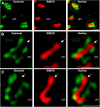
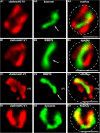
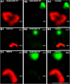
Distribution of dynamin in photoreceptor ribbon synapses at high resolution (immunolabeling of 0.5 μm thin sections). A, 0.5 μm thin sections of the mouse retina were double immunolabeled with polyclonal antibodies against RIBEYE (U2656) and mouse monoclonal antibodies against dynamin (hudy-1). Dashed circles in A denote single immunolabeled presynaptic photoreceptor terminals. Images shown in A were obtained by conventional imaging. B, C, 2D-SR-SIM images of 0.5 μm thin sections from the mouse retina that were double immunolabeled with rabbit polyclonal antibodies against RIBEYE (U2656) and mouse monoclonal antibodies against dynamin (hudy-1). The dynamin immunosignal is highly enriched around the synaptic ribbon (arrow). OPL, Outer plexiform layer. Scale bars, 1 μm.
Figure 3.
Figure 3.
Localization of dynamin in relation to other synaptic proteins of the presynaptic photoreceptor terminal. A, B, The 0.5 μm thin sections from mouse retina were triple immunolabeled with rabbit polyclonal antibodies against PSD-95 (L667) (A)/ or VGLUT1 (B), mouse monoclonal antibodies against dynamin (hudy-1) (A, B), and DyLight 650-direct labeled primary antibodies against RIBEYE(B)/CtBP2 (A, B). In A, the PSD-95 immunosignals label the presynaptic plasma membrane of the presynaptic terminals (Koulen et al., 1998; Aartsen et al., 2009), thus demarcating the extension of a single presynaptic terminal (arrowheads in A). RIBEYE and dynamin are located close to each other at the distal end of the photoreceptor terminal that is facing the INL (A). In B, presynaptic terminals were immunolabeled with antibodies against VGLUT1, a marker protein of glutamatergic synaptic vesicles. Single-photoreceptor presynaptic terminals are indicated by the white dashed circles in B. Similar as in A, RIBEYE and dynamin are located close to each other at the distal border of the immunolabeled glutamatergic vesicles of the presynaptic terminal that faces the INL. ONL, Outer nuclear layer; OPL, outer plexiform layer; INL, inner nuclear layer; sr, synaptic ribbon. Arrows next to INL and ONL point into the direction of the respective layer. Scale bars, 1 μm.
Figure 4.
Figure 4.
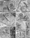
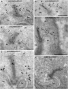
Postembedding immunogold labeling of photoreceptor synapses of the mouse retina with dynamin antibodies. A–H, Ultrathin sections immunolabeled with mouse monoclonal antibodies against dynamin (hudy-1). Binding of the primary antibodies was detected with goat anti-mouse antibodies conjugated to 10 nm gold particles. The dynamin immunogold label is highly enriched at the presynaptic plasma membrane surrounding the synaptic ribbon (arrowheads in A–H). This dynamin immunogold label at the periactive zone was particularly strong at the lateral presynaptic plasma membrane pouches opposite to dendritic tips of postsynaptic horizontal cells (ho). I, A control incubation in which only secondary antibody (but no primary antibody) was applied. No immunosignal was observed under these incubations, further stressing the specificity of the immunolabeling results. A quantitative minor portion of dynamin immunolabel was found in a cytosolic localization within the presynaptic terminal (dashed circles in C and E). This minor portion could result from either labeling of endomembranes or tangential views of dynamin on periactive zone of lateral pouches above the section plane. Please note that a postembedding protocol was used for immunolabeling. In postembedding protocols, no osmium tetroxide can be used. Therefore, lipid-rich membrane compartments (i.e., synaptic vesicles) remain invisible with postembedding methods. pr, Presynaptic terminal; sr, synaptic ribbon; pm, extrasynaptic plasma membrane (outside of the presynaptic plasma membrane invagination with no contact to the postsynaptic cavity; see also Fig. 17); nu, nucleus. Black arrowheads point to dynamin immunogold particles close to the synaptic ribbons. Scale bars: A–E, G, I, 500 nm; F, H, 200 nm.
Figure 5.
Figure 5.
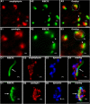
A, B, Distribution of amphiphysin and syndapin in photoreceptor ribbon synapses at high resolution (conventional imaging). A, B, The 0.5 μm thin sections of the mouse retina were double immunolabeled with polyclonal antibodies against amphiphysin and mouse monoclonal antibodies against RIBEYE(B)-domain/CtBP2 (in A), and polyclonal antibodies against syndapin and mouse monoclonal antibodies against RIBEYE(B)-domain/CtBP2 (in B). Arrows point to immunolabeled synaptic ribbons. Dashed circles in A and B denote single-immunolabeled presynaptic terminals/synaptic ribbon complexes of rod photoreceptors. C, D, Multicolor, 2D-SR-SIM analyses of the distribution of amphiphysin and syndapin in photoreceptor synapses. In C and D, 0.5 μm thin sections of the mouse retina were triple immunolabeled with Alexa Fluor 488 directly labeled mouse monoclonal antibody against RIBEYE(B)-domain/CtBP2, rabbit polyclonal antibody against amphiphysin (C) or syndapin (D) and mouse monoclonal antibody against dynamin (C, D). Arrows point to immunolabeled synaptic ribbons. OPL, Outer plexiform layer. Scale bars, 1 μm.
Figure 6.
Figure 6.
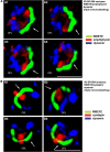
Multicolor 3D-SR-SIM of RIBEYE, dynamin, and syndapin/amphiphysin in the active zone of single rod photoreceptor synapses. In A, triple-immunolabeling 3D-SR-SIM analyses were performed with antibodies against RIBEYE, dynamin, and amphiphysin; in B, triple-immunolabeling 3D-SR-SIM analyses were performed with antibodies against RIBEYE, dynamin, and syndapin (A1–A4 and B1–B4, respectively), denoting different lateral views of the same single-immunolabeled synaptic ribbon of a rod photoreceptor synapse. Arrows denote the immunolabeled synaptic ribbon. OPL,Outer plexiform layer. Scale bars, 1 μm.
Figure 7.
Figure 7.
Localization of endophilin in photoreceptor synapses of the mouse retina. A, The 0.5 μm thin sections of the mouse retina were double immunolabeled with rabbit polyclonal antibodies against endophilin and mouse monoclonal antibodies against RIBEYE(B)-domain/CtBP2. B, The 0.5 μm thin sections of the mouse retina were double immunolabeled with rabbit polyclonal antibodies against endophilin and mouse monoclonal antibodies against dynamin. C, D, The 0.5 μm thin sections of the mouse retina were double immunolabeled with rabbit polyclonal antibodies against endophilin and mouse monoclonal antibodies against the synaptic vesicle protein 2 (panSV2; detecting all SV2 isoforms). E, The 0.5 μm thin sections of the mouse retina were double immunolabeled with rabbit polyclonal antibodies against endophilin and mouse monoclonal antibodies against VGLUT1. Dashed circles in B, D, and E denote single presynaptic photoreceptor terminals. Endophilin is diffusely distributed throughout the presynaptic terminal and is not particularly enriched around the synaptic ribbon. All micrographs were obtained by conventional imaging. ONL, Outer nuclear layer; OPL, outer plexiform layer; INL, inner nuclear layer. Scale bars: A, 10 μm; B, 5 μm; C, 12 μm; D, E, 1 μm.
Figure 8.
Figure 8.
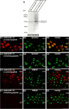
Pre-absorption control experiments for the immunolabeling analyses. A–H, Double immunolabeling of 0.5 μm thin mouse retinal sections with the indicated antibodies preabsorbed with either their specific peptide used for immunization (B, D, F, H) or with an unrelated control peptide (A, C, E, G). To visualize ribbon synapses, sections were coimmunolabeled with either rabbit polyclonal antibodies against RIBEYE (U2656 in A and B) or mouse monoclonal antibodies against RIBEYE(B)-domain/CtBP2. Preabsorption with the specific peptide completely blocked the respective immunosignals at the synaptic ribbon (B, D, F, and H), whereas the control peptide had no influence on the immunosignals (A, C, E, and G), showing the specificity of the immunolabeling results. ONL, Outer nuclear layer; OPL, outer plexiform layer; INL, inner nuclear layer. Scale bars, 10 μm.
Figure 9.
Figure 9.
The Ca2+-binding phosphatase calcineurin, a Ca2+ sensor of endocytosis, is enriched at the synaptic ribbon. A, The 0.5 μm thin sections of the mouse retina were double immunolabeled with affinity-purified rabbit polyclonal antibodies against calcineurin and mouse monoclonal antibodies against RIBEYE(B)domain/CtBP2 (conventional imaging). Calcineurin is highly enriched at the synaptic ribbons (arrowheads in A). B, The insets show a single-immunolabeled synaptic ribbon. ONL, Outer nuclear layer; OPL, outer plexiform layer. Scale bars, 10 μm.
Figure 10.
Figure 10.
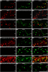
High-magnification analyses of CHC-V1 in relation to bassoon, RIBEYE, and dynamin in single-photoreceptor synapses. A, The 0.5 μm thin sections of the mouse retina were double immunolabeled with rabbit polyclonal antibodies against CHC-V1 and mouse monoclonal antibodies against bassoon. In B and C, sections were double immunolabeled with rabbit polyclonal antibodies against CHC-V1 and mouse monoclonal antibodies against RIBEYE(B)-domain/CtBP2. In D, sections were double immunolabeled with rabbit polyclonal antibodies against CHC-V1 and mouse monoclonal antibodies against dynamin. CHC-V1 is located very close to both RIBEYE and bassoon but does not overlap. In contrast, the CHC-V1 immunosignals overlap with the dynamin immunosignal at the active zone of photoreceptor ribbon synapses to a large extent (D). A, B, and D were obtained by conventional imaging at high magnification; C is a maximum projection of a z-stack obtained by 2D-SR-SIM. Dashed circles in A–D denote single presynaptic photoreceptor terminals; clathrinHC-V1, CHC-V1; OPL, outer plexiform layer. Scale bars, 1 μm.
Figure 11.
Figure 11.
Localization of CHC-V1 in the presynaptic rod photoreceptor terminal in relation to PSD-95 and VGLUT1. A, B, The 0.5 μm thin sections from mouse retina were triple immunolabeled with mouse monoclonal antibodies against PSD-95 (A) or VGLUT1 (B), rabbit polyclonal antibodies against CHC-V1 (abcam) (A, B) and DyLight 650 direct labeled primary antibodies against RIBEYE(B)/CtBP2 (A, B). The PSD-95 immunosignals in A demarcate the plasma membrane of photoreceptor presynaptic terminals in the OPL (A, arrowheads). In B, presynaptic terminals were immunolabeled with antibodies against the vesicular transporter VGLUT1, a marker protein of glutamatergic synaptic vesicles. The dashed circle in B denotes a single-immunolabeled presynaptic photoreceptor terminal. RIBEYE and CHC-V1 are located close to each other at the distal end of the photoreceptor terminal that is facing the INL (A, B). ONL, Outer nuclear layer; clathrinHC-V1, CHC-V1; OPL, outer plexiform layer; INL, inner plexiform layer. Scale bars, 1 μm.
Figure 12.
Figure 12.
Postembedding immunogold labeling of photoreceptor synapses from the mouse retina with antibodies against CHC-V1. A–E, Ultrathin sections of the mouse retina were immunolabeled with mouse monoclonal antibodies against CHC-V1 (catalog #21679, abcam). Binding of the primary antibodies was detected with goat anti-rabbit antibodies conjugated to 10 nm gold particles. A strong CHC-V1 immunogold label (arrowheads) was observed at the plasma membrane in close proximity to the synaptic ribbon (sr). The immunogold labeling experiments confirm the previously shown immunofluorescence labeling data and demonstrate the enrichment of CHC-V1 in the periactive zone of the photoreceptor ribbon synapse. Arrowheads indicate the CHC-V1 enrichment in the periactive zone. Only a very minor fraction of CHC-V1 was found in the presynaptic cytosol (dashed circle). F is a negative control in which the primary antibody was omitted. clathrinHC-V1, CHC-V1; pr, presynaptic photoreceptor terminal; ho, dendritic tips of postsynaptic horizontal cells; pm, plasma membrane. Scale bars: A–F, 250 nm.
Figure 13.
Figure 13.
A, Western blot analyses of two different clathrin heavy chains, variant 1 and variant 2 (after separation in 5% acrylamide SDS-PAGE running gels). The running position of the immunoreactive bands detected by the different antibodies against the CHC-V1 (lane 1) and antibodies against clathrin heavy chain variant 2 (lane 2) differ slightly. CHC-V1 is slightly smaller than CHC-V2 in Western blot analyses (after separation in 5% acrylamide SDS-PAGE running gels). B–E, Pre-absorption control experiments for the antibodies against CHC-V1 and CHC-V2 (immunolabeling analyses). Double immunolabeling of 0.5 μm thin mouse retinal sections with the indicated antibodies preabsorbed with either their specific peptide used for immunization (C, E) or with an unrelated control peptide (B, D). In parallel, sections were incubated with monoclonal anti-dynamin antibodies (B, C) or RIBEYE (D, E) as labeling positive controls. The specific peptides completely blocked the respective clathrin heavy chain immunosignals (C, E), whereas the control peptide had no influence of the clathrin heavy chain immunosignals (B, D) showing the specificity of the immunolabeling signals. ONL, Outer nuclear layer; OPL, outer plexiform layer; clathrinHC-V1, CHC-V1; clathrinHC-V2, CHC-V2. Scale bars: B–E, 5 μm.
Figure 14.
Figure 14.
Two different clathrin heavy chain variants are found in photoreceptor terminals at different locations (immunofluorescence analyses). A, The 0.5 μm thin sections of the mouse retina were double immunolabeled with rabbit polyclonal antibodies against CHC-V1 and mouse monoclonal antibodies against CHC-V2. The immunosignals for the two variants of clathrin heavy chain do not overlap and are located a large distance from each other (A). In B and C, sections were double immunolabeled with polyclonal antibodies against bassoon (B) and RIBEYE (C) to relate the localization of CHC-V2 to the localization of these proteins in the presynaptic photoreceptor terminal. Bassoon and RIBEYE are localized in a large distance from immunolabeled CHC-V2. For further localization data of CHC-V2 in relation to other proteins of the presynaptic terminal, see also Figure 15. ONL, Outer nuclear layer; OPL, outer plexiform layer; clathrinHC-V1, CHC-V1; clathrinHC-V2, CHC-V2. Scale bars, 1 μm.
Figure 15.
Figure 15.
Localization of CHC-V2 (clathrinHC-V2) in the presynaptic photoreceptor terminal in relation to PSD-95 and VGLUT1. In A and B, 0.5 μm thin sections of the mouse retina were triple immunolabeled with rabbit polyclonal antibodies against CHC-V2 and mouse monoclonal antibodies against either PSD-95 (A) or VGLUT1 (B). The synaptic ribbon was visualized with a DyLight 650-labeled primary antibody against RIBEYE(B)-domain/CtBP2. The PSD-95 immunosignals demarcate the plasma membrane of the entire photoreceptor presynaptic terminal (arrowheads). The CHC-V2 antibody labeled a spot-like structure in the presynaptic terminal (arrows) that—in contrast to CHC-V1—was localized in a large distance from the synaptic ribbon. A virtually identical immunolabeling pattern was obtained if the sections were triple immunolabeled with a monoclonal antibody (X22, abcam) against CHC-V2, a rabbit polyclonal antibody against PSD-95, and the DyLight 650 directly labeled antibody against RIBEYE(B)-domain/CtBP2 (data not shown). Identical results were obtained if presynaptic vesicles were immunolabeled with rabbit polyclonal antibodies against VGLUT1 and if CHC-V2 was immunolabeled with the monoclonal clathrin heavy chain antibody X22 (abcam) (data not shown). Similarly, as in A and B, the localization of the synaptic ribbon was determined by DyLight 650-labeled primary antibody against RIBEYE(B)-domain/CtBP2 in these incubations (data not shown). B, The CHC-V2 was located in a spot-like manner (arrow in B2) at the entry of the presynaptic terminal. CHC-V2 was separated from the synaptic ribbon by the bulk of glutamatergic synaptic vesicles that were immunolabeled by VGLUT1 antibodies (B). This distribution of CHC-V2 is in contrast to the distribution of CHC-V1 (see Figs. 10, 11). In contrast to CHC-V2, CHC-V1 is highly enriched around the synaptic ribbon (compare with Fig. 11A,B). Dashed circles in B denote a single presynaptic photoreceptor terminal in the outer plexiform layer (OPL). clathrinHC-V2, CHC-V2; clathrinHC-V1, CHC-V1. Scale bars, 1 μm.
Figure 16.
Figure 16.
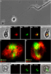
Imaging of synaptic ribbon-associated endocytosis in isolated mouse photoreceptors. Isolated mouse photoreceptors (shown in A) were incubated for 2 min with sulforhodamine (SR101), which is taken up by fluid-phase endocytosis. Afterward, SR101-loaded photoreceptors were fixed and immnunolabeled with antibodies against RIBEYE(B)-domain/CtBP2. A hotspot of SR101 uptake is found in close association with the synaptic ribbon (B–D; and data not shown). B was obtained by conventional imaging; C and D are maximum projections of z-stacks from confocal imaging. Pretreatment with dynasore, a specific inhibitor of dynamin (100 μm), completely inhibited the synaptic ribbon-associated uptake of SR101 (red channel) (E and data not shown). Scale bars, 1 μm.
Figure 17.
Figure 17.
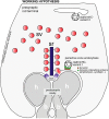
Simplified, schematic summary of the immunolocalization data presented in the manuscript. Key players of endocytic membrane traffic, including dynamin, dynamin-binding proteins, and CHC-V1, are enriched in a periactive zone of photoreceptor synapses. Besides CHC-V1, CHC-V2 is also present in the presynaptic photoreceptor terminal. In contrast to CHC-V1, CHC-V2 is located a large distance from the periactive zone, possibly on an endosomal compartment in the proximal region of the presynaptic terminal. Endosomal-like membrane compartments have been previously observed by transmission electron microscopy in this part of the photoreceptor terminal (Ripps et al., 1976; Schacher et al., 1976; Schaeffer and Raviola, 1978; Cooper and McLaughlin, 1983). The drawing of the photoreceptor terminal is modified based on a drawing of Gray and Pease (1971). sr, Synaptic ribbon; sv, synaptic vesicles; clathrinHC-V1, clathrin heavy chain variant 1, CHC-V1; clathrinHC-V2, CHC-V2; CaV, voltage-gated calcium channels of the photoreceptor active zone; h, postsynaptic dendritic tip of a horizontal cell; b, postsynaptic dendritic tip of an invaginating bipolar cell; bsn, bassoon.

Similar articles

Cited by

References

    1. Aartsen WM, Arsanto JP, Chauvin JP, Vos RM, Versteeg I, Cardozo BN, Bivic AL, Wijnholds J. PSD95beta regulates plasma membrane Ca2+-pump localization at the photoreceptor synapse. Mol Cell Neurosci. 2009;41:156–165. doi: 10.1016/j.mcn.2009.02.003. - DOI - PubMed
    1. Alpadi K, Magupalli VG, Käppel S, Köblitz L, Schwarz K, Seigel GM, Sung CH, Schmitz F. RIBEYE recruits Munc119, the mammalian ortholog of the Caenorhabditis elegans protein unc119, to synaptic ribbons of photoreceptor synapses. J Biol Chem. 2008;283:26461–26467. doi: 10.1074/jbc.M801625200. - DOI - PMC - PubMed
    1. Amstutz B, Gastaldelli M, Kälin S, Imelli N, Boucke K, Wandeler E, Mercer J, Hemmi S, Greber UF. Subversion of CtBP1-controlled macropinocytosis by human adenovirus serotype3. EMBO J. 2008;27:956–969. doi: 10.1038/emboj.2008.38. - DOI - PMC - PubMed
    1. Andresen BT, Rizzo MA, Shome K, Romero G. The role of phosphatidic acid in the regulation of the Ras/MEK/Erk signalling cascade. FEBS Lett. 2002;531:65–68. doi: 10.1016/S0014-5793(02)03483-X. - DOI - PubMed
    1. Babai N, Bartoletti TM, Thoreson WB. Calcium regulates vesicle replenishment at the cone ribbon synapse. J Neurosci. 2010;30:15866–15877. doi: 10.1523/JNEUROSCI.2891-10.2010. - DOI - PMC - PubMed

Publication types

MeSH terms

Substances