. 2013 Jul;246(7):571-9.
doi: 10.1007/s00232-013-9571-6. Epub 2013 Jun 19.

β1-adrenergic receptor recycles via a membranous organelle, recycling endosome, by binding with sorting nexin27

Affiliations

β1-adrenergic receptor recycles via a membranous organelle, recycling endosome, by binding with sorting nexin27

Takatoshi Nakagawa et al. J Membr Biol. 2013 Jul.

Abstract

In cardiomyocytes, β1-adrenergic receptor (β1-AR) plays an important role in regulating cardiac functions. Upon continuous ligand stimulation, β1-AR is internalized and mostly recycled back to the plasma membrane (PM). The recycling endosome (RE) is one of the membranous organelles involved in the protein recycling pathway. To determine whether RE is involved in the internalization of β1-AR upon ligand stimulation, we evaluated the localization of β1-AR after stimulation with a β-agonist, isoproterenol (Iso), in β1-AR-transfected COS-1 cells. After 30 min of Iso treatment and cell surface labeling with the appropriate antibodies, β1-AR was internalized from PM and translocated into the perinuclear region, the same location as the transferrin receptor, an RE marker. We then evaluated whether sorting nexin 27 (SNX27) participated in the β1-AR recycling pathway. When β1-AR and SNX27 were coexpressed, β1-AR coimmunoprecipitated with SNX27. In addition, shRNA-mediated silencing of SNX27 compromised β1-AR recycling and enhanced its delivery into lysosome. Overall, β1-AR on PM was internalized into RE upon Iso stimulation and recycled by RE through binding with SNX27 in COS-1 cells.


Figures
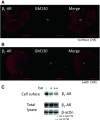
Fig. 1
Fig. 1
Steady-state localization and internalization of β1-AR upon Iso stimulation in COS-1 cells. β1-AR cells were treated without (a) or with (b) cycloheximide (CHX, 50 μg/ml) for 4 h and stained with anti-HA or anti-GM130 Abs. Images were captured using the LSM500. c Internalization of β1-AR from the cell surface. β1-AR cells were biotinylated using 0.1 mg/ml NHS-sulfo-biotin on ice for 30 min, quenched with 0.1 M glycine, washed with prewarmed DMEM thrice and then incubated with 2.5 × 10−7 (+) or 10−6 M (++) of Iso for 2 h. Biotinylated proteins concentrated with avidin-agarose were subjected to SDS-PAGE followed by Western blotting (upper panel cell surface proteins probed with anti-β1-AR Ab; middle and lower panels total proteins probed with anti-β1-AR Ab or anti-β-actin Ab, respectively). Bar 20 μm
Fig. 2
Fig. 2
Effects of Iso stimulation on the internalization of β1-AR into RE in COS-1 cells. β1-AR cells were plated onto glass coverslips. Cells were incubated with or without 10−6 M Iso for 30 min in the presence of anti-HA Ab. Cells were then fixed with 4 % paraformaldehyde followed by 0.1 % Triton X-100 (for 10 min in each step). Anti-TfnR Ab was added and incubated for 1 h at room temperature. Abs against HA or TfnR were probed with Alexa546-labeled anti-rabbit or Alexa488-labeled anti-mouse Abs, respectively. A laser scanning microscope (LSM500) was used to capture the images (upper panel images without Iso; lower panel images with Iso for 30 min)
Fig. 3
Fig. 3
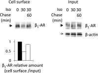
Recycling of internalized β1-AR to the cell surface after Iso stimulation in COS-1 cells. Cells expressing β1-AR were incubated with 10−6 M Iso. After 30 min of incubation, Iso was removed and the cells were further incubated for 60 min. To quantify the amount of β1-AR on the cell surface, cells were then biotinylated. Biotinylated proteins were subjected to SDS-PAGE and Western blot analyses. Cell surface biotinylated proteins probed with anti-β1-AR Ab; Input total proteins probed with β1-AR (upper) or β-actin Ab (lower). Lower panel Quantitative analyses of β1-AR on the cell surface relative to total β1-AR were densitometrically analyzed using Quantity One (Bio-Rad). The ratio of β1-AR on the cell surface relative to total β1-AR normalized with β-actin is shown. Most of the β1-AR that was once internalized by Iso treatment was recovered to the cell surface at 60 min after the removal of Iso (arrow)
Fig. 4
Fig. 4
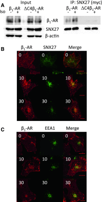
Role of SNX27 in β1-AR recycling after Iso stimulation in COS-1 cells. Coimmunoprecipitation of β1-AR or ∆C4β1-AR with SNX27. COS-1 cells were transfected with β1-AR or ∆C4β1-AR and SNX27 (myc-tagged). Cells were stimulated with or without 10−6 M Iso for 10 min, lysed and coimmunoprecipitated with anti-Myc Ab. Precipitated proteins were separated by SDS-PAGE and immunoblotted with anti-β1-AR or Myc Abs. Lysates (Input) were also probed with the same Abs as well as anti-β-actin Ab. Localization of β1-AR and SNX27 (b) or EE (c). β1-AR was transfected into COS-1 cells with or without SNX27 (b, c, respectively). Transfected cells were plated onto glass coverslips, treated with 10−6 M Iso for 10 and 30 min in the presence of anti-HA Ab and coimmunostained with anti-Myc Ab (b) or anti-EEA1 Ab (c) as described in “Materials and Methods” images were taken with the LSM500 microscope. Bar 20 μm
Fig. 5
Fig. 5
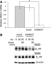
Effects of SNX27 silencing on β1-AR recycling after Iso stimulation in COS-1 cells. a Evaluation of shSNX27 in COS-1 cells. shSNX27 was transfected into COS-1 cells, and 48 h later, total cellular RNA was extracted, reverse-transcribed and subjected to real-time RT-PCR analysis. SNX27 expression levels in transfected cells were compared with those in nontransfected and empty vector–transfected COS-1 cells. b Effects of SNX27 silencing on β1-AR recycling. β1-AR cells were transfected with shSNX27 or an empty vector (mock), and 48 h later, cells were treated without or with 10−6 M Iso for 30 min (0 and 30, respectively). After incubation, cells were washed to remove Iso and further incubated for 60 min (chase) (Iso 30, Chase 60). Cells were then biotinylated and lysed. Biotinylated proteins were collected with avidin-agarose and subjected to SDS-PAGE, followed by Western blot analysis using anti-β1-AR and β-actin Abs. The p value was calculated using a Dunnett’s test. *p < 0.05
Fig. 6
Fig. 6
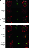
Effects of SNX27 silencing on the subcellular localization of β1-AR after Iso stimulation in COS-1 cells. Either empty vector (a) or shSNX27 (b) was transfected into β1-AR cells, and 24 h later, cells were plated onto glass coverslips. After 48 h, cells were treated without or with 10−6 M Iso for 30 min (−Iso and Iso 30 min, respectively). After incubation, cells were washed to remove Iso and further incubated for 60 min (chase) (Iso 30 and Chase 60 min) to follow the cell surface recovery of β1-AR. During Iso incubation, anti-HA Abs were added to label cell surface proteins. After incubation, cells were fixed and the internalized β1-AR (labeled with anti-HA Ab) was costained with anti-TfnR Ab. Bar 20 μm
Fig. 7
Fig. 7
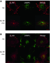
Effects of SNX27 silencing on the degradation of β1-AR after Iso stimulation in COS-1 cells. Empty vector (a) or shSNX27 (b) was transfected into β1-AR cells, and 24 h later, cells were plated onto glass coverslips. After 48 h, cells were treated without or with 10−6 M Iso for 30 min (−Iso and Iso 30 min, respectively). During incubation with Iso, anti-HA Ab was added to label cell surface proteins. After incubation, cells were fixed and the internalized β1-AR (labeled with anti-HA Ab) was costained with anti-LAMP1 Ab. Bar 20 μm
Fig. 8
Fig. 8
Scheme of a possible role of SNX27 in β1-AR intracellular traffic. Continuous ligand stimulation induces the internalization of β1-AR from the cell surface. β1-AR is first transported to EE, where SNX27 mainly localizes, and sorted into either pathway, recycling or degradation. The selection might depend on binding with SNX27. Namely, it is possible that β1-AR is recycled to PM through RE by binding with SNX27 in EE, while it is sorted into lysosome for degradation without binding with SNX27

Similar articles

Cited by

References

    1. Ang AL, Taguchi T, Francis S, Folsch H, Murrells LJ, Pypaert M, Warren G, Mellman I. Recycling endosomes can serve as intermediates during transport from the Golgi to the plasma membrane of MDCK cells. J Cell Biol. 2004;167:531–543. doi: 10.1083/jcb.200408165. - DOI - PMC - PubMed
    1. Balana B, Maslennikov I, Kwiatkowski W, Stern KM, Bahima L, Choe S, Slesinger PA. Mechanism underlying selective regulation of G protein-gated inwardly rectifying potassium channels by the psychostimulant-sensitive sorting nexin 27. Proc Natl Acad Sci USA. 2010;108:5831–5836. doi: 10.1073/pnas.1018645108. - DOI - PMC - PubMed
    1. Cai L, Loo LS, Atlashkin V, Hanson BJ, Hong W. Deficiency of sorting nexin 27 (SNX27) leads to growth retardation and elevated levels of N-methyl-d-aspartate receptor 2C (NR2C) Mol Cell Biol. 2011;31:1734–1747. doi: 10.1128/MCB.01044-10. - DOI - PMC - PubMed
    1. Cao TT, Deacon HW, Reczek D, Bretscher A, von Zastrow M. A kinase-regulated PDZ-domain interaction controls endocytic sorting of the beta2-adrenergic receptor. Nature. 1999;401:286–290. doi: 10.1038/45816. - DOI - PubMed
    1. Cullen PJ. Endosomal sorting and signalling: an emerging role for sorting nexins. Nat Rev Mol Cell Biol. 2008;9:574–582. doi: 10.1038/nrm2427. - DOI - PubMed

LinkOut - more resources