

Functional OCT4-specific CD4+ and CD8+ T cells in healthy controls and ovarian cancer patients
- PMID: 23762805
- PMCID: PMC3667911
- DOI: 10.4161/onci.24271
Functional OCT4-specific CD4+ and CD8+ T cells in healthy controls and ovarian cancer patients
Abstract
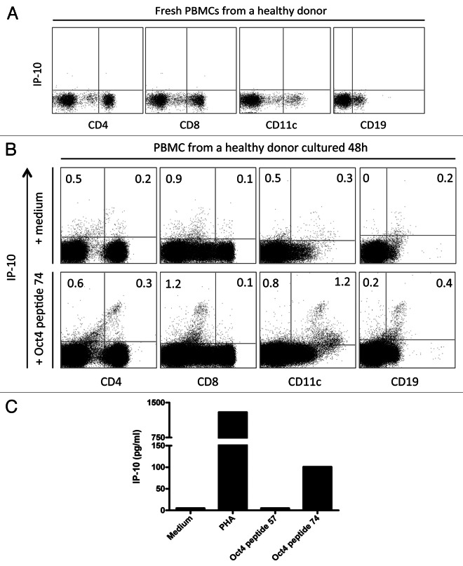
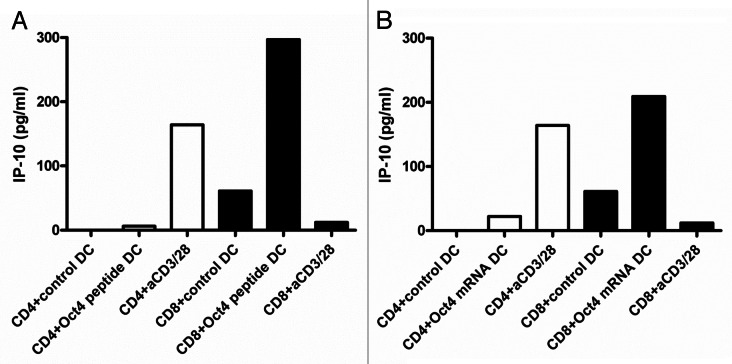
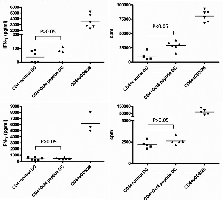
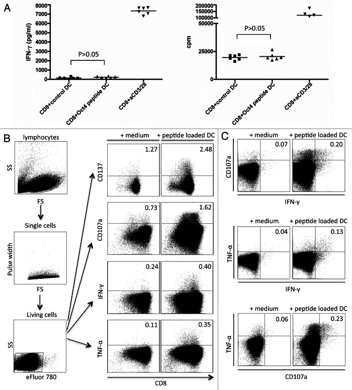
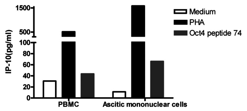
The identification of growth and differentiation pathways that are responsible for the proliferation and survival of cancer stem cells (CSCs) has opened avenues for the discovery of novel therapeutic targets. In the initial phase of an anticancer immune response, T cells specific for tumor-associated antigens develop in patients and, at least under selected circumstances, are able to eliminate malignant cells. However, it remains unknown whether CSC-specific T cells are also operational. We found naturally occurring multifunctional CD4+ and CD8+ T cells specific for the stem cell marker OCT4 among the peripheral blood mononuclear cells (PBMCs) of both healthy individuals and ovarian cancer patients. Moreover, lymphocytes isolated from the ascites of patients affected by ovarian malignancies also contained OCT4-specific T cells. OCT4-reactive CD4+ T cells did not produce interferon γ (IFNγ) and IFNγ-inducible protein 10 (IP-10) but were capable of proliferation upon stimulation with dendritic cells (DCs) loaded with an OCT4-derived peptide or OCT4 mRNA. OCT4-reactive CD8+ cells did not proliferate in response to a similar challenge, yet produced IP-10 as well as sufficient amounts of IFNγ to induce IP-10 . Furthermore, CD8+ cytotoxic T cells were able to release their lysosomal components, as indicated by the mobilization of CD107a. These results demonstrate the existence of anti-CSC specific T cells in ovarian cancer patients.
Keywords: CD4; CD8; IP-10; ovarian cancer; proliferation; stem cell markers.
Figures
Similar articles
-
Exosomes in the ascites of ovarian cancer patients: origin and effects on anti-tumor immunity.Oncol Rep. 2011 Mar;25(3):749-62. doi: 10.3892/or.2010.1119. Epub 2010 Dec 22. Oncol Rep. 2011. PMID: 21181093
-
[Lymphocyte activation markers in patients with ovarian cancer].Ginekol Pol. 2012 Oct;83(10):737-43. Ginekol Pol. 2012. PMID: 23383558 Polish.
-
Breast cancer stem cell RNA-pulsed dendritic cells enhance tumor cell killing by effector T cells.Oncol Lett. 2020 Mar;19(3):2422-2430. doi: 10.3892/ol.2020.11338. Epub 2020 Jan 23. Oncol Lett. 2020. PMID: 32194742 Free PMC article.
-
Effect of human bone marrow mesenchymal stromal cells on cytokine production by peripheral blood naive, memory, and effector T cells.Stem Cell Res Ther. 2015 Jan 5;6(1):3. doi: 10.1186/scrt537. Stem Cell Res Ther. 2015. PMID: 25559824 Free PMC article.
-
Dendritic cell gene therapy.Surg Oncol Clin N Am. 2002 Jul;11(3):645-60. doi: 10.1016/s1055-3207(02)00027-3. Surg Oncol Clin N Am. 2002. PMID: 12487060 Review.
Cited by
-
Ovarian cancer stem cells: Critical roles in anti-tumor immunity.Front Genet. 2022 Nov 10;13:998220. doi: 10.3389/fgene.2022.998220. eCollection 2022. Front Genet. 2022. PMID: 36437919 Free PMC article. Review.
-
Monitoring cancer stem cells: insights into clinical oncology.Onco Targets Ther. 2016 Feb 11;9:731-40. doi: 10.2147/OTT.S96645. eCollection 2016. Onco Targets Ther. 2016. PMID: 26929644 Free PMC article.
-
Immune evasion by cancer stem cells.Regen Ther. 2021 Mar 11;17:20-33. doi: 10.1016/j.reth.2021.02.006. eCollection 2021 Jun. Regen Ther. 2021. PMID: 33778133 Free PMC article. Review.
-
Immune Curbing of Cancer Stem Cells by CTLs Directed to NANOG.Front Immunol. 2018 Jun 19;9:1412. doi: 10.3389/fimmu.2018.01412. eCollection 2018. Front Immunol. 2018. PMID: 29971070 Free PMC article.
-
Tumor microenvironment of cancer stem cells: Perspectives on cancer stem cell targeting.Genes Dis. 2023 Jul 19;11(3):101043. doi: 10.1016/j.gendis.2023.05.024. eCollection 2024 May. Genes Dis. 2023. PMID: 38292177 Free PMC article. Review.
References
-
- Bamias A, Koutsoukou V, Terpos E, Tsiatas ML, Liakos C, Tsitsilonis O, et al. Correlation of NK T-like CD3+CD56+ cells and CD4+CD25+(hi) regulatory T cells with VEGF and TNFalpha in ascites from advanced ovarian cancer: Association with platinum resistance and prognosis in patients receiving first-line, platinum-based chemotherapy. Gynecol Oncol. 2008;108:421–7. doi: 10.1016/j.ygyno.2007.10.018. - DOI - PubMed
Publication types
LinkOut - more resources
Full Text Sources
Other Literature Sources
Research Materials
Miscellaneous