. 2013 Jun 6;8(6):e64877.
doi: 10.1371/journal.pone.0064877. Print 2013.

Akt-signal integration is involved in the differentiation of embryonal carcinoma cells

Affiliations

Akt-signal integration is involved in the differentiation of embryonal carcinoma cells

Bo Chen et al. PLoS One. .

Abstract

The mechanism by which Akt modulates stem cell homeostasis is still incompletely defined. Here we demonstrate that Akt phosphorylates special AT-rich sequences binding protein 1 (SATB1) at serine 47 and protects SATB1 from apoptotic cleavage. Meanwhile, Akt phosphorylates Oct4 at threonine 228 and Klf4 at threonine 399, and accelerates their degradation. Moreover, PI3K/Akt signaling enhances the binding of SATB1 to Sox2, thereby probably impairing the formation of Oct4/Sox2 regulatory complexes. During retinoic acid (RA)-induced differentiation of mouse F9 embryonal carcinoma cells (ECCs), the Akt activation profile as well as its substrate spectrum is strikingly correlated with the down-regulation of Oct4, Klf4 and Nanog, which suggests Akt activation is coupled to the onset of differentiation. Accordingly, Akt-mediated phosphorylation is crucial for the capability of SATB1 to repress Nanog expression and to activate transcription of Bcl2 and Nestin genes. Taken together, we conclude that Akt is involved in the differentiation of ECCs through coordinated phosphorylations of pluripotency/differentiation factors.


Conflict of interest statement

Competing Interests: The authors have declared that no competing interests exist.

Figures

Figure 1
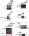
Figure 1. Akt phosphorylates SATB1 at serine 47 in a PI3K-dependent manner.
(A) Akt phosphorylates GST-SATB1 1–204 and GST-SATB1 1-495. Purified GST fusion proteins were subjected to in vitro kinase assay in the presence of immunoprecipitated Akt. The samples were resolved on 10% SDS-PAGE and blotted with anti-phospho-Akt substrate, anti-phospho-Akt (T308) and anti-Myc. (B) The phosphorylation site resides on SATB1 within an N-terminal fragment spanning amino acids 1–51. (C) Akt phosphorylates GST-SATB1, but not GST-SATB1 S47A or GST-SATB1 S47D. (D) Ectopically expressed Myc-SATB1 is phosphorylated by Akt. Myc-SATB1 was co-expressed with Myc-Akt (WT), Myc-Akt (Myr) or Myc-Akt (DN) in HEK293T cells. Whole-cell lysates were subjected to immunoblotting with anti-phospho-Akt substrate, anti-Myc and anti-Actin. (E) The anti-phospho-SATB1 (S47) raised in rabbit specifically recognizes the phosphorylated SATB1 at serine 47. (F) HEK293T cells were transfected with empty vector or Myc-SATB1, 24 h post-transfection, cells were rinsed twice with PBS and replenished with fresh DMEM with no FBS supplemented. After additional 24 h, cells were treated with LY294002 (20 µM) for 2 h, followed by 20 min stimuli with or without IGF-1 (50 ng/ml) or FBS (20%). Whole-cell lysates were subjected to western blotting. (G) HEK293T cells were treated as indicated. Whole cell lysates were subjected to western blotting with antibodies of anti-phospho-SATB1 (S47) and anti-Myc. HC, heavy chain; WCL, whole cell lysate; Myr, myristoylation.
Figure 2
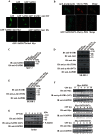
Figure 2. Akt shields SATB1 from apoptotic cleavage.
(A) Evenly distributed signals were observed in the majority of HEK293A cells transfected with GFP-SATB1, whereas punctate structures were detected in a small fraction of cells (white arrowhead). Akt (Myr) decreased the ratio of cells with dot-like signals. (B) In cells with dot-like structures, GFP-SATB1S47A colocalized with Cherry-PML, whereas GFP-SATB1S47D did not. (C) Expression of SATB1 was analyzed in MCF-7 cells stably integrated with empty vector, wild-type SATB1, SATB1S47A or SATB1S47D, respectively. (D) Expression of SATB1 and Akt activation were analyzed in stable SK-BR-3 cell lines. (E) SATB1 stability and Akt activation were documented in SK-BR-3 cells carrying wild-type SATB1 together with Akt (Myr) or Akt (DN). (F) HEK293A cells were transfected with Myc-SATB1, Myc-SATB1S47A or Myc-SATB1S47D, respectively, and treated with CPT for 0, 2, 4, 6, 8 or 10 h. The cell lysates were subjected to immunobloting with anti-SATB1, anti-phospho-SATB1 (S47) and anti-GAPDH. (G) Jurkat cells were treated with CPT as in (F). The cell lysates were subjected to immunoblotting with anti-SATB1, anti-phospho-SATB1 (S47) and anti-GAPDH.
Figure 3
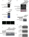
Figure 3. Akt phosphorylates Oct4 and accelerates its degradation.
(A) Oct4 phosphorylation by Akt is ATP-dependent. (B) Akt phosphorylates GST-Oct4, but not GST, GST-Oct4T228A, GST-Oct4T228D or GST-Oct4T228E. Under the same reaction condition, mutation of serine 229 to alanine or aspartic acid significantly reduced the phosphorylation efficiency. (C) The anti-phospho-Oct4 antibody (T228) was raised in rabbit and its specificity was tested using in vitro kinase assay. (D) Akt phosphorylates wild-type Oct4, rather than its mutants at threonine 228. Flag-GFP-Oct4 or its mutants was ectopically expressed together with Myc-Akt (Myr) in HEK293T cells. Immunoprecipitates with anti-Flag were subjected to immunoblotting with anti-phospho-Akt substrate. (E) Expression of GFP-tagged Oct4 and its mutants in HEK293A cells. (F) Half-life of Oct4 and its mutants in transfected HEK293A cells treated with CHX (15 µg/ml). (G) HEK293A cells were transfected with Flag-GFP-Oct4 together with Myc-Akt (WT), Myc-Akt (Myr) or Myc-Akt (DN) and were treated with DMSO or MG132 (20 µM) for 5 h. Immunoprecipitates with anti-Flag were subjected to immunoblotting with anti-phospho-Akt substrate and anti-Flag.
Figure 4
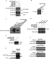
Figure 4. Akt phosphorylates Klf4 at threonine 399 and increases its degradation.
(A) Akt associates with and phosphorylates Klf4. Reciprocal immunoprecipitations were carried out in HEK293A cells transfected as indicated. (B and C) Akt phosphorylates Klf4 at threonine 399, rather than threonine 397. (D) Akt phosphorylates Flag-Klf4 in transfected HEK293A cells. Immunoprecipitates with anti-Flag were subjected to immunoblotting with anti-phospho-Akt substrate and anti-Flag. (E) Klf4T399E is poorly expressed in transiently transfected HEK293A cells. Flag-Klf4, Flag-Klf4T399A or Flag-Klf4T399E was transfected together with Flag-Wwp2. Cell lystaes were subjected to immunoblotting with anti-Flag. (F) Half-life of Flag-Klf4 and its mutants in transfected HEK293A cells treated with CHX (15 µg/ml). (G) Inhibition of Akt activation by PI3K inhibitor LY294002 reduces Klf4 ubiquitination. Flag-tagged Klf4 and His-tagged Ubiquitin were co-expressed in HEK293A cells. Cells were serum starved and treated with LY294002 (20 µM) for 5 h, followed by 5 h with or without MG132 (20 µM). Immunoprecipitates with anti-Flag were subjected to immunoblotting with anti-Flag and anti-Ubiquitin. WCL, whole cell lysates.
Figure 5
Figure 5. SATB1 binding to Sox2 is enhanced by Akt signaling.
(A) SATB1 associates with Sox2, but not Oct4 or Klf4. (B) Endogenous interaction between SATB1 and Sox2. SATB1 was immuprecipitated from F9 cell lysates, immuprecipitations were subjected to immunblotting with anti-SATB1 and anti-Sox2. (C) Mapping of SATB1 domain required for Sox2 binding using GST pull-down assay. (D) A schematic representation of Flag-GFP-tagged Sox2 constructs is shown. (E) Mapping of Sox2 domains that are necessary for SATB1 binding. (F) SATB1S47A sequesters much less Sox2 than wild-type SATB1 and SATB1S47D. (G) Inhibition of Akt signaling disrupts SATB1/Sox2 interaction. HEK293T cells were co-transfected with Myc-SATB1 and Flag-Sox2, serum-starved and treated as indicated.
Figure 6
Figure 6. Akt-mediated phosphorylation is critical for the regulatory role of SATB1 in F9 cell differentiation and Nanog expression.
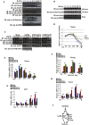
(A) The profiles of Akt activity and its substrates match the change of pluripotency factors during RA-induced F9 cell differentiation process. F9 cells were seeded on petri dishes, induced with RA (1 µM) and harvested at 0, 1, 2, 4, 6, 8, 12, 24, 48 or 72 h. Cell lysates were subjected to immunoblotting with antibodies of anti-Oct4, anti-Nanog, anti-SATB1, anti-Klf4, anti-Akt1, anti-phospho-Akt (S473), anti-phospho-Akt (T308), anti-phospho-Akt substrate and anti-GAPDH. (B) The F9 stable cell lines were induced in the presence of RA as in (A) and harvested at 0, 2, 4, 8, 12, 24, 48 or 72 h, Immunoprecipitates with anti-Oct4 were subjected to immunoblotting with anti-Oct4 and anti-phospho-Oct4 (T228). (C) SATB1 and SATB1S47D are more efficient than SATB1S47A with respect to Nanog repression. The F9 stable cell lines were induced in the presence of RA as in (A) and harvested at 12, 24, 48 or 72 h. Cell lysates were subjected to immunoblotting with anti-Nanog, anti-Oct4, anti-Klf4, anti-SATB1, anti-phospho-SATB1 and anti-GAPDH. (D) A schematic representation of the dynamic change of Nanog expression in Figure 6C is shown. (E) The F9 stable cell lines were induced as in (C) and quantitative RT-PCR was performed to analyze the transcription level of Nanog. Results are from three independent experiments. (F) The F9 stable cell lines were induced with RA for 0, 12 or 24h and SATB1 occupancy on Nanog locus was documented using ChIP assay. (G and H) The F9 stable cell lines were induced as in (C) and quantitative RT-PCR was performed for Bcl2 and Nestin, two differentiation genes. (I) A working model for Akt-involved pluripotency/differentiation switch. See Discussion for details. The error bars in (E), (F), (G) and (H) represent mean ± SD from three independent experiments. Student’s t-test was performed between wild-type SATB1 and SATB1S47A groups (*p<0.05; **p<0.01).

Similar articles

Cited by

References

    1. Yilmaz OH, Valdez R, Theisen BK, Guo W, Ferguson DO, et al. (2006) Pten dependence distinguishes haematopoietic stem cells from leukaemia-initiating cells. Nature 441: 475–482. - PubMed
    1. Zhang J, Grindley JC, Yin T, Jayasinghe S, He XC, et al. (2006) PTEN maintains haematopoietic stem cells and acts in lineage choice and leukaemia prevention. Nature 441: 518–522. - PubMed
    1. Ito K, Bernardi R, Morotti A, Matsuoka S, Saglio G, et al. (2008) PML targeting eradicates quiescent leukaemia-initiating cells. Nature 453: 1072–1078. - PMC - PubMed
    1. Chen C, Liu Y, Liu R, Ikenoue T, Guan KL, et al. (2008) TSC-mTOR maintains quiescence and function of hematopoietic stem cells by repressing mitochondrial biogenesis and reactive oxygen species. J Exp Med 205: 2397–2408. - PMC - PubMed
    1. Matsuoka S, Oike Y, Onoyama I, Iwama A, Arai F, et al. (2008) Fbxw7 acts as a critical fail-safe against premature loss of hematopoietic stem cells and development of T-ALL. Genes Dev 22: 986–991. - PMC - PubMed

Publication types

MeSH terms

Grants and funding

This work was supported by the National Basic Research Program of China (2011CB965203 to WD and YH); the National High Technology Research and Development Program of China (2011AA020118 to WD); and the intra-institutional funding program (2010PY05 to WD). The funders had no role in study design, data collection and analysis, decision to publish, or preparation of the manuscript.