The 2009 pandemic (H1N1) viruses isolated from pigs show enhanced pathogenicity in mice
- PMID: 23758678
- PMCID: PMC3686621
- DOI: 10.1186/1297-9716-44-41
The 2009 pandemic (H1N1) viruses isolated from pigs show enhanced pathogenicity in mice
Abstract
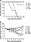
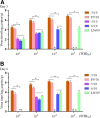
Since the emergence of the 2009 pandemic (H1N1) virus (2009/H1N1) in April 2009, cases of transmission from humans to pigs have been reported frequently. In our previous studies, four 2009/H1N1 variants were isolated from pigs. To better understand the phenotypic differences of the pig isolates compared with the human isolate, in this study mice were inoculated intranasally with different 2009/H1N1 viruses, and monitored for morbidity, mortality, and viral replication, cytokine production and pathological changes in the lungs. The results show that all isolates show effective replication in lungs, but varying in their ability to cause morbidity. In particular, the strains of A/swine/Nanchang/3/2010 (H1N1) and A/swine/Nanchang/F9/2010 (H1N1) show the greatest virulence with a persisting replication in lungs and high lethality for mice, compared with the human isolate A/Liaoning /14/2009 (H1N1), which shows low virulence in mice. Furthermore, the lethal strains could induce more severe lung pathological changes and higher production of cytokines than that of other strains at an early stage. Amino acid sequence analysis illustrates prominent differences in viral surface glycoproteins and polymerase subunits between pig isolates and human strains that might correlate with their phenotypic differences. These studies demonstrate that the 2009/H1N1 pig isolates exhibit heterogeneous infectivity and pathogencity in mice, and some strains possess an enhanced pathogenicity compared with the human isolate.
Figures


Similar articles
-
Pathogenesis of pandemic influenza A (H1N1) and triple-reassortant swine influenza A (H1) viruses in mice.J Virol. 2010 May;84(9):4194-203. doi: 10.1128/JVI.02742-09. Epub 2010 Feb 24. J Virol. 2010. PMID: 20181710 Free PMC article.
-
Two amino acid residues in the N-terminal region of the polymerase acidic protein determine the virulence of Eurasian avian-like H1N1 swine influenza viruses in mice.J Virol. 2024 Oct 22;98(10):e0129324. doi: 10.1128/jvi.01293-24. Epub 2024 Aug 30. J Virol. 2024. PMID: 39212447 Free PMC article.
-
Experimental infection with a Thai reassortant swine influenza virus of pandemic H1N1 origin induced disease.Virol J. 2013 Mar 16;10:88. doi: 10.1186/1743-422X-10-88. Virol J. 2013. PMID: 23497073 Free PMC article.
-
Comparative virulence of wild-type H1N1pdm09 influenza A isolates in swine.Vet Microbiol. 2015 Mar 23;176(1-2):40-9. doi: 10.1016/j.vetmic.2014.12.021. Epub 2014 Dec 31. Vet Microbiol. 2015. PMID: 25601799
-
The R251K Substitution in Viral Protein PB2 Increases Viral Replication and Pathogenicity of Eurasian Avian-like H1N1 Swine Influenza Viruses.Viruses. 2020 Jan 2;12(1):52. doi: 10.3390/v12010052. Viruses. 2020. PMID: 31906472 Free PMC article.
Cited by
-
PB2-588I enhances 2009 H1N1 pandemic influenza virus virulence by increasing viral replication and exacerbating PB2 inhibition of beta interferon expression.J Virol. 2014 Feb;88(4):2260-7. doi: 10.1128/JVI.03024-13. Epub 2013 Dec 11. J Virol. 2014. PMID: 24335306 Free PMC article.
-
Comparison of the virulence of three H3N2 canine influenza virus isolates from Korea and China in mouse and Guinea pig models.BMC Vet Res. 2018 May 2;14(1):149. doi: 10.1186/s12917-018-1469-1. BMC Vet Res. 2018. PMID: 29716608 Free PMC article.
-
Sus scrofa miR-204 and miR-4331 Negatively Regulate Swine H1N1/2009 Influenza A Virus Replication by Targeting Viral HA and NS, Respectively.Int J Mol Sci. 2017 Apr 3;18(4):749. doi: 10.3390/ijms18040749. Int J Mol Sci. 2017. PMID: 28368362 Free PMC article.
-
Effect of AcHERV-GmCSF as an Influenza Virus Vaccine Adjuvant.PLoS One. 2015 Jun 19;10(6):e0129761. doi: 10.1371/journal.pone.0129761. eCollection 2015. PLoS One. 2015. PMID: 26090848 Free PMC article.
References
-
- Dawood FS, Jain S, Finelli L, Shaw MW, Lindstrom S, Garten RJ, Gubareva LV, Xu X, Bridges CB, Uyeki TM. Emergence of a novel swine-origin influenza A (H1N1) virus in humans. N Engl J Med. 2009;360:2605–2615. - PubMed
-
- Forgie SE, Keenliside J, Wilkinson C, Webby R, Lu P, Sorensen O, Fonseca K, Barman S, Rubrum A, Stigger E, Marrie TJ, Marshall F, Spady DW, Hu J, Loeb M, Russell ML, Babiuk LA. Swine outbreak of pandemic influenza A virus on a Canadian research farm supports human-to-swine transmission. Clin Infect Dis. 2011;52:10–18. doi: 10.1093/cid/ciq030. - DOI - PMC - PubMed
-
- Sreta D, Tantawet S, Na Ayudhya SN, Thontiravong A, Wongphatcharachai M, Lapkuntod J, Bunpapong N, Tuanudom R, Suradhat S, Vimolket L, Poovorawan Y, Thanawongnuwech R, Amonsin A, Kitikoon P. Pandemic (H1N1) 2009 virus on commercial swine farm, Thailand. Emerg Infect Dis. 2010;16:1587–1590. doi: 10.3201/eid1610.100665. - DOI - PMC - PubMed
Publication types
MeSH terms
Substances
LinkOut - more resources
Full Text Sources
Other Literature Sources
Miscellaneous

