. 2013 May 28:6:14.
doi: 10.3389/fnmol.2013.00014. eCollection 2013.

Alcohol and NMDA receptor: current research and future direction

Affiliations

Alcohol and NMDA receptor: current research and future direction

Raman Chandrasekar. Front Mol Neurosci. .

Abstract

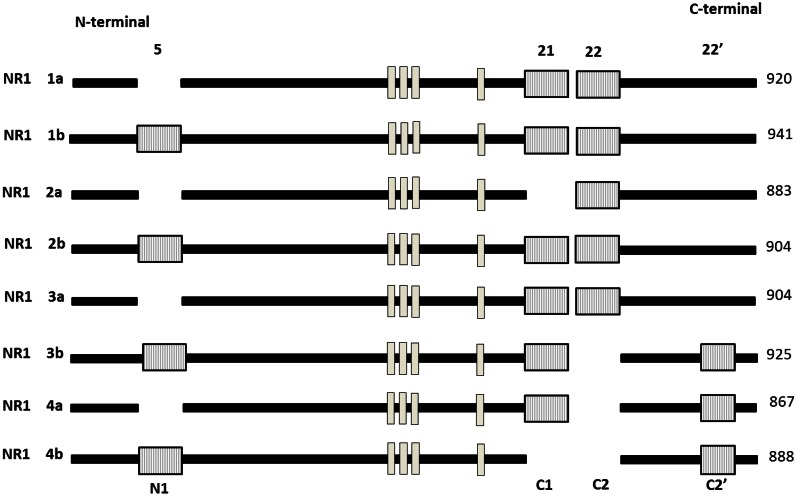
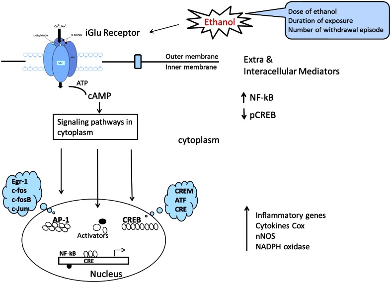
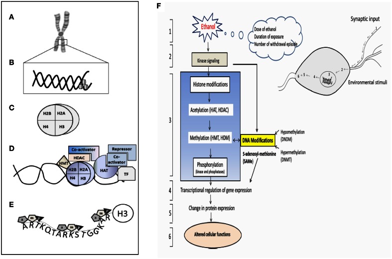
The brain is one of the major targets of alcohol actions. Most of the excitatory synaptic transmission in the central nervous system is mediated by N-methyl-D-aspartate (NMDA) receptors. However, one of the most devastating effects of alcohol leads to brain shrinkage, loss of nerve cells at specific regions through a mechanism involving excitotoxicity, oxidative stress. Earlier studies have indicated that chronic exposure to ethanol both in vivo and in vitro, increases NR1 and NR2B gene expression and their polypeptide levels. The effect of alcohol and molecular changes on the regulatory process, which modulates NMDAR functions including factors altering transcription, translation, post-translational modifications, and protein expression, as well as those influencing their interactions with different regulatory proteins (downstream effectors) are incessantly increasing at the cellular level. Further, I discuss the various genetically altered mice approaches that have been used to study NMDA receptor subunits and their functional implication. In a recent countable review, epigenetic dimension (i.e., histone modification-induced chromatin remodeling and DNA methylation, in the process of alcohol related neuroadaptation) is one of the key molecular mechanisms in alcohol mediated NMDAR alteration. Here, I provide a recount on what has already been achieved, current trends and how the future research/studies of the NMDA receptor might lead to even greater engagement with many possible new insights into the neurobiology and treatment of alcoholism.

Keywords: NMDA receptor; RNA-binding protein; alcohol; epigenetic; fetal cortical neurons; glutamate; splice variant; transcription.


Figures

Figure 1
Figure 1
(A) Classification of glutamate receptor (B) Phylogenetic analysis of all Glu receptor protein membrane. Sequence similarity of ionotropic and metabatropic gluatamate receptor family members. Branch length reflects distances between sequences. The bar indicated the normalized distance score derived from the pairwise sequence similarity score according to Feng and Doolittle, . PLC, Phospholipase C; AC, Adenylaste cylase; downwards arrow, decrease; upwards arrow, increase concentration. Adapted from Kew and Kemp (2005).
Figure 2
Figure 2
Schematic diagram of an individual subunit of iGlu receptor. (A) Proposed membrane topology of an individual iGluR subunit (NR1/NR2). (B) The ligand-binding region in the iGluR is formed by two separate extra cellular loops containing the S1 and S2 domains. There are three hydrophobic trans membrane domains, TM1, TM2, and TM3 which fully span the membrane. A re-entrant membrane loop forms the pore that lines an ion channel in iGlu receptors. The amino terminal domain (N) and the ligand binding domain are located in extracellular space. The carboxy-terminal (C) domains situate intracellular and regulatory activity. The model was adapted and modified from Dingledine et al. (1999), Koo and Hampson (2010).
Figure 3
Figure 3
Schematic representation of the relation sequence conservation with in functional domains of NMDA receptor (GluR) with bacterial periplasmic receptor. (A) Inotropic glutamate receptor (B) metabotropic receptor (C) bacterial periplasmic binding protein or/GAGA receptor. Orange, PDZ binding domain [according to Christopherson et al. (2001) and Xia et al. (2000)]; black, PKC; green, CaMKII [according to Strack et al. (2000)]; Red, Src binding domain [according to Schumann et al. (2009) and Nakazawa et al. (2001)]; Lavender, proximal endocytosis motif; Delphinium, Zing binding domain; TM1–TM7, Trans membrane domain 1–7; LAOBP, lysine-arginine-ornithine binding protein; LIVBP, Leucine/isoleucine/ornithine binding protein region are predicated accordingly Ryan et al. (2008).
Figure 4
Figure 4
Comparative sequence analysis of the trans membrane domain (M1,M3,M4) region of NMDA receptor subunits, Glu receptor with AMPA. Upward arrow indicates the trans membrane region end; *indicates the consensus residue of NR1/NR2 subunits. [Adapted from Higuera (2009)].
Figure 5
Figure 5
Schematic representation of modular exon structure of the eight functional NR1 splice variants. Absence of (1–4A) or presence of (1–4B) of a 21 amino acid sequence close to the N-terminal region also illustrated. The length of the mature protein is given in amino acid (aa) in right side. There are three different deletions at the C-terminal end. Exon 5 (63 bp), exon 21 (111 bp) featuring an ER retention signal, exon 22 (356 bp), 22′ alternative C-terminal (66 bp) featuring a divalent motif for enhanced ER export. Trans membrane domain (TM 1–4) are shown in white box. [Adapted and modified from Hynd et al. (2004)].
Figure 6
Figure 6
Molecular structure of the NR1/NR2 subunit pairs shows a continuous molecular view. A model of the mouse NR1/NR2 subunit was derived by homology modeling and is depicted as a stereo view transparent grid surface ribbon model (A,C). Position in NR1 subunit was highlighted green (Met818, Leu819) and blue (Gly638, Phe639) residue are possible binding region of M3 and M4 domain. (B) Position in NR2 subunit M3, M4 domain interact with ethanol sensitivity are illustrated in red (Met823/Leu824) and silver color (Phe636/Phe637) binding site (D). NR1 (Accession no. NM_01177657.1) and NR2 (Accession No. NM_008170.2) subunit 3D structure prediction by using PHYRE2 automatic (http://www.sbg.bio.ic.ac.uk/phyre2) server. Ethanol binding active residues marked according to Ren et al. (2012) by using PyMol program (www.pymol.org).
Figure 7
Figure 7
Schematic representation of signal transduction route where by ligands at the cell surface interact with, thereby activate, membrane NMDAR and result in altered gene expression. This converts ATP to cAMP, the convergence of comprehensive signaling transduction in cytoplasm after ethanol influence is directed toward a reduction in CREB signaling and an increase in NF-κB signaling. Finally CREB is able to regulate the transcription of downstream target genes and leads to imbalance between procytokine-oxidative stress and pro-survival gene transcription. [The model was adapted and modified from Johnston (2003) and Cao et al. (2012)].
Figure 8
Figure 8
Schematic representation of the epigenetic modification and a hypothetical complex transcription. (A) Chromosome, (B) Nucleosome (DNA), (C) Histone, (D) Different components involving enzymes, co-activator, repressor and transcription factors essential for the histone modification. (E) H3 histone tail and their major component of epigenetic factors acetylation (a), methylation (m), and phosphorylation (p). (F) Schematic diagram shows pathway involved in the ethanol-induced epigenetic changes and role of methylation and leading to the expression of genes and proteins. (1) Ethanol exposure of nerve cells (environmental stimuli), (2) these external stimuli result in changes in downstream genes and kinase signaling pathway. (3) Histone Modification (Acetylation, methylation and phosphorylation) and their responsible enzymes. (4) Transcriptional regulation and gene expression, (5) translation (protein expression), (6) altered cellular function. HAT- histrone acetyl transferase; HDAC- histone deacetylase; HDM-histone demethylase; HMT-histone methyl transferase. The model was adapted and modified from Shukla et al. (2008).
Figure 9
Figure 9
The diagram summaries the ethanol induced to NMDA receptor subunits transcriptional regulatory binding protein family expression. Available literature characterizing the effect of chronic ethanol on transcription factors binding elements involving gene regulation.
Figure 10
Figure 10
Schematic flow chart depicts, complex mRNA life cycle. In generally the expression of biologically active proteins in eukaryotic cells is governed by multiple events: chromatin structure, transcriptional initiation, processing and modification of mRNA transcripts (mRNA is synthesized as precursor heteronuclear RNA: hnRNA and change into mature mRNA molecules), transport of mRNA into cytoplasm (hnRNA processing occurs largely in the nucleus and is accompanied by the binding of mRNA with numerous RNA binding proteins that form messenger ribonucleoprotiens; mRNP), stability or decay of mRNA transcripts, initiation and elongation of mRNA translation, co/post-translation modification and intercellular transport and degradation of the expression protein. The model was adapted and modified from Adeli (2011).

Similar articles

Cited by

References

    1. Abdolmaleky H. M., Cheng K. H., Faraone S. V., Wilcox M., Glatt S. J., Gao F., et al. (2006). Hypomethylation of MB-COMT promoter is a major risk factor for schizophrenia and bipolar disorder. Hum. Mol. Genet. 15, 3132–3145 10.1093/hmg/ddl253 - DOI - PMC - PubMed
    1. Acosta G., Hasenkamp W., Daunais J. B., Friedman D. P., Grant K. A., Hemby S. E. (2010). Ethanol self-administration modulation of NMDA receptor subunit and related synaptic protein mRNa expression in preforontal cortical fields in cynomolgus kineys. Brain Res. 1318, 144–154 10.1016/j.brainres.2009.12.050 - DOI - PMC - PubMed
    1. Adeli K. (2011). Translational control mechanisms in metabolic regulation: critical role of RNA biding protins, microRNAs, and cytoplasmic RNA granules. Am. J. Physiol. Endocrinol. Metab. 301, E1051–E1064 10.1152/ajpendo.00399.2011 - DOI - PubMed
    1. Alarcon J. M., Malleret G., Touzani K., Vronskaya S., Ishii S., Kandel E. R., et al. (2004). Chromatin acetylation, memory, and LTP are impaired in CBP−/− mice: a model for the cognitive deficit in Rubinstein-Taybi syndrome and its amelioration. Neuron 42, 947–959 10.1016/j.neuron.2004.05.021 - DOI - PubMed
    1. Amir R. E., Van den Veyver I. B., Wan M., Tran C. Q., Francke U., Zoghbi H. Y. (1999). Rett syndrome is caused by mutations in X-linked MECP2, encoding methyl-CpG-binding protein 2. Nat. Genet. 23, 185–188 10.1038/13810 - DOI - PubMed

LinkOut - more resources