. 2013 Aug;41(15):7401-19.
doi: 10.1093/nar/gkt512. Epub 2013 Jun 9.

Mapping the LINE1 ORF1 protein interactome reveals associated inhibitors of human retrotransposition

Affiliations

Mapping the LINE1 ORF1 protein interactome reveals associated inhibitors of human retrotransposition

John L Goodier et al. Nucleic Acids Res. 2013 Aug.

Erratum in

Abstract

LINE1s occupy 17% of the human genome and are its only active autonomous mobile DNA. L1s are also responsible for genomic insertion of processed pseudogenes and >1 million non-autonomous retrotransposons (Alus and SVAs). These elements have significant effects on gene organization and expression. Despite the importance of retrotransposons for genome evolution, much about their biology remains unknown, including cellular factors involved in the complex processes of retrotransposition and forming and transporting L1 ribonucleoprotein particles. By co-immunoprecipitation of tagged L1 constructs and mass spectrometry, we identified proteins associated with the L1 ORF1 protein and its ribonucleoprotein. These include RNA transport proteins, gene expression regulators, post-translational modifiers, helicases and splicing factors. Many cellular proteins co-localize with L1 ORF1 protein in cytoplasmic granules. We also assayed the effects of these proteins on cell culture retrotransposition and found strong inhibiting proteins, including some that control HIV and other retroviruses. These data suggest candidate cofactors that interact with the L1 to modulate its activity and increase our understanding of the means by which the cell coexists with these genomic 'parasites'.


Figures
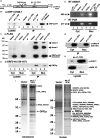
Figure 1.
Figure 1.
pc-L1-1FH immunoprecipitates basal L1 RNP complexes from 293T cell lysates after α-FLAG agarose purification. (A) Structure of FLAG-HA-tagged pc-L1-1FH cloned in vector pcDNA6 myc/his B. RT: ORF2 reverse transcriptase domain; EN: endonuclease domain; PCMV: CMV promoter; BGH An: bovine growth hormone polyadenylation signal. (B) FLAG-tagged ORF1p expressed from the construct pc-L1-1FH binds α-FLAG agarose independent of RNase digestion (lanes 5 and 8), but untagged ORF1p (construct pc-L1-RP) will not bind (lane 6). (C and D) Detection of L1 proteins in the RNP IP. Lanes 1–4: input lysates; lanes 6–9: immunoprecipitates; lanes 1, 2, 6 and 7: cytoplasmic fractions; lanes 3, 4, 8 and 9: nuclear fractions. (C) FLAG-HA-tagged ORF1p, detected by α-FLAG antibody. Putative ORF1p dimer and trimer bands are visible in IP samples (lanes 7 and 9). The reason for their absence in lysate samples is unclear (lanes 2 and 4). IP purification factors were determined for cytoplasmic (lanes 2 versus 7, 26-fold) and nuclear (lanes 4 versus 9, 42-fold) fractions and are presented in Supplementary Table S3. Lane labels are at the bottom of panel D. (D) ORF2p detected by α-ORF2-N (154–167) antibody (lanes 7 and 9). ORF2p in nuclear lysate samples is below the level of detection (lanes 3 and 4). (E) ORF2p reverse transcriptase activity detected in both nuclear and cytoplasmic IP reactions containing pc-L1-1FH (lanes 3 and 5), but not in reactions with the empty vector (lanes 2 and 4). RT- control: the RT incubation step was omitted and 2 µl of pc-L1-1FH immunoprecipitate was added directly to the PCR reaction. No PCR product was detected (lanes 1 and 6). The assay is described in Kulpa and Moran (51). (F) L1 RNA detected by RT–PCR (lanes 3 and 5). RT-: RT enzyme was omitted from the cDNA synthesis step using pc-L1-1FH immunoprecipitates (lanes 1 and 6). (G) FLAG-HA-tagged ORF2p is detected in nuclear and cytoplasmic extracts after IP of pc-L1-2FH. (H) The purity of nuclear and cytoplasmic whole-cell lysate fractions is shown by western blotting. α-HDAC1 is a strictly nuclear protein (54) and α-MEK1/2 is cytoplasmic (55). (I) Immunoprecipitated samples resolved on silver-stained polyacrylamide gels. To support protein identification data from complex IP samples, selected prominent band regions were excised for additional MS sequencing. Both cytoplasmic (left) and nuclear (right) IP fractions are shown.
Figure 2.
Figure 2.
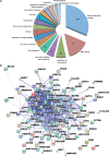
Proteins identified with the L1 form a tight network of interactions dominated by RNA-binding proteins. (A) Pie chart of results of DAVID (Database for Annotation, Visualization and Integrated Discovery) analysis showing selected functional categories for the 96 candidate proteins (42). Protein counts for each category are shown within the slices. Protein names are listed in Supplementary Table S4. (B) STRING (Search tool for the retrieval of interacting genes/proteins)-derived network of known protein–protein interactions among the 96 candidate proteins. The confidence view is shown in Jensen et al. (43).
Figure 3.
Figure 3.
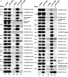
Ectopically expressed and endogenous proteins associate with L1 complexes in multiple cell lines. (A) V5-, 6xMyc- or GFP-tagged proteins exogenously expressed in 293T cells specifically co-immunoprecipitate with pc-L1-1FH, but not empty vector (pcDNA6 myc/his B) [IP: α-FLAG affinity gel, western blotting (WB): α-V5, α-Myc or α-GFP]. IP reactions were in the presence or absence of 15 μg/ml RNase (lanes 3–5). Lysate input samples are also shown (lanes 1 and 2). Several protein panels are reproduced from Goodier et al. (29). GFP-mIGF2BP1 is derived from mouse. The bottom-most panel is representative of tagged ORF1p in the input and IP fractions and confirms that RNase treatment does not affect ORF1p immunoprecipitation on α-FLAG agarose. Molecular weights shown include the epitope tag. The protein standard is See Blue Plus 2 (Invitrogen). (B) Co-IP of endogenous ORF1p from 2102Ep cells by selected V5-tagged proteins (IP: α-V5/IgG affinity gel). Upper rows: detection of ORF1p (WB: α-ORF1 AH40). Asterisk indicates proteins that strongly co-IP endogenous ORF1p. ‘o’ marks proteins that clearly associate with ORF1p on gel overexposure. Lower rows confirm successful IP of the test proteins (WB: α-V5). Exposure times are not necessarily the same for each lane. Input lysate fractions are shown in Supplementary Figure S1. (C) Co-IP of selected endogenous proteins by pc-L1-1FH from 293T cells (IP: α-FLAG affinity gel, WB: various antibodies). The antibody name is followed by the expected protein molecular weight. NCL has an expected weight of 77 kDa, but observed molecular weight of ∼100 kDa. As previously reported (27), an antibody against DDX39B [UAP56; (50)] detects a dominant band of 55 kDa in cytoplasmic lysates, and a smaller isoform of 49 kDa (the expected size for DDX39B) that co-IPs with tagged ORF1p. In total, 12 antibodies were tested; α-PCBP2 (Abnova), α-FBL (Santa Cruz) and α-TOP1 (Spring) failed to detect their endogenous targets in pc-L1-1FH immunoprecipitates. The bottom-most panel shows efficient immunoprecipitation of tagged ORF1p detected by α-FLAG antibody.
Figure 3.
Figure 3.
Ectopically expressed and endogenous proteins associate with L1 complexes in multiple cell lines. (A) V5-, 6xMyc- or GFP-tagged proteins exogenously expressed in 293T cells specifically co-immunoprecipitate with pc-L1-1FH, but not empty vector (pcDNA6 myc/his B) [IP: α-FLAG affinity gel, western blotting (WB): α-V5, α-Myc or α-GFP]. IP reactions were in the presence or absence of 15 μg/ml RNase (lanes 3–5). Lysate input samples are also shown (lanes 1 and 2). Several protein panels are reproduced from Goodier et al. (29). GFP-mIGF2BP1 is derived from mouse. The bottom-most panel is representative of tagged ORF1p in the input and IP fractions and confirms that RNase treatment does not affect ORF1p immunoprecipitation on α-FLAG agarose. Molecular weights shown include the epitope tag. The protein standard is See Blue Plus 2 (Invitrogen). (B) Co-IP of endogenous ORF1p from 2102Ep cells by selected V5-tagged proteins (IP: α-V5/IgG affinity gel). Upper rows: detection of ORF1p (WB: α-ORF1 AH40). Asterisk indicates proteins that strongly co-IP endogenous ORF1p. ‘o’ marks proteins that clearly associate with ORF1p on gel overexposure. Lower rows confirm successful IP of the test proteins (WB: α-V5). Exposure times are not necessarily the same for each lane. Input lysate fractions are shown in Supplementary Figure S1. (C) Co-IP of selected endogenous proteins by pc-L1-1FH from 293T cells (IP: α-FLAG affinity gel, WB: various antibodies). The antibody name is followed by the expected protein molecular weight. NCL has an expected weight of 77 kDa, but observed molecular weight of ∼100 kDa. As previously reported (27), an antibody against DDX39B [UAP56; (50)] detects a dominant band of 55 kDa in cytoplasmic lysates, and a smaller isoform of 49 kDa (the expected size for DDX39B) that co-IPs with tagged ORF1p. In total, 12 antibodies were tested; α-PCBP2 (Abnova), α-FBL (Santa Cruz) and α-TOP1 (Spring) failed to detect their endogenous targets in pc-L1-1FH immunoprecipitates. The bottom-most panel shows efficient immunoprecipitation of tagged ORF1p detected by α-FLAG antibody.
Figure 4.
Figure 4.
snRNAs, scRNAs (left) and selected mRNAs (right) are detected in L1 RNP immunoprecipitates by RT–PCR. Results are for whole-cell lysates (lanes 1–4) and immunoprecipitates (lanes 5–8) from empty vector (pcDNA6 myc/his B; lanes 1, 3, 5 and 7) and pc-L1-1FH (lanes 2, 4, 6 and 8) transfected cells. RT enzyme was omitted (lanes 1, 2, 5 and 6) or included (lanes 3, 4, 7 and 8).
Figure 5.
Figure 5.
Many L1-associated proteins co-localize with EGFP-ORF1p in cytoplasmic granules of unstressed 293T cells. (A–W) Construct ORF1-EGFP-L1-RP was co-transfected with V5-tagged proteins in all cases except (B) FL-CSDA, (G) RFP-HNRNPA1 and (H) mouse GFP-mIGF2BP1 (the latter being co-transfected with pc-L1-1FH, which was detected by α-FLAG antibody). Only overlaid confocal micrographs are shown. (X) Endogenous LARP1 protein co-localizes with ORF1-EGFP-L1-RP in 293T cells. (Y) Endogenous ORF1p and PCBP2 co-localize in some cytoplasmic granules of 2102Ep cells (shown by arrows). (Z) Endogenous ORF1p and fibrillarin (FBL) co-localize in nucleoli of 2102Ep cells. ORF1p is typically found in nucleoli of only a minor percentage of cells (53). Enlargement of two nucleoli are shown. In Y and Z endogenous, ORF1p is detected by α-ORF1 AH40.1 antibody.
Figure 6.
Figure 6.
Some L1-associated proteins strongly inhibit L1 retrotransposition in 293T cells. (A) The 99-PUR-RPS-EGFP was co-transfected in 293T cells with empty vector (pcDNA3) or test constructs expressing tagged proteins. Five days later, percentages of EGFP-positive cells were determined by flow cytometry. Each plasmid pair was transfected in four replicate wells, and results are normalized to pcDNA3 vector control (black bar). Proteins are ordered by their effect on retrotransposition. (B) To control for any off-target effects, test constructs were co-transfected with pCEP-EGFP, a plasmid that constitutively expresses EGFP. Four days later, cells were assayed for gain or loss of fluorescent cells (panel below). Fluorescence is normalized to pcDNA3 control (table, top row). Readings of ≤80% are marked below with ‘+’. Standard deviation for four replicates is shown (bottom row). These results were then used to adjust the retrotransposition levels of Figure 1A by dividing by the average of the pCEP-EGFP expression readings. P-values were calculated by two-tailed t-test and are indicated above each histogram bar (*P < 0.05, **P < 0.01, ***P < 0.001). The inset table summarizes the number of proteins that fall into each retrotransposition percent range. (C) Results of MultiTox-Fluor Multiplex Cytotoxicity assay (Promega) for potential cell toxicity caused by overexpression of test proteins. Test constructs were transfected in 96-well plates and assayed at 3 days. The histogram shows ratios of live to dead cell readings normalized to empty vector control.

Similar articles

Cited by

References

    1. De Koning APJ, Gu W, Castoe TA, Batzer MA, Pollock DD. Repetitive elements may comprise over two-thirds of the human genome. PLoS Genet. 2011;7:e1002384. - PMC - PubMed
    1. Luan DD, Korman MH, Jakubczak JL, Eickbush TH. Reverse transcription of R2Bm RNA is primed by a nick at the chromosomal target site: a mechanism for non-LTR retrotransposition. Cell. 1993;72:595–605. - PubMed
    1. Lander ES, Linton LM, Birren B, Nusbaum C, Zody MC, Baldwin J, Devon K, Dewar K, Doyle M, FitzHugh W, et al. Initial sequencing and analysis of the human genome. Nature. 2001;409:860–921. - PubMed
    1. Zhang Z, Harrison PM, Liu Y, Gerstein M. Millions of years of evolution preserved: a comprehensive catalog of the processed pseudogenes in the human genome. Genome Res. 2003;13:2541–2558. - PMC - PubMed
    1. Wang H, Xing J, Grover D, Hedges DJ, Han K, Walker JA, Batzer MA. SVA elements: a hominid-specific retroposon family. J. Mol. Biol. 2005;354:994–1007. - PubMed

Publication types

MeSH terms