. 2013 Jun 6;153(6):1354-65.
doi: 10.1016/j.cell.2013.04.052.

Visualizing GroEL/ES in the act of encapsulating a folding protein

Affiliations

Visualizing GroEL/ES in the act of encapsulating a folding protein

Dong-Hua Chen et al. Cell. .

Abstract

The GroEL/ES chaperonin system is required for the assisted folding of many proteins. How these substrate proteins are encapsulated within the GroEL-GroES cavity is poorly understood. Using symmetry-free, single-particle cryo-electron microscopy, we have characterized a chemically modified mutant of GroEL (EL43Py) that is trapped at a normally transient stage of substrate protein encapsulation. We show that the symmetric pattern of the GroEL subunits is broken as the GroEL cis-ring apical domains reorient to accommodate the simultaneous binding of GroES and an incompletely folded substrate protein (RuBisCO). The collapsed RuBisCO folding intermediate binds to the lower segment of two apical domains, as well as to the normally unstructured GroEL C-terminal tails. A comparative structural analysis suggests that the allosteric transitions leading to substrate protein release and folding involve concerted shifts of GroES and the GroEL apical domains and C-terminal tails.


Figures

Figure 1
Figure 1. The GroEL protein folding cycle involves a series of allosteric transitions within the chaperonin complex
Non-native substrate proteins enter the GroEL reaction cycle by binding to the open trans ring of an asymmetric GroEL-GroES complex, pulling the trans ring into the high-affinity “T” state (A; for cycle details, see Cliff et al., 2006; Horwich and Fenton, 2009; Lin and Rye, 2006). Protein encapsulation is initiated by highly cooperative binding of ATP to the trans ring, populating the R1 state (B), a conformational state of the GroEL ring with high affinity for the non-native protein but not yet for GroES. The R2 state (C) retains substantial, though weakened, affinity for the non-native protein, binds GroES and encapsulates the substrate protein (D). Transitions into or between the R1 and R2 states are also linked to disassembly of the GroEL-GroES complex on the opposite ring. The ATP-bound GroEL-GroES complex has a high affinity for GroES in the R3 state (E), which releases the non-native substrate protein into the enclosed cis cavity, to initiate folding. Hydrolysis of ATP within the cis ring triggers a transition of the complex to at least one additional conformational state (F).
Figure 2
Figure 2. The structure of the EL43Py398A-GroES-ATP complex determined at 8.9 Å resolution by cryo-EM with C7 symmetry imposed
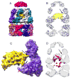
(A) Side view of the EL43Py398A-GroES-ATP density map displayed at a contour level of 1.3 σ. Individual GroEL subunits are shown in different colors; GroES is magenta. All other density maps shown in this study are displayed at a contour level of 1.0 σ (unless otherwise noted). (B) Close-up view of a single EL43Py398A cis-ring subunit (contour level of 1.5 σ) overlapped with a rigid-body, flexibly refined fit of the GroEL-GroES-ADP crystal structure (PDB ID: 1AON; magenta) using the program DireX (Schröder et al., 2007). The stem loop containing Cys 43 and the GroEL C-terminus are labeled with arrows. (C) A medial slice of the density map shown in (A), with the density rendered transparent and superimposed on a rigid-body, flexibly refined fit of the GroEL-GroES-ADP crystal structure. In (C), extra density is visible at the tips of the equatorial stem loops of each GroEL subunit (amino acids 34–52; black dashed circles). The observed densities beyond amino acid 525 in the C-terminal tails are indicated by red arrows. (D) The additional stem-loop density for each subunit is shown (inside of black dashed circle), viewed from above, at a slice level indicated by the dashed blue line in (A). The seven stem loops are labeled 1–7, respectively. A single N-1-pyrene maleimide dye molecule (green) was rigid-body fit into the density at the tip of one stem loop in the EL43Py398A-GroES-ATP complex using Chimera. (E) View of the cis-ring equatorial domain near the subunit C-termini, viewed from above, at the slice level indicated by the black dashed line in (A). Substantial density (large red arrow) is visible in the region of the subunit C-termini, well beyond the last crystallographically resolved residue (small red arrow). The position of the GroEL subunit N-terminus is indicated by the blue arrow.
Figure 3
Figure 3. The structure of the EL43Py398A-GroES-ATP complex containing non-native RuBisCO within the cis cavity determined at 9.2 Å by cryo-EM without imposed symmetry
(A) A side view of the density map of EL43Py398A-RuBisCO-GroES-ATP complex (contour level 1.23 σ) shown colored as in Figure 2, with density from the encapsulated, non-native RuBisCO monomer shown in gold. (B) A medial slice of the EL43Py398A-RuBisCO-GroES-ATP complex with the density rendered transparent and overlapped with a rigid-body, flexibly refined fit of the GroEL-ADP-GroES crystal structure (PDB ID: 1AON) to the cryo-EM map. Additional density around the GroEL equatorial domain stem loops makes direct contact with the non-native RuBisCO monomer (dashed black circles). The RuBisCO is also in contact with the lower region of the apical domain of one cis-ring GroEL subunit in the region of F281 (black arrow). (C) A close-up view of one cis-ring GroEL subunit in direct contact (long black arrow) with the non-native RuBisCO monomer (gold; contour level of 1.05 σ). The GroEL subunit stem loop (short black arrow) and C-terminus (red arrow) are indicated. (D) A medial slice of the variance map derived for the EL43Py398A-RuBisCO-GroES-ATP complex (red; see Experimental Procedures) is shown overlapped with the average map of the complex (gray; orientation as in panel B), calculated from 100 3D reconstructions of the complex computed during the variance calculations. The largest variations in the density map are from the non-native RuBisCO monomer and cavity-facing regions of the GroEL equatorial domains, most likely the C-termini of the cis and trans rings.
Figure 4
Figure 4. The C7 symmetry of the GroEL cis-ring is broken in the EL43Py398A-RuBisCO-GroES-ATP complex near points of contact between the non-native RuBisCO and the GroEL cavity wall
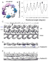
(A) Cis-ring apical domains of the EL43Py398A-RuBisCO-GroES-ATP structure are shown as in Figure 3A, viewed from the top of the cis ring. A gap (black arrow) in the ring density is observed between subunit 2 (purple) and subunit 3 (dark cyan). (B) The cross correlation coefficient between the map of the cis-ring apical domains and a symmetric reference indicates subunit 4 is closer to subunit 3, which is approximately 9 degrees off its C7 symmetrical position, leaving a gap between subunits 3 and 2 (black arrow). (C) The gap (black arrow) between two neighboring GroEL subunits is shown in an unwrapped, planar display from the outside of the 9.2-Å density map of EL43Py398A-RuBisCO-GroES-ATP, as viewed from the side. (D) Top-view slice of the planar map, through the lower region of the cis ring apical domains (panel C, blue dashed line) shows interactions between the non-native RuBisCO monomer and the lower aspect of the GroEL apical domains of subunits 2 and 4 (blue circles). (E) Top-view slice of the planar map through the upper section of the equatorial domains (panel C, dashed black line) indicates contacts with the GroEL subunits near the stem-loop region of the equatorial domain. The isosurface threshold for (D and E) is 0.9σ.
Figure 5
Figure 5. The structure of the EL398A-GroES-ATP complex containing RuBisCO within the cis cavity determined at 15.9 Å by cryo-EM reconstruction without imposed symmetry
(A) A side view of the EL398A-RuBisCO-GroES-ATP density map. (B) Medial slice of the EL398A-RuBisCO-GroES-ATP complex indicates direct contact between the folding RuBisCO monomer and the C-terminal and stem-loop region of one cis-ring GroEL subunit (black arrow).
Figure 6
Figure 6. Removal of the GroEL C-terminal tails results in premature substrate protein release and reduced encapsulation efficiency
(A) Experimental schematic: non-native substrate protein (blue) is bound to the open trans ring of a GroEL ADP bullet complex in the presence of excess GroES. Encapsulation is initiated by the addition of ATP. ATP binding and turnover is limited to a single round by addition of hexokinase and glucose within 10 sec of ATP addition. Complexed and free substrate proteins are separated by gel filtration chromatography with an in-line fluorescence detector. (B) Example of an encapsulation experiment using GFP as the substrate protein. The positions of encapsulated GFP (GroEL-GroES complex) and released GFP (free GFP) are indicated with arrows, for both wild-type GroEL (wtGroEL) and the Δ526 truncation mutant (Δ526). Encapsulation is quantitated for three independent substrates: (C) GFP (normalized fluorescence peak area; n = 6), (D) rhodanese (normalized SDS-PAGE band intensity by densitometry; n = 4) and (E) RuBisCO (fluorescently labeled; n = 6). The reduction in encapsulation of non-native substrate protein by Δ526 GroEL relative to wtGroEL is robust: P = 6.5 ×10⁻⁹ for GFP, P = 0.0007 for rhodanese, and P = 0.0007 for RuBisCO (paired t-test; error bars are one standard deviation; see methods for additional details).
Figure 7
Figure 7. The transition from the R2 to the R3 state in the presence RuBisCO involves large structural rearrangements of both the cis and trans rings
(A–C)Atomic models of the GroEL-GroES complex (PDB ID: 1AON) were refined against density maps of the empty EL43Py398A-GroES-ATP (R2ATP; Figure 2A) and EL398A-GroES-ATP (R3ATP; (Ranson et al., 2006)) complexes. (D-F) Atomic models of the GroEL-GroES complex (PDB ID: 1AON) were refined against density maps of the EL43Py398A-RuBisCO-GroES-ATP complex (R2ATP + sub; Figure 3A) and the EL398A-RuBisCO-GroES-ATP (R3ATP + sub; Figure 5A). (A) Side view of the EL43Py398A-GroES-ATP density map: structural shifts associated with movement from an empty R2 complex to an empty R3 complex are illustrated with a field of difference vectors (blue lines and dots) to indicate the change in Cα-positions from R2 (start) to R3 (end; square). Vector lengths are scaled by a factor of 2 to improve visibility. (B) View of structural changes in the cis apical domains and (C) trans ring apical domains. (D) Side view of the EL43Py398A-RuBisCO-GroES-ATP density map: structural shifts indicating the differences between the substrate-occupied R2 and R3 complexes. (E) View of structural changes in the cis apical domains, and (F) the trans ring apical domains. For (B), (C), (E) and (F) the viewing direction and selected slice density are indicated by the black arrow and horizontal lines on the GroEL-GroES density map shown in the inset, to the lower right. In all cases, strongly restrained flexible model refinement was carried out with DireX. The designations R2 and R3 reference the functional allosteric states of the GroEL ring, illustrated in Figure 1.

Similar articles

Cited by

References

    1. Badcoe IG, Smith CJ, Wood S, Halsall DJ, Holbrook JJ, Lund P, Clarke AR. Binding of a chaperonin to the folding intermediates of lactate dehydrogenase. Biochemistry. 1991;30:9195–9200. - PubMed
    1. Baker ML, Ju T, Chiu W. Identification of secondary structure elements in intermediate-resolution density maps. Structure. 2007;15:7–19. - PMC - PubMed
    1. Braig K, Otwinowski Z, Hegde R, Boisvert D, Joachimiak A, Horwich A, Sigler P. The crystal structure of the bacterial chaperonin GroEL at 2.8 A. Nature. 1994;371:578–586. - PubMed
    1. Burston SG, Ranson NA, Clarke AR. The origins and consequences of asymmetry in the chaperonin reaction cycle. J Mol Biol. 1995;249:138–152. - PubMed
    1. Chen D, Song J, Chuang D, Chiu W, Ludtke S. An expanded conformation of single-ring GroEL-GroES complex encapsulates an 86 kDa substrate. Structure. 2006;14:1711–1722. - PubMed

Publication types

MeSH terms