. 2013 Aug;94(2):367-76.
doi: 10.1189/jlb.0313118. Epub 2013 Jun 6.

B7-1/B7-2 blockade overrides the activation of protective CD8 T cells stimulated in the absence of Foxp3+ regulatory T cells

Affiliations

B7-1/B7-2 blockade overrides the activation of protective CD8 T cells stimulated in the absence of Foxp3+ regulatory T cells

James M Ertelt et al. J Leukoc Biol. 2013 Aug.

Abstract

Although T cell activation has been classically described to require distinct, positive stimulation signals that include B7-1 (CD80) and B7-2 (CD86) costimulation, overriding suppression signals that avert immune-mediated host injury are equally important. How these opposing stimulation and suppression signals work together remains incompletely defined. Our recent studies demonstrate that CD8 Teff activation in response to cognate peptide stimulation is actively suppressed by the Foxp3(+) subset of CD4 cells, called Tregs. Here, we show that the elimination of Treg suppression does not bypass the requirement for positive B7-1/B7-2 costimulation. The expansion, IFN-γ cytokine production, cytolytic, and protective features of antigen-specific CD8 T cells stimulated with purified cognate peptide in Treg-ablated mice were each neutralized effectively by CTLA-4-Ig that blocks B7-1/B7-2. In turn, given the efficiency whereby CTLA-4-Ig overrides the effects of Treg ablation, the role of Foxp3(+) cell-intrinsic CTLA-4 in mitigating CD8 Teff activation was also investigated. With the use of mixed chimera mice that contain CTLA-4-deficient Tregs exclusively after the ablation of WT Foxp3(+) cells, a critical role for Treg CTLA-4 in suppressing the expansion, cytokine production, cytotoxicity, and protective features of peptide-stimulated CD8 T cells is revealed. Thus, the activation of protective CD8 T cells requires positive B7-1/B7-2 costimulation even when suppression by Tregs and in particular, Treg-intrinsic CTLA-4 is circumvented.

Keywords: Treg; bacterial; costimulation; cytotoxic T cells.


Figures

Figure 1.
Figure 1.. CTLA-4-Ig mitigates expansion and IFN-γ production for peptide-stimulated CD8 cells in Treg-ablated mice.
(A) Schematic illustrating our experimental approach. Foxp3DTR or Foxp3WT mice are treated initially with DT and CTLA-4-Ig or isotype control antibody and adoptively transferred CD90.1+ OVA-specific CD8 cells (Day −1). The following day (Day 0), OVA257–264 peptide is administered and the expansion and activation of OVA-specific CD8 cells enumerated 5 days thereafter (Day 5). Rx,.(B) Representative plots and cumulative data showing percent and total number of OVA-specific CD90.1+ among CD8 splenocytes after OVA257–264 peptide administration in Treg-ablated or Treg-sufficient mice treated with CTLA-4-Ig or isotype control antibody. (C) Representative plots and cumulative data showing percent and total number OVA-specific CD90.1+ among CD8 splenocytes, 5 days after OVA257–264 peptide stimulation or no peptide control in isotype or CTLA-4-Ig-treated B6 or CD80−/− CD86−/−-recipient mice. (D) Representative plots and cumulative data showing percent and total number of IFN-γ+ among OVA-specific CD90.1+ cells, 5 days after peptide administration and ex vivo stimulation with OVA257–264 peptide (line histogram) or no stimulation controls (shaded histograms) in each group of mice. These data are combined from three independent experiments, each with similar results containing three to four mice/group. Bar, mean ± 1 sd; **P < 0.01; ***P < 0.001.
Figure 2.
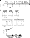
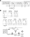
Figure 2.. CTLA-4-Ig eliminates cytotoxicity of peptide-stimulated CD8 T cells.
(A) Schematic illustrating our experimental approach. Foxp3DTR or Foxp3WT mice are treated initially with DT and CTLA-4-Ig or isotype control antibody (Day −1) and stimulated with OVA257–264 peptide the following day (Day 0). Five days thereafter, a 1:1 ratio of CFSE-labeled OVA257–264 peptide, pulsed or unpulsed control CD90.1+ target cells, were administered and the recovery of each subset enumerated 8 h thereafter. (B) Pretransfer and post-transfer ratio of OVA257–264-pulsed (CFSElow) and unpulsed control (CFSEhigh) target cells in each group of mice. Representative histograms show CFSE levels among CD90.1+ splenocytes (upper) and composite analysis of percent lysis of OVA257–264-pulsed compared with unpulsed control cells recovered from each group of mice (lower). These data are combined from two independent experiments, each with similar results containing three to four mice/group. Bar, mean ± 1 sd; ***P < 0.001.
Figure 3.
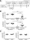
Figure 3.. CTLA-4-Ig blocks peptide-stimulated protection against Lm infection.
(A) Schematic illustrating our experimental approach. Foxp3DTR or Foxp3WT mice are treated initially with DT and CTLA-4-Ig or isotype control antibody (Day −1) and stimulated with OVA257–264 peptide the following day (Day 0). Five days thereafter, mice are infected with Lm-OVA and the number of recoverable CFUs enumerated 3 days thereafter (Day 8). (B) Number of recoverable Lm-OVA CFUs in the spleen and liver for Treg-ablated or Treg-sufficient mice treated with CTLA-4-Ig (■) or isotype control (◽) antibody. (C) Number of recoverable Lm-OVA CFUs in the spleen and liver for B6 mice stimulated with OVA257–264 peptide or no peptide controls. These data are combined from three independent experiments, each with similar results containing three to four mice/group. Bar, mean ± 1 sd. LOD, Limits of detection; **P < 0.01; ***P < 0.001.
Figure 4.
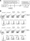
Figure 4.. Tregs with targeted defects in defined molecules reconstitute Foxp3DTR mixed chimera mice.
(A) Schematic illustrating our experimental approach. Foxp3DTR mice on the CD45.1 background are sublethally irradiated and reconstituted with BM cells from Foxp3WT mice on the CD45.2 background, containing defects in CTLA-4 and IL-10 or combined defects in CTLA-4 and IL-10. (B) Representative plots showing percent Foxp3+ and Foxp3 among CD4 splenocytes and relative CD45.1 expression by these cells (Foxp3+ cells, line histograms; Foxp3 cells, shaded histograms) after reconstitution with donor Foxp3WT CD45.2+ BM cells from each group of mice before (upper panels) or after (lower panels) the initiation of DT treatment. These data are representative of three independent experiments, each with similar results.
Figure 5.
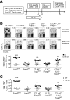
Figure 5.. Treg CTLA-4 suppresses peptide-stimulated CD8 T cell expansion and IFN-γ production.
(A) Schematic illustrating our experimental approach. Each group of mixed chimera mice containing unique Foxp3WT Tregs and WT Foxp3DTR Tregs is initiated on DT treatment (Day −7) and adoptively transferred CD90.1+ OVA-specific CD8 cells, 6 days thereafter (Day −1). The following day (Day 0), the OVA257–264 peptide is administered and the expansion and activation of OVA-specific CD8 cells enumerated 5 days thereafter (Day 5). (B) Representative plots and cumulative data show percent and total number of OVA-specific CD90.1+ among CD8 splenocytes after peptide administration in each group of mice containing exclusively the indicated subset of Foxp3WT Tregs (after DT treatment) or mixed with WT Foxp3DTR Tregs (before DT treatment). (C) Total number IFN-γ+ OVA-specific CD90.1+ cells after ex vivo stimulation with OVA257–264 peptide for the mice described in B. These data are combined from three independent experiments, each with similar results containing three to four mice/group. Bar, mean ± 1 sd; **P < 0.01; ***P < 0.001.
Figure 6.
Figure 6.. Treg CTLA-4 suppresses cytotoxicity of peptide-stimulated CD8 T cells.
(A) Schematic illustrating our experimental approach. Each group of mixed chimera mice containing unique Foxp3WT Tregs and WT Foxp3DTR Tregs is initiated on DT treatment (Day −7) and stimulated with OVA257–264 peptide, 7 days thereafter (Day 0). Five days later, a 1:1 ratio of CFSE-labeled OVA257–264 peptide-pulsed or unpulsed control CD90.1+ target cells is administered and the recovery of each subset enumerated 8 h thereafter. (B) Pretransfer and post-transfer ratio of OVA257–264-pulsed (CFSElow) and unpulsed (CFSEhigh) target cells in each group of mice. CFSE levels among CD90.1+ splenocytes (upper histograms) and composite analysis of percent lysis of OVA257–264-pulsed compared with unpulsed control cells recovered from each group of mice (lower graph). These data are combined from two independent experiments, each with similar results containing three to four mice/group. Bar, mean ± 1 sd; ***P < 0.001.
Figure 7.
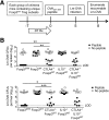
Figure 7.. Treg CTLA-4 impedes peptide-stimulated protection against Lm infection.
(A) Schematic illustrating our experimental approach. Each group of mixed chimera mice containing unique Foxp3WT Tregs and WT Foxp3DTR Tregs is initiated on DT treatment (Day −7) and stimulated with OVA257–264 peptide, 7 days thereafter (Day 0). Five days later, mice are infected with Lm-OVA and the number of recoverable CFUs enumerated 3 days thereafter (Day 8). (B) Number of recoverable Lm-OVA CFUs in the spleen and liver for each group of mice containing only the indicated subset of Foxp3WT Tregs, with OVA257–264 peptide stimulation or no peptide controls. These data are combined from three independent experiments, each with similar results containing three to four mice/group. Bar, mean ± 1 sd; *P < 0.05; **P < 0.01; ***P < 0.001 difference between each group of peptide-stimulated mice.

Similar articles

Cited by

References

    1. Curtsinger J. M., Mescher M. F. (2010) Inflammatory cytokines as a third signal for T cell activation. Curr. Opin. Immunol. 22, 333–340 - PMC - PubMed
    1. Haring J. S., Badovinac V. P., Harty J. T. (2006) Inflaming the CD8+ T cell response. Immunity 25, 19–29 - PubMed
    1. Iwasaki A., Medzhitov R. (2010) Regulation of adaptive immunity by the innate immune system. Science 327, 291–295 - PMC - PubMed
    1. Kanneganti T. D., Lamkanfi M., Nunez G. (2007) Intracellular NOD-like receptors in host defense and disease. Immunity 27, 549–559 - PubMed
    1. Rowe J. H., Ertelt J. M., Way S. S. (2012) Foxp3(+) regulatory T cells, immune stimulation and host defence against infection. Immunology 136, 1–10 - PMC - PubMed

Publication types

MeSH terms