. 2013:2013:920612.
doi: 10.1155/2013/920612. Epub 2013 Apr 29.

Dissecting Major Signaling Pathways throughout the Development of Prostate Cancer

Affiliations

Dissecting Major Signaling Pathways throughout the Development of Prostate Cancer

Henrique B da Silva et al. Prostate Cancer. 2013.

Abstract

Prostate cancer (PCa) is one of the most common malignancies found in males. The development of PCa involves several mutations in prostate epithelial cells, usually linked to developmental changes, such as enhanced resistance to apoptotic death, constitutive proliferation, and, in some cases, to differentiation into an androgen deprivation-resistant phenotype, leading to the appearance of castration-resistant PCa (CRPCa), which leads to a poor prognosis in patients. In this review, we summarize recent findings concerning the main deregulations into signaling pathways that will lead to the development of PCa and/or CRPCa. Key mutations in some pathway molecules are often linked to a higher prevalence of PCa, by directly affecting the respective cascade and, in some cases, by deregulating a cross-talk node or junction along the pathways. We also discuss the possible environmental and nonenvironmental inducers for these mutations, as well as the potential therapeutic strategies targeting these signaling pathways. A better understanding of how some risk factors induce deregulation of these signaling pathways, as well as how these deregulated pathways affect the development of PCa and CRPCa, will further help in the development of new treatments and prevention strategies for this disease.


Figures
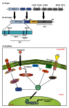
Figure 1
Figure 1
Androgen receptor (AR) signaling in prostate cancer. (a) Schematic representation of the AR gene, highlighting some major AR mutations and their exon localization. (b) Schematic representation of AR protein structure with indication of its functional domains. (c) AR-mediated signaling pathway. The androgen-receptor (AR) signaling pathway begins with the translocation of the testosterone to the cytoplasm, where it can be converted to dihydrotestosterone (DHT) and then promote the receptor dimerization and its further migration to the nucleus. A variety of signals, including PTEN-dependent downregulation, can also merge to AR stabilization and further activation (as indicated).
Figure 2
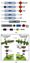
Figure 2
The NF-κB signaling and prostate cancer. (a) Domain structure of NF-κB family members and its direct modulators IκB and IKK. The last two NF-κB members p50 and p52 are derived from the C-terminal processing of p105 and p100, respectively. All NF-κB family members contain an N-terminal Rel-homology domain (RHD) that governs the DNA binding, protein dimerization, and interaction to IκB. The Rel subfamily, RelA, RelB, and c-Rel, also contain a C-terminal transcriptional activation domain (TAD) and the subunit RelB has an additional leucine zipper (LZ) domain at the N-terminus. The IκB family mainly consists of IκBα, IκBβ, IκBγ, IκBε, and BCL-3 proteins (p100 also operates as an IκB-like protein in the non-canonical pathway). The IκB proteins contain ankyrin-repeat motifs (ANK) in their C-terminal region that interact with the RHD of NF-κB proteins and then prevent their nuclear translocation and DNA binding. The IκB kinase (IKK) complex is primarily composed of the two catalytic subunits IKK1 (or IKKα) and IKK2 (or IKKβ) and the scaffolding protein NEMO (or IKKγ). IKK1 and IKK2 are structurally related and both contain an LZ domain and a helix-loop-helix region (HLH), with a C-terminal portion containing a NEMO binding domain (NBD). NEMO has an alpha helical region along with two coiled-coil (CC) regions and a putative zinc finger (ZF) domain. (b) The TNF-dependent NF-κB signaling pathway. The canonical pathway in normal cells is used as an example for the signaling through TNF receptor. The activated IKK complex phosphorylates IκB that is then degraded by the proteasome. Upon degradation of IκB, the subunits of NF-κB are released and the complex is free to migrate to the nucleus. The canonical NF-κB pathway in prostate tumor cells is often constitutively activated, potentially due to increased levels of specific receptors like TNF receptors (TNFRs), which dramatically increase IκB degradation and the translocation of NF-κB dimers to the nucleus to activate κB-responsive genes involved in the development and progression of the tumor. Additionally, undetermined tyrosine kinase subpathways lead to NIK activation, which induces constitutive IKK activity and then constitutive NF-κB activation in androgen receptor-negative prostate cancer cell lines.
Figure 3
Figure 3
The PI3K/AKT signaling in prostate cancer. PI3K/AKT can induce enhanced activation of cancer cells by direct downstream effects. At the same time, this pathway (and downstream target genes) might affect the action of ERKs, which could lead to inhibition of AR-dependent activation, thus favoring an AR-independent growth. Conversely, AR pathway target genes can limit the PI3K/AKT pathway, favoring an AR-dependent tumor growth. A deregulated PI3K pathway (usually due to mutated or null PTEN) can also inhibit the Ras/MEK/ERK pathway, through enhanced activation of AKT. PI3K/AKT can also enhance the presence of stable metalloproteinase receptors (MT1-MMP), which favors invasive and metastatic phenotypes for these tumor cells. TGFβ signaling (through TGFβ3 ligation) can have a dual role in PI3K/AKT in PCa cells; for instance, benign cell lines enhance the expression of AKT and subsequent activation of this pathway following TGFβ3 engagement; malignant cell lines enhance PTEN expression in response to TGFβ3 engagement. Finally, N-cadherin enhanced expression in PCa cells leads to enhanced production of CCL2, which avoids autophagy in part through PI3K/AKT pathway.
Figure 4
Figure 4
The JAK/STAT signaling in prostate cancer. (1) The JAK/STAT pathway has been found constitutively activated in PCa cells, leading to induction of tumor cell proliferation and apoptosis inhibition mediated by STAT3 activation. (2) BRCA1/2 is required for DNA repair in normal cells. However, in PCa, BRCA1 can bind STAT3 to promote JAK/STAT3 activation. (3) AR is a well-characterized cross-talk pathway in PCa. When activated, AR can bind to STAT3 leading to the activation of JAK/STAT cascade, being important in the induction of cell proliferation and apoptosis inhibition. (4) Under stress conditions, ATF3 is activated and plays a crucial role in the maintenance of cell integrity and homeostasis. ATF3 does so by interacting with AR, leading to inhibition of androgen signaling and, consequently, the inhibition of cell proliferation. However, ATF3 is downregulated in PCa cells, suggesting that this pathway provides an important mechanism of defense against cancer. (5) Similarly, C/EBPδ is required to inhibit cell proliferation by binding to STAT3. Nevertheless, C/EBPδ is typically downregulated in PCa, and, therefore, it could be used as an strategy in the development of therapeutic drugs against PCa growth.
Figure 5
Figure 5
Overview of ERK and PI3K activation and their crosstalk. The binding of the ligand to RTK dimerizes and activates the receptor, leading to the recruitment of multiple Grb2 and Shp2 molecules, which further leads to the binding of a second anchoring protein Gab1 to the complex and to the activation of Son of Sevenless (SOS). This event leads to the activation of Raf/Ras/MEK/ERK pathway. Once phosphorylated, ERKs also phosphorylate a great number of substrates present in both nucleus and cytoplasm. In the nucleus, ERK phosphorylates a series of transcription factors including Elk1, c-Fos, p53, Ets1/2, and c-Jun, each one acting as regulators of cell proliferation, differentiation, and morphogenesis. The recruitment and activation of Grb2 and Shp2 also leads to the recruitment of another docking protein, Gab1. Once phosphorylated, Gab1 recruits PI3K to the membrane, where it phosphorylates the inositol ring of PIP-2 into PIP-3. PIP-3 facilitates the phosphorylation of AKT, which in turn regulates the activity of p53 and BAD. Blue and red arrows indicate up- and downregulated proteins in PCa, respectively.
Figure 6
Figure 6
The TGF-β/SMAD signaling pathway and its implication in prostate cancer. When a TGF-β ligand binds to the constitutively active type II receptor, this complex associates with the type I receptor, forming a tetrameric receptor. The type II receptor phosphorylates and activates the type I receptor, which allows the recruitment of R-SMADs. The activated type I receptor then phosphorylates the MH2 domain of R-SMAD, activating it. Activated R-SMADs form complexes with SMAD4, which is then translocated to the nucleus. In the nucleus, SMAD complexes interact with nuclear proteins to activate or repress the transcription of target genes. Furthermore, BMP-10 can signal through SMAD-independent pathways and inhibit cell growth, invasiveness, and migration. TGF-β can also promote androgen receptor (AR) translocation into the nucleus and AR-dependent gene transcription. AR can combine with SMAD4 and regulate TGF-β-mediated apoptosis. According to the TGF-β central dogma, in normal epithelium or early-stage cancer cells, TGF-β acts as a tumor suppressor, by inhibiting cell growth, invasiveness, and motility and promoting apoptosis. In more advanced cancer cells, TGF-β has tumor-promoting functions; it promotes proliferation, invasion, and motility of cells and inhibits apoptosis. Green arrows indicate potentially up-regulated proteins in PCa.
Figure 7
Figure 7
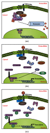
The Wnt signaling and its implications in the development of prostate cancer. (a) In an inactive state, the protein β-catenin is sequestered in a complex in the presence of Axin, GSK3β, CK1α, and APC. This complex allows ubiquitination of β-catenin and its subsequent degradation in a proteasome-dependent manner, maintaining this pathway inactive in the absence of Wnt. (b) After binding of Wnt to Frizzled receptor complex (which includes the adaptor molecules LRP5/LRP6), this allows the recruitment of Dishevelled (DVL) and Axin; the recruitment of Axin disrupts the inactivation complex and releases β-catenin, which translocates to the nucleus and functions as a transcription factor, inducing expression of several genes related to proliferation, such as c-myc and cyclin D1. (c) In the PCa environment, β-catenin can combine with AR proteins, whose levels are typically increased in PCa, enhancing their function as transcription factors and leading to increased gene expression of pro-survival and proliferative factors.

Similar articles

Cited by

References

    1. Verras M, Sun Z. Roles and regulation of Wnt signaling and β-catenin in prostate cancer. Cancer Letters. 2006;237(1):22–32. - PubMed
    1. Mullins JK, Loeb S. Environmental exposures and prostate cancer. Urologic Oncology. 2012;30:216–219. - PubMed
    1. Siegel R, DeSantis C, Virgo K, et al. Cancer treatment and survivorship statistics, 2012. CA: A Cancer Journal for Clinicians. 2012;62:220–241. - PubMed
    1. Maluf FC, Smaletz O, Herchenhorn D. Castration-resistant prostate cancer: systemic therapy in 2012. Clinics. 2012;67:389–394. - PMC - PubMed
    1. Center MM, Jemal A, Lortet-Tieulent J, et al. International variation in prostate cancer incidence and mortality rates. European Urology. 2012;61:1079–1092. - PubMed

LinkOut - more resources