Review
. 2013:68:467-87.
doi: 10.1007/978-94-007-6552-8_16.

Entry of enveloped viruses into host cells: membrane fusion

Affiliations
Review

Entry of enveloped viruses into host cells: membrane fusion

Vicente Más et al. Subcell Biochem. 2013.

Abstract

Viruses are intracellular parasites that hijack the cellular machinery for their own replication. Therefore, an obligatory step in the virus life cycle is the delivery of the viral genome inside the cell. Enveloped viruses (i.e., viruses with a lipid envelope) use a two-step procedure to release their genetic material into the cell: (i) they first bind to specific surface receptors of the target cell membrane and then, (ii) they fuse the viral and cell membranes. This last step may occur at the cell surface or after internalization of the virus particle by endocytosis or by some other route (e.g., macropinocytosis). Remarkably, the virus-cell membrane fusion process goes essentially along the same intermediate steps as other membrane fusions that occur for instance in vesicular fusion at the nerve synapsis or cell-cell fusion in yeast mating. Specialized viral proteins, fusogens, promote virus-cell membrane fusion. The viral fusogens experience drastic structural rearrangements during fusion, liberating the energy required to overcome the repulsive forces that prevent spontaneous fusion of the two membranes. This chapter describes the different types of viral fusogens and their mode of action, as are currently known.


Figures
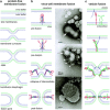
Fig. 16.1
Fig. 16.1
Steps of the membrane fusion process. (a) Diagram of the fusion steps between two protein-free lipid bilayers. From top to bottom: the lipids (represented by heads and tails) of the two bilayers are initially curved into nipple-like structures that approach the two membranes. This is followed by formation of the stalk in which the two proximal leaflets are fused. This stalk is then expanded forming a hemifusion diaphragm in which lipids of the distal leaflets of the bilayers are now in direct contact. Finally, rupture of the hemifusion diaphragm leads to formation of the fusion pore. (b) Diagram of the virus-cell fusion process: As an example, fusion mediated by the influenza HA is illustrated. Left panels: HA is a homotrimer that initially binds to the cell surface (not shown) by interactions of each subunit head with sialic acid. Then, the virus is internalized by endocytosis. For simplicity, only 1 HA trimer in the pre-fusion conformation is shown, anchored into the viral membrane. After endosome acidification, the HA globular head falls apart, allowing refolding of the molecule to produce three long α-helices. The fusion peptides, placed at the N-terminal end of each α-helix, insert into the target membrane. This intermediate, dubbed pre-hairpin, refolds to bring the two membranes into proximity leading to formation of the lipid stalk followed by formation of the hemifusion diaphragm (not shown). Finally, the fusion pore is formed by the concerted action of several HA molecules that adopt a very stable post-fusion conformation. Right panels: The upper panel shows a mixture of an influenza virus particle (strain X31, H3N2, white arrow) and liposomes (some of them indicated by white arrowheads) made with lipids commonly found in cell membranes, incubated at neutral pH. Note the glycoprotein spikes (mostly haemagglutinin, HA) sticking out of the viral membrane in contrast with the smooth surface of liposomes. The middle panel shows the same virus/liposome mixture after incubation for 5–10 s at pH 5.0 followed by neutralization. Note binding of liposomes to the virus surface and initiation of virus-liposome fusion. The lower panel shows the virus/liposome mixture after incubation for 5 min at pH 5.0 followed by neutralization. Note that the virus has fused with several liposomes, yielding a large vesicle with viral glycoproteins disperse throughout the surface and with a small liposome still in the process of fusion. The HA spikes also have changed morphology after exposure to low pH and fusion. (Courtesy of L.J. Calder and S.A. Wharton, Division of Virology, MRC National Institute for Medical Research, London, UK). (c) Diagram of vesicle fusion at the synaptic junction: Initially one of the SNARE proteins (synaptobrevin, blue) is inserted into the vesicle membrane, while three other SNAREs (two SNAP 25, green and one syntaxin, red) are inserted in the plasma membrane. After an initial interaction, refolding of the SNAREs leads to formation of a bundle of four parallel α-helices that drives approximation of the two membranes and formation of the stalk and hemifusion intermediates (not shown). Completion of the SNARE complex results in formation of the fusion pore. In the two lower panels of parts (b) and (c), two HAs and two SNARE complexes are shown surrounding the fusion pore, although the actual number of molecules involved in fusion pore formation is likely to be higher
Fig. 16.2
Fig. 16.2
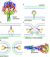
Membrane fusion mediated by a class I fusion protein (Paramyxovirus). The atomic structures of the pre-fusion form of Parainfluenza virus type 5 (PIV5) [22] (upper left) and the post-fusion form of Respiratory Syncytial Virus (RSV) [53] (lower right) F proteins are shown as ribbons. The same protein regions are highlighted with identical colors in the two conformations. (a–d) Diagram of the fusion process denoting: (a) the pre-fusion paramyxovirus F protein trimer inserted in the viral membrane before activation, (b) formation of the pre-hairpin structure which includes refolding of the long central HRA α-helices (blue) with the fusion peptide (red) inserted into the cell membrane, (c) collapse of the pre-hairpin to approach the two membranes, and (d) formation of the fusion pore and stabilization of the F trimer in the post-fusion conformation. In the last two steps, two F protein molecules are represented to indicate the cooperation needed to drive the fusion process
Fig. 16.3
Fig. 16.3
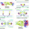
Membrane fusion mediated by a class II fusion protein (Flavivirus). Ribbon representation of the atomic structures of the dengue virus E protein dimer in the pre-fusion conformation [54] (upper left) and the E protein trimer in the post-fusion conformation [55] (lower right). Domains I, II and III of the E glycoprotein are colored red, yellow and blue, respectively. (a–d) Diagram of the fusion process denoting: (a) the structure of the flavivirus E glycoprotein dimer already in the endosome before activation, (b) dissociation of the E protein subunits, refolding of the fusion domain (yellow) and insertion of the fusion loop (green) into the endosomal membrane, (c) formation and refolding of the E protein trimer to approach the two membranes, and (d) formation of the fusion pore and stabilization of the E trimer in the post-fusion conformation. In the last two steps, two E protein molecules are represented to indicate the cooperation needed to drive the fusion process
Fig. 16.4
Fig. 16.4
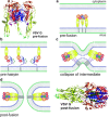
Membrane fusion mediated by a class III fusion protein (Vesicular Stomatitis Virus, VSV). Ribbon representations of the VSV glycoprotein (G), in the pre- (upper left) and post-fusion (lower right) conformations. Domains are colored similarly in all images. The fusion domain is colored in yellow and the fusion loops in green. (a–d) Diagram of the fusion process denoting: (a) the structure of the VSV G glycoprotein trimer already in the endosome before activation, (b) dissociation of the G protein subunits, refolding of the fusion domain (yellow) and insertion of the fusion loop (green) into the endosomal membrane, (c) formation and refolding of the G protein trimer to approach the two membranes, and (d) formation of the fusion pore and stabilization of the G primer in the post-fusion conformation. In the last two steps, two G protein molecules are represented to indicate the cooperation needed to drive the fusion process

Similar articles

Cited by

References

References and Further Reading

    1. Chernomordik LV, Kozlov MM. Mechanics of membrane fusion. Nat Struct Mol Biol. 2008;15:675–683. doi: 10.1038/nsmb.1455. - DOI - PMC - PubMed
    1. Yang L, Huang HW. Observation of a membrane fusion intermediate structure. Science. 2002;297:1877–1879. doi: 10.1126/science.1074354. - DOI - PubMed
    1. Chanturiya A, Chernomordik LV, Zimmerberg J. Flickering fusion pores comparable with initial exocytotic pores occur in protein-free phospholipid bilayers. Proc Natl Acad Sci U S A. 1997;94:14423–14428. doi: 10.1073/pnas.94.26.14423. - DOI - PMC - PubMed
    1. Harrison SC. Viral membrane fusion. Nat Struct Mol Biol. 2008;15:690–698. doi: 10.1038/nsmb.1456. - DOI - PMC - PubMed
    1. Munoz-Barroso I, Durell S, Sakaguchi K, Appella E, Blumenthal R. Dilation of the human immunodeficiency virus-1 envelope glycoprotein fusion pore revealed by the inhibitory action of a synthetic peptide from gp41. J Cell Biol. 1998;140:315–323. doi: 10.1083/jcb.140.2.315. - DOI - PMC - PubMed
Further Reading
    1. Palfreyman MT, Jorgensen EM (2009) In vivo analysis of membrane fusion. In: Encyclopedia of Life Sciences (ELS). John Wiley & Sons, Chichester. doi:10.1002/9780470015902.a0020891
    1. Grove J, Marsh M. The cell biology of receptor-mediated virus entry. J Cell Biol. 2011;7:1071–1082. doi: 10.1083/jcb.201108131. - DOI - PMC - PubMed
    1. Helenius A. Virus entry and uncoating. In: Knipe DM, Howley PM, editors. Fields virology. 5. Philadelphia: Lippincott, Williams and Wilkins; 2007.
    1. Also especially recommended for further reading are references [4, 9, 15, 42, 44] listed above

Publication types

LinkOut - more resources