. 2013 Jul;140(13):2711-23.
doi: 10.1242/dev.089987. Epub 2013 May 29.

Deficient FGF signaling causes optic nerve dysgenesis and ocular coloboma

Affiliations

Deficient FGF signaling causes optic nerve dysgenesis and ocular coloboma

Zhigang Cai et al. Development. 2013 Jul.

Abstract

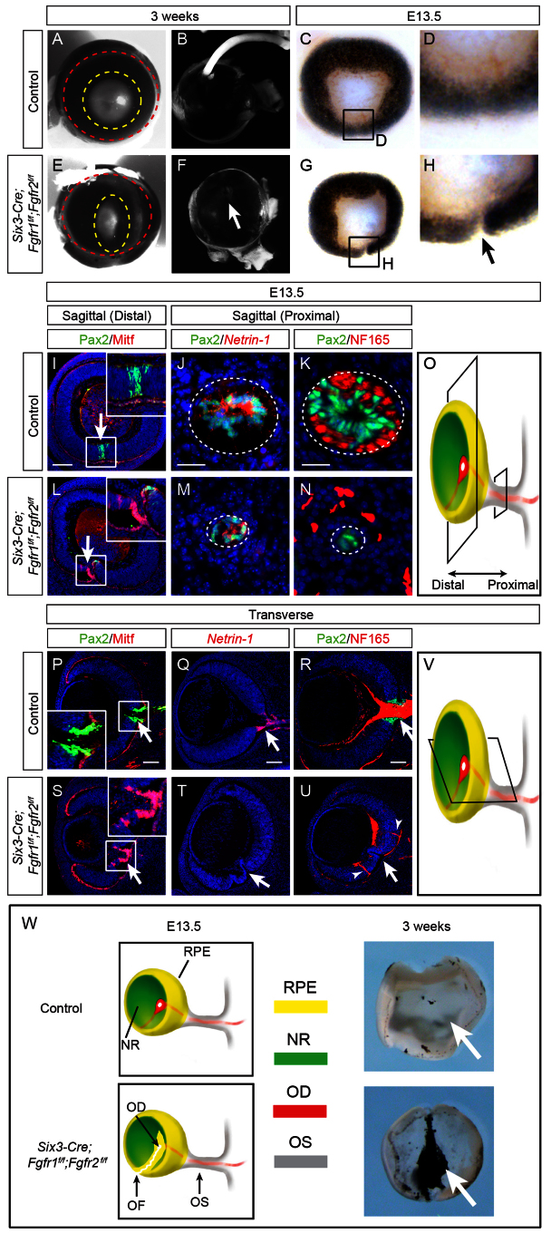
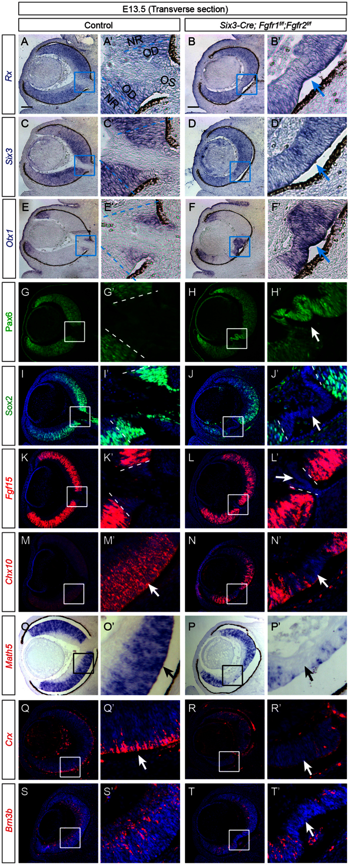
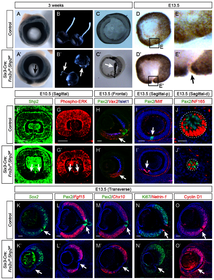
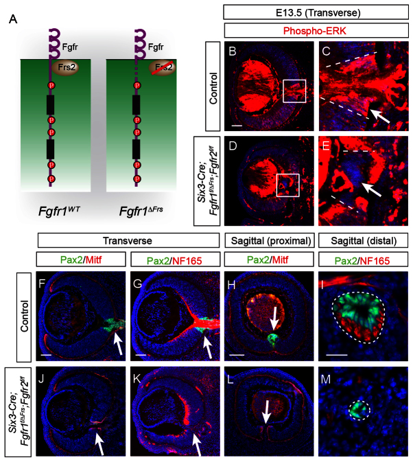
FGF signaling plays a pivotal role in eye development. Previous studies using in vitro chick models and systemic zebrafish mutants have suggested that FGF signaling is required for the patterning and specification of the optic vesicle, but due to a lack of genetic models, its role in mammalian retinal development remains elusive. In this study, we show that specific deletion of Fgfr1 and Fgfr2 in the optic vesicle disrupts ERK signaling, which results in optic disc and nerve dysgenesis and, ultimately, ocular coloboma. Defective FGF signaling does not abrogate Shh or BMP signaling, nor does it affect axial patterning of the optic vesicle. Instead, FGF signaling regulates Mitf and Pax2 in coordinating the closure of the optic fissure and optic disc specification, which is necessary for the outgrowth of the optic nerve. Genetic evidence further supports that the formation of an Frs2α-Shp2 complex and its recruitment to FGF receptors are crucial for downstream ERK signaling in this process, whereas constitutively active Ras signaling can rescue ocular coloboma in the FGF signaling mutants. Our results thus reveal a previously unappreciated role of FGF-Frs2α-Shp2-Ras-ERK signaling axis in preventing ocular coloboma. These findings suggest that components of FGF signaling pathway may be novel targets in the diagnosis of and the therapeutic interventions for congenital ocular anomalies.

Keywords: Coloboma; FGF; Frs2α; Mitf; Mouse; Optic disc; Optic fissure; Optic nerve; Pax2; Ras; Shp2.


Figures

Fig. 1.
Fig. 1.
Ocular coloboma and optic nerve aplasia in the Six3-Cre;Fgfr1flox/flox;Fgfr2flox/flox mice. (A-H) The adult Six3-Cre;Fgfr1flox/flox;Fgfr2flox/flox animals displayed a teardrop-shaped iris (circled in yellow) and a loss of optic nerve (ON) (arrow in F). At E13.5, the optic fissure was completely closed in the control eye, but it remained open in the Six3-Cre;Fgfr1flox/flox;Fgfr2flox/flox embryos (arrow in H). (I-O) Sagittal sections of the E13.5 mutant eyes revealed ectopic Mitf expression in the open optic fissure (arrow in L), the hypoplastic optic stalk (circled in M) and a loss of optic nerve as indicated by lack of NF165 staining (circled in N). A sagittal section scheme is shown in O. (P-V) Transverse sections showed that the presumptive optic disc in Fgfr1/2 mutant lost Pax2 and netrin 1 expression, but gained Mitf expression (S and T, arrows). As a result, NF165-stained retinal ganglion cell axons were misrouted (U, arrowheads) and optic nerve was not formed (U, arrow). The transverse section through the optic disc region is shown in V. (W) (Left) Summary of the ocular phenotypes in the E13.5 Six3-Cre;Fgfr1flox/flox; Fgfr2flox/flox embryos. Adapted, with permission, from Bharti et al. (Bharti et al., 2006). The control eyeball is composed of an outside RPE layer (yellow) and an inside neural retina (green) layer, which fuse at the ventral optic fissure (faint red), but remained open in the posterior optic disc (red). In the Six3-Cre;Fgfr1flox/flox;Fgfr2flox/flox mutants, the ventral neural retina along the optic fissure were transformed to RPE, preventing the closure of the eyeball. A similar transformation of optic disc to RPE prevented the formation of optic nerve. NR, neural retina; OD, optic disc; OF, optic fissure; ON, optic nerve; OS, optic stalk; RPE, retinal pigmented epithelium. (Right) Three-week-old eye balls were dissected to remove RPE covering the back of the eye, revealing abnormal pigmentation in mutant optic disc and optic fissure (arrows). Scale bars: 100 μm in I,L; 25 μm in J,K,M,N; 100 μm in P-U.
Fig. 2.
Fig. 2.
Defective retina genesis in the Six3-Cre;Fgfr1flox/flox;Fgfr2flox/flox mutants. (A-F′) At E13.5, ablation of Fgfr1 and Fgfr2 resulted in the expansion of Rx, Six3 and Otx1 into presumptive optic stalk. (G-J′) In Fgfr1/2 mutants, the expression of Pax6 expanded from the neural retina and RPE into the presumptive optic disc region, where the normal expression of Sox2 was lost. (K-T′) The neural retina makers Fgf15, Chx10, Math5, Crx and Brn3b were excluded from presumptive optic disc and neural retina adjacent to the optic fissure. Dashed lines indicate the boundary of optic disc. Arrows indicate the presumptive optic disc/stalk (A-L′) and neural retina (M-T′) in mutants. NR, neural retina; OD, optic disc; OS, optic stalk. Scale bars: 100 μm.
Fig. 3.
Fig. 3.
The Six3-Cre;Fgfr1flox/flox;Fgfr2flox/flox mutant optic vesicle exhibit normal axial patterning but defective optic fissure development. (A-F′) At E11.5, the loss of Fgfr1 and Fgfr2 signaling did not affect the expression of midline Shh, dorsal Bmp4, ventral Bmp7 and their downstream effectors Gli1 and phospho-Smad1. (G-L′) Levels of the dorsal-ventral polarity markers Tbx5, Vax2, Radldh3 and Nlz2, and the temporal-nasal markers BF1 and BF2 were unchanged in Fgfr1/2 mutants. (M-R′) Although levels of the optic vesicle patterning genes Rx, Six3, Pax6 and Otx1 were also unaffected, levels of the neural retinal marker Chx10 and the optic disc marker Pax2 were downregulated in ventral retina, whereas the RPE maker Mitf was ectopically expressed in the ventral optic fissure in Fgfr1/2 mutants. Scale bars: 100 μm.
Fig. 4.
Fig. 4.
Fgfr1/2 ablation disrupts ERK signaling. (A-D,G-J) In the E10.5 Six3-Cre;Fgfr1flox/flox;Fgfr2flox/flox embryos, phospho-ERK staining was lost in the ventral optic vesicle (G, arrows), the developing optic disc (H, arrows), the ventral optic stalk (I, arrow) and optic chiasm (J, arrow). The optic stalk and optic chiasm were outlined in white. (E,F,K,L) The control optic disc marked by Pax2 at E13.5 displayed strong phospho-ERK staining, which was abolished in the Fgfr1/2 mutants. (M-Q′) Consistent with the deficient ERK signaling, the cell cycle regulator cyclin D1 and proliferation marker Ki67 were lost in the mutant optic disc region. (R-S′,U-V′) Increased cell death in the mutant optic disc region was shown by cleaved caspase 3 and TUNEL staining. Broken lines mark the boundaries of the optic disc. (O,T) The ratio of Ki-67 and TUNEL-positive cells versus DAPI-positive cells were measured in the indicated region and analyzed using Student’s t test. n=8 for each genotype, *P<0.05. **P<0.01. Scale bars: 100 μm.
Fig. 5.
Fig. 5.
Concomitant ablation of Frs2a and Shp2 caused coloboma defects. (A-E′) The Six3-Cre;Frs2aflox/flox;Shp2flox/flox mutants exhibited ocular coloboma (A′, arrow), optic nerve hypoplasia/aplasia (B′, arrows) and ectopic pigmentation (C′, arrow) at 3 weeks, and an open optic fissure at E13.5 (E′, arrow). (F-J′) Shp2 and phospho-ERK staining was downregulated along the ventral optic fissure in the E10.5 Frs2a/Shp2 mutants. At E13.5, Vax2 expression was unchanged in the ventral retina (H′, arrow), but the optic fissure was transformed to Mitf-positive RPE (I′, arrow) and the neurofilament marker NF165 was diminished in the optic stalk (circled in J′). (K-O′) The retinal progenitor markers Sox2, Fgf15 and Chx10 were lost along optic fissure in the ventral neural retina (K′,L′,M′, arrows). The optic disc region lost the cell proliferation marker Ki67, cyclin D1 and the optic disc marker netrin 1 (N′,O′, arrows). Scale bars: 100 μm in F-J′; 25 μm in K-O′.
Fig. 6.
Fig. 6.
Deletion of the Shp2-docking site on Frs2α disrupted ERK signaling in retinal development. (A) Frs2α-Shp2 interaction. In control retina, Frs2α recruits Shp2 to activate downstream Ras-ERK signaling for optic fissure and optic disc development. Mutating the two phospho-tyrosine residues in Frs2α abrogates the Shp2 docking site in the Frs2α2F allele, but leaves the Shp2-independent function of Frs2α intact. This allows a direct test of Frs2α-Shp2 interaction in regulating retinal development. (B-P) The Six3-Cre;Frs2α2F/flox;Shp2flox/flox mutants phenocopied the Six3-Cre;Frs2αflox/flox;Shp2flox/flox mutants in the loss of Shp2 and phospho-ERK in central retina around the optic disc region (C,D,F,G, arrows). Both mutants gained ectopic Mitf and lost the optic disc marker Pax2 and netrin 1 (I,J,M, arrows). As a result, the optic nerve was not formed in either mutant (O,P, arrows). Scale bar: 100 μm.
Fig. 7.
Fig. 7.
The Frs2α-binding site on Fgfr1 is required for ERK signaling and the development of the optic disc and optic fissure. (A) The Fgfr1ΔFrs allele retains all phospho-tyrosine residues, but lost the domain for interacting with Frs2. (B-E) ERK phosphorylation was lost in the deformed optic disc region in the Six3-Cre;Fgfr1ΔFrs2/flox;Fgfr2flox/flox retina (E, arrow). (F-M) The Six3-Cre;Fgfr1ΔFrs2/flox;Fgfr2flox/flox mutant exhibited ectopic Mitf along the optic fissure (J and L, arrows) and lost Pax2 at optic disc (K, arrow). The Pax2-labeled mutant optic stalk is hypoplastic without NF165-stained optic nerve (circled in M). Scale bars: 100 μm in B-E; 25 μm in F-M.
Fig. 8.
Fig. 8.
Retinal FGF-Frs2α-Shp2 FGF signaling can be substituted by constitutive Kras signaling. (A) Kras rescue experiments. In the absence of upstream Fgfr-Frs2-Shp2 signaling, the constitutively activated KrasG12D mutant can still activate ERK signaling to promote optic fissure and optic disc development. (B-K) Despite of loss of Shp2 staining (B,G, arrows), phospho-ERK staining was recovered in the Six3-Cre;Frs2αflox/flox;Shp2flox/flox;LSL-KrasG12D mutant retina (C,H, arrows). This led to the formation of Pax2- and netrin 1-expressing optic disc and NF165-expressing optic nerve (D-F,I-K, arrows). (L-S) Similarly, the Six3-Cre;Fgfr1flox/flox; Fgfr2flox/flox;LSL-KrasG12D mutant eye also displayed increased ERK phosphorylation (L,P, arrows), formation of optic disc (M-O, arrows), fusion of optic fissure (Q, arrow) and establishment of optic nerve. Scale bars: 100 μm in B-I,L-Q; 25 μm in J,K,R,S.

Similar articles

Cited by

References

    1. Alur R. P., Vijayasarathy C., Brown J. D., Mehtani M., Onojafe I. F., Sergeev Y. V., Boobalan E., Jones M., Tang K., Liu H., et al. (2010). Papillorenal syndrome-causing missense mutations in PAX2/Pax2 result in hypomorphic alleles in mouse and human. PLoS Genet. 6, e1000870 - PMC - PubMed
    1. Ascaso F. J., Del Buey M. A., Huerva V., Latre B., Palomar A. (1993). Noonan’s syndrome with keratoconus and optic disc coloboma. Eur. J. Ophthalmol. 3, 101–103 - PubMed
    1. Bao Z.-Z. (2008). Intraretinal projection of retinal ganglion cell axons as a model system for studying axon navigation. Brain Res. 1192, 165–177 - PMC - PubMed
    1. Barbieri A. M., Broccoli V., Bovolenta P., Alfano G., Marchitiello A., Mocchetti C., Crippa L., Bulfone A., Marigo V., Ballabio A., et al. (2002). Vax2 inactivation in mouse determines alteration of the eye dorsal-ventral axis, misrouting of the optic fibres and eye coloboma. Development 129, 805–813 - PubMed
    1. Bäumer N., Marquardt T., Stoykova A., Spieler D., Treichel D., Ashery-Padan R., Gruss P. (2003). Retinal pigmented epithelium determination requires the redundant activities of Pax2 and Pax6. Development 130, 2903–2915 - PubMed

Publication types

MeSH terms

Substances

LinkOut - more resources