. 2013;9(5):e1003384.
doi: 10.1371/journal.ppat.1003384. Epub 2013 May 23.

Human papillomavirus (HPV) upregulates the cellular deubiquitinase UCHL1 to suppress the keratinocyte's innate immune response

Affiliations

Human papillomavirus (HPV) upregulates the cellular deubiquitinase UCHL1 to suppress the keratinocyte's innate immune response

Rezaul Karim et al. PLoS Pathog. 2013.

Abstract

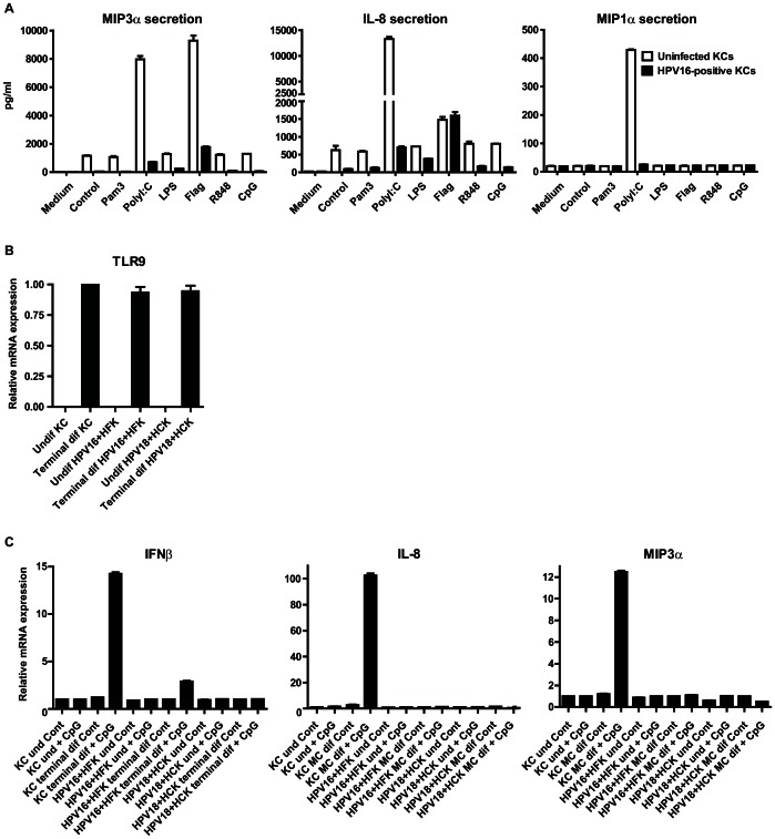
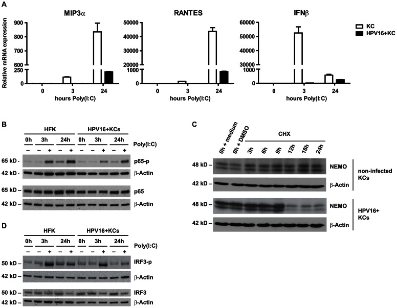
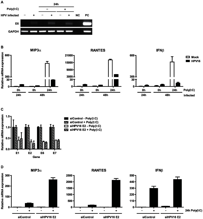
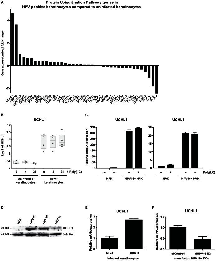
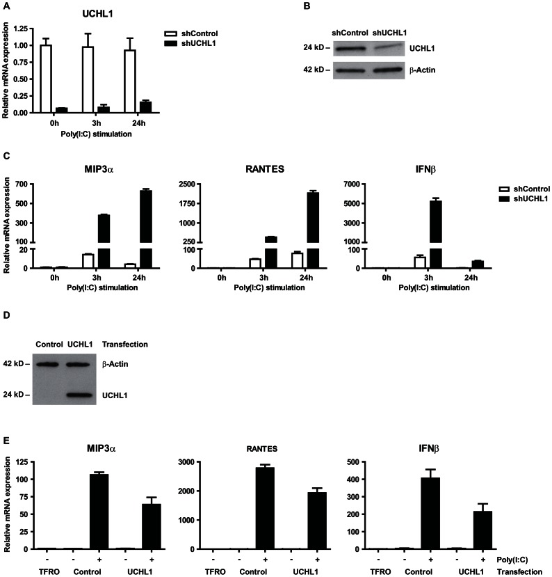
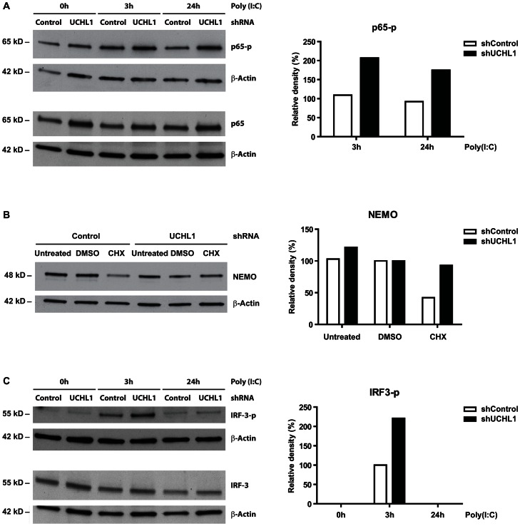
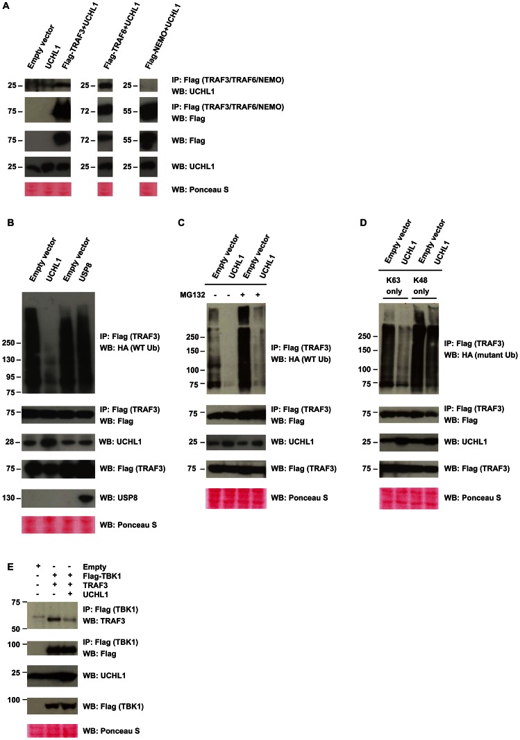
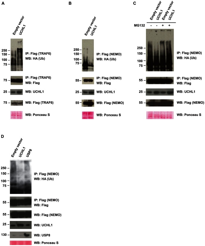
Persistent infection of basal keratinocytes with high-risk human papillomavirus (hrHPV) may cause cancer. Keratinocytes are equipped with different pattern recognition receptors (PRRs) but hrHPV has developed ways to dampen their signals resulting in minimal inflammation and evasion of host immunity for sustained periods of time. To understand the mechanisms underlying hrHPV's capacity to evade immunity, we studied PRR signaling in non, newly, and persistently hrHPV-infected keratinocytes. We found that active infection with hrHPV hampered the relay of signals downstream of the PRRs to the nucleus, thereby affecting the production of type-I interferon and pro-inflammatory cytokines and chemokines. This suppression was shown to depend on hrHPV-induced expression of the cellular protein ubiquitin carboxyl-terminal hydrolase L1 (UCHL1) in keratinocytes. UCHL1 accomplished this by inhibiting tumor necrosis factor receptor-associated factor 3 (TRAF3) K63 poly-ubiquitination which lead to lower levels of TRAF3 bound to TANK-binding kinase 1 and a reduced phosphorylation of interferon regulatory factor 3. Furthermore, UCHL1 mediated the degradation of the NF-kappa-B essential modulator with as result the suppression of p65 phosphorylation and canonical NF-κB signaling. We conclude that hrHPV exploits the cellular protein UCHL1 to evade host innate immunity by suppressing PRR-induced keratinocyte-mediated production of interferons, cytokines and chemokines, which normally results in the attraction and activation of an adaptive immune response. This identifies UCHL1 as a negative regulator of PRR-induced immune responses and consequently its virus-increased expression as a strategy for hrHPV to persist.


Conflict of interest statement

The authors have declared that no competing interests exist.

Figures

Figure 1
Figure 1. The presence of high risk human papillomavirus interferes with pattern recognition receptor (PRR) signaling of keratinocytes.
(A) Cytokine production of non-differentiated uninfected or HPV16+ keratinocytes after stimulation with different indicated PRR stimuli as measured by ELISA. (B) TLR9 expression as measured by qRT-PCR on total RNA samples from undifferentiated (und) and terminally differentiated (terminal dif) uninfected KCs, and HPV16 and HPV18 positive KC cultures. (C) IFNβ, IL-8 and MIP3α expression levels in unstimulated or CpG ODN-stimulated uninfected KCs, and two different HPV (16 or 18) positive KC cultures as examined by qRT-PCR. KCs were either left undifferentiated (und) or terminally differentiated (terminal dif) after which they were stimulated with CpG (10 µg/ml) for 7 hours. (BC) Gene expression was normalized using GAPDH mRNA expression levels.
Figure 2
Figure 2. Canonical NF-κB signaling is impaired upstream of the transcription factor p65.
(A) Poly(I∶C) induced cytokine expression in HPV16+ KCs compared to non-infected KCs. MIP3a, RANTES and IFNβ expression was measured by qRT-PCR. Gene expression was normalized using GAPDH mRNA levels and standardized against 0 h of stimulation with poly(I∶C). (B) Poly(I∶C) stimulated phosphorylation levels of p65 in HPV16+ KCs compared to non-infected KCs. Total p65 levels and p65 phosphorylation status were determined in whole cell extracts by western blotting. β-actin served as loading control. (C) NEMO degradation in HPV16+ KCs compared to non-infected KCs. Monolayer cultures were treated with 100 µM cycloheximide (CHX) and harvested after 0, 3, 6, 9, 12, 18 and 24 hours. Whole cell extracts were analyzed by western blotting using antibodies against NEMO and β-actin (control for protein degradation). (D) Poly(I∶C) stimulation-induced phosphorylation levels of IRF3 in hrHPV+ KCs compared to KCs. Total IRF3 levels and IRF3 phosphorylation status were determined in whole cell extracts by western blotting. β-actin served as loading control.
Figure 3
Figure 3. Expression of human papillomaviral transcripts are required to impair cytokine expression of poly(I∶C) stimulated keratinocytes.
(A, B) Cytokine expression at the initial stage of HPV16 infection. Primary basal layer human foreskin keratinocytes were infected with native HPV16. (A) Viral early gene E6 expression was analyzed 1 and 2 (24 h poly(I∶C)) days after infection by PCR. NC: negative control, PC: positive control, HPV16+ KCs. (B) MIP3a, RANTES and IFNβ expression was measured by qRT-PCR. Gene expression was normalized against GAPDH mRNA levels and standardized against the 0 h poly(I∶C) stimulated non-infected cells. Similar results were observed in two independent experiments. (C, D) Poly(I∶C)-induced cytokine expression in HPV+ KCs transfected with control siRNA (siControl) or siRNA targeting HPV16 E2 (siHPV16 E2). E1, E2, E6, E7 (C) as well as MIP3a, RANTES, and IFNβ (D) expression was analyzed by qRT-PCR. Gene expression was normalized against GAPDH mRNA levels and standardized against no poly(I∶C) siControl. For all three genes the response to poly(I∶C) was significantly higher when HPV16 E2 was suppressed (p<0.001, one-way ANOVA).
Figure 4
Figure 4. HPV induces expression of UCHL1 in keratinocytes.
(A) Summary of all differentially expressed genes within the Protein Ubiquitination Pathway. Differentially expressed genes between four uninfected KC and four hrHPV+ KC cultures with adjusted p-value≤0.05 identified 24 hours after poly(I∶C) stimulation by microarray analysis (log2 ratios) are shown. (B) UCHL1 microarray gene expression values (log2 intensities) after 0, 4, and 24 hours of poly(I∶C) stimulation in four primary KCs and four hrHPV+ KCs (circles). The box represents the 25th and 75th percentiles, the median is indicated with a horizontal line within the box, and the whiskers represent the minimum and maximum. (C) UCHL1 expression in HPV16+ human foreskin keratinocytes (HFK; left panel) and HPV16+ human vaginal keratinocytes (HVK; right panel) when compared to uninfected KCs. KCs were either left unstimulated or stimulated with poly(I∶C) for 24 hrs. UCHL1 expression was normalized against GAPDH. (D) UCHL1 protein levels in HPV16+ human foreskin keratinocytes (HPV16) and HPV16+ or HPV18+ human vaginal keratinocytes (HVK16 or HVK18, respectively) when compared to non-infected KCs (HFK) as detected by western blotting (WB) in whole cell extracts. β-actin served as loading control. (E) UCHL1 expression at the initial stage of HPV16 infection. Primary basal layer human foreskin keratinocytes were infected with native HPV16 (HPV16 infected keratinocytes) or not (Mock). UCHL1 mRNA expression was analyzed by qRT-PCR 2 days after infection. Gene expression was normalized against GAPDH mRNA levels and standardized against the non-infected cells. Similar results were observed in two independent experiments. (F) UCHL1 expression in HPV+ KCs transfected with control siRNA (siControl) or siRNA targeting HPV16 E2 (siHPV16 E2). UCHL1 expression was analyzed by qRT-PCR. Gene expression was normalized against GAPDH mRNA levels and standardized against siControl. Similar results were observed in more than 3 independent experiments.
Figure 5
Figure 5. UCHL1 is responsible for suppressing poly(I∶C) mediated gene activation of IFN-I and proinflammatory cytokines in hrHPV-infected KC.
(AC) UCHL1 knock-down effect of poly(I∶C) mediated gene expression of IFN-I and proinflammatory cytokines. HPV16+ keratinocytes were transduced with lentiviral vectors expressing shRNA against control mRNA (TurboGFP; shControl) or targeting mRNA of UCHL1 (shUCHL1). Cells were either left unstimulated, or were stimulated with poly(I∶C) for 3 or 24 hrs. (A) UCHL1 mRNA expression was analyzed by qRT-PCR and (B) UCHL1 protein levels were analyzed by western blotting in whole cell extracts, β-actin served as loading control. (C) MIP3α, RANTES and IFNβ mRNA expression was analyzed by qRT-PCR. Gene expression was normalized against GAPDH mRNA levels and standardized against 0 h of stimulation with poly(I∶C). (D, E) UCHL1 overexpression effect on the activation of poly(I∶C) mediated gene expression of IFNβ and proinflammatory cytokines. Uninfected keratinocytes were transfected with a vector harboring the UCHL1 gene, an empty control or only received the transfection agent (TFRO). Cells were either left unstimulated, or were stimulated with poly(I∶C) for 24 hrs. (D) UCHL1 protein levels were upregulated in the UCHL1-transfected cells as detected by western blotting in whole cell extracts, β-actin served as loading control. (E) MIP3α and RANTES mRNA expression was analyzed by qRT-PCR. Gene expression was normalized against GAPDH mRNA levels and standardized against the TFRO at 0 h of stimulation with poly(I∶C).
Figure 6
Figure 6. UCHL1 reduces phosphorylation levels of IRF3 and p65 and degrades NEMO in hrHPV-positive KC.
(A) UCHL1 knock down effect on poly(I∶C) stimulated p65 phosphorylation in HPV16+ keratinocytes. Monolayer cultures of shControl or shUCHL1-expressing HPV16+ KCs were stimulated for 0, 3 or 24 hours with Poly(I∶C). Whole cell extracts were analyzed by western blotting for p65, p65-p and β-Actin (as loading control). The relative expression of p65-p was quantified by measuring its density and by normalizing it to that of β-Actin. The expression levels of p65-p in the 0 h Poly(I∶C) cells were set to 100% for both shControl and shUCHL1 cells. The p65-p levels in the 3 h and 24 h Poly(I∶C) cells were calculated against the levels measured at 0 h Poly(I∶C) (right panel). (B) NEMO protein levels after knock down of UCHL1 in HPV16+ KCs. Monolayer cultures of shControl or shUCHL1-expressing HPV16+ KCs were treated with 100 µM cycloheximide (CHX) for 16 hours. Whole cell extracts were analyzed by western blot using antibodies against NEMO and β-Actin (control for protein content). The relative expression of NEMO was quantified by measuring its density and by normalizing it to that of β-Actin. The expression of NEMO in the DMSO control was set to 100% (right panel). (C) UCHL1 knock down effect on poly(I∶C) stimulated IRF3 phosphorylation in HPV16+ keratinocytes. Similar to A, however cell extracts were analyzed by western blotting using antibodies against IRF3, IRF3-p and β-Actin (as loading control). The relative expression of IRF3-p was quantified by measuring its density and by normalizing it to that of β-Actin. The expression of IRF3-p in the 3 h Poly(I∶C) control cells (no knock down of UCHL1) was set to 100% (right panel).
Figure 7
Figure 7. Interaction of UCHL1 with the PRR downstream signaling molecule TRAF3.
(A) UCHL1 directly interacts with TRAF3 and TRAF6 but not NEMO. HEK293T cells were co-transfected as indicated and the respective TRAF3, TRAF6 or NEMO proteins were immunoprecipitated using Flag antibody, and co-precipitating UCHL1 was detected by WB. As a control a WB analysis for Flag was performed indicating that both TRAF3 and NEMO were present. The bottom three panels show a WB analysis of Flag and UCHL1 of non- immunoprecipitated lysate and a Ponceau S stained loading control for WB. (B) UCHL1, but not the control ubiquitin-specific protease 8 (USP8) mediates deubiquitination of TRAF3. HEK293T cells were co-transfected with Flag-TRAF3, HA-tagged wild-type ubiquitin (WT-Ub), and with either empty vector, WT UCHL1 or USP8. TRAF3 was immunoprecipitated with Flag antibody and WB was done with HA or Flag antibodies (top panels). The bottom four panels show a WB analysis of Flag, UCHL1, and USP8 of non- immunoprecipitated lysate and a Ponceau S stained loading control for WB. (C) Deubiquitination but not degradation of TRAF3 by UCHL1. HEK293T cells were co-transfected with Flag-TRAF3, HA-tagged wild-type ubiquitin (WT-Ub), and with either empty vector or WT UCHL1. Cells were left untreated or treated with proteasome blocker MG132. TRAF3 was immunoprecipitated with Flag antibody and WB was done with HA or Flag antibodies (top two panels). (D) UCHL1 mainly removes K63-linked poly-ubiquitin chains of TRAF3. HEK293T cells expressing Flag-TRAF3, HA-tagged mutant ubiquitin either K63 Only or K48 Only, and WT UCHL1 were immunoprecipitated with Flag antibody and analyzed by HA or Flag antibodies (top two panels). (E) UCHL1 lowers TRAF3-TBK1 complex formation. HEK293T cells were co-transfected and TBK1 was immunoprecipitated using Flag antibody, and co-precipitating TRAF3 or TBK1 was detected by WB (top two panels).
Figure 8
Figure 8. Interaction of UCHL1 with the PRR downstream signaling molecules TRAF6 and NEMO.
(A) UCHL1 overexpression results in a modest poly-ubiquitination of TRAF6. HEK293T cells were co-transfected with Flag-TRAF6, HA-tagged WT-Ub, and with either empty vector or WT UCHL1. TRAF6 was immunoprecipitated with Flag antibody and western Blotting (WB) was done with HA or Flag antibodies (top two panels). The bottom three panels show a WB analysis of UCHL1 and Flag of non-immunoprecipitated lysate and a Ponceau S stained loading control for WB. (B) The effect of UCHL1 on NEMO. HEK293T cells were co-transfected with Flag-NEMO, HA-tagged WT-Ub, and with either empty vector or WT UCHL1. NEMO was immunoprecipitated with Flag antibody and WB was done with HA or Flag antibodies (top two panels). (C) The overexpression of UCHL1 mediates the degradation of NEMO. HEK293T cells were co-transfected with Flag-NEMO, HA-tagged WT-Ub, and with either empty vector or WT UCHL1. Cells were left untreated or were treated with MG132, NEMO was immunoprecipitated with Flag antibody and WB was done with HA or Flag antibodies (top two panels). (D) USP8 does not deubiquitinate NEMO. HEK293T cells were co-transfected with Flag-NEMO, HA-tagged wild-type ubiquitin (WT-Ub) and UCHL1 or USP8. NEMO was immunoprecipitated with Flag antibody and WB was done with HA antibodies (top panel). The bottom four panels show a WB analysis of Flag, UCHL1, and USP8 of non-immunoprecipitated lysate and a Ponceau S stained loading control for WB.

Similar articles

Cited by

References

    1. zur Hausen H (2002) Papillomaviruses and cancer: from basic studies to clinical application. Nat Rev Cancer 2: 342–350. - PubMed
    1. Doorbar J (2006) Molecular biology of human papillomavirus infection and cervical cancer. Clin Sci (Lond) 110: 525–541. - PubMed
    1. Frazer IH (2009) Interaction of human papillomaviruses with the host immune system: a well evolved relationship. Virology 384: 410–414. - PubMed
    1. Richardson H, Kelsall G, Tellier P, Voyer H, Abrahamowicz M, et al. (2003) The natural history of type-specific human papillomavirus infections in female university students. Cancer Epidemiol Biomarkers Prev 12: 485–490. - PubMed
    1. Takeuchi O, Akira S (2009) Innate immunity to virus infection. Immunol Rev 227: 75–86. - PMC - PubMed

Publication types

MeSH terms