. 2013 May 28:13:261.
doi: 10.1186/1471-2407-13-261.

N-glycosylation of ICAM-2 is required for ICAM-2-mediated complete suppression of metastatic potential of SK-N-AS neuroblastoma cells

Affiliations

N-glycosylation of ICAM-2 is required for ICAM-2-mediated complete suppression of metastatic potential of SK-N-AS neuroblastoma cells

Joseph M Feduska et al. BMC Cancer. .

Abstract

Background: Cell adhesion molecules (CAMs) are expressed ubiquitously. Each of the four families of CAMs is comprised of glycosylated, membrane-bound proteins that participate in multiple cellular processes including cell-cell communication, cell motility, inside-out and outside-in signaling, tumorigenesis, angiogenesis and metastasis. Intercellular adhesion molecule-2 (ICAM-2), a member of the immunoglobulin superfamily of CAMs, has six N-linked glycosylation sites at amino acids (asparagines) 47, 82, 105, 153, 178 and 187. Recently, we demonstrated a previously unknown function for ICAM-2 in tumor cells. We showed that ICAM-2 suppressed neuroblastoma cell motility and growth in soft agar, and induced a juxtamembrane distribution of F-actin in vitro. We also showed that ICAM-2 completely suppressed development of disseminated tumors in vivo in a murine model of metastatic NB. These effects of ICAM-2 on NB cell phenotype in vitro and in vivo depended on the interaction of ICAM-2 with the cytoskeletal linker protein α-actinin. Interestingly, ICAM-2 did not suppress subcutaneous growth of tumors in mice, suggesting that ICAM-2 affects the metastatic but not the tumorigenic potential of NB cells. The goal of the study presented here was to determine if the glycosylation status of ICAM-2 influenced its function in neuroblastoma cells.

Methods: Because it is well documented that glycosylation facilitates essential steps in tumor progression and metastasis, we investigated whether the glycosylation status of ICAM-2 affected the phenotype of NB cells. We used site-directed mutagenesis to express hypo- or non-glycosylated variants of ICAM-2, by substituting alanine for asparagine at glycosylation sites, and compared the impact of each variant on NB cell motility, anchorage-independent growth, interaction with intracellular proteins, effect on F-actin distribution and metastatic potential in vivo.

Results: The in vitro and in vivo phenotypes of cells expressing glycosylation site variants differed from cells expressing fully-glycosylated ICAM-2 or no ICAM-2. Most striking was the finding that mice injected intravenously with NB cells expressing glycosylation site variants survived longer (P ≤ 0.002) than mice receiving SK-N-AS cells with undetectable ICAM-2. However, unlike fully-glycosylated ICAM-2, glycosylation site variants did not completely suppress disseminated tumor development.

Conclusions: Reduced glycosylation of ICAM-2 significantly attenuated, but did not abolish, its ability to suppress metastatic properties of NB cells.


Figures
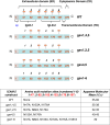
Figure 1
Figure 1
Schematic structure of ICAM-2 and ICAM-2 variants. Schematic representation of the structures of ICAM-2 and hypo-glycosylated ICAM-2 glycosylation site variants (gsv). ICAM-2 wild type (WT) contains six glycosylated amino acids in its extracellular domain (ED), at residues 47, 82, 105, 153, 176 and 187. The ED also contains two immunoglobulin-like domains (designated IgLD-1 and -2). Short transmembrane (TD) and cytoplasmic (CD) domains are also present. The N-terminus of the nascent protein contains a 21-amino acid signal peptide (sp) that is not present in the 254 amino acid mature protein. Variants were constructed by substituting alanines (A) for asparagines (N) at the indicated amino acid residues. For example, gsv1,4,5 contains alanines at mutation sites 1, 4, and 5 (amino acids 47, 153, and 176); therefore, gsv1,4,5 is not able to be glycosylated at these three sites. Variant gsv1-6 has alanines at all six glycosylation sites, and is non-glycosylated when expressed in SK-N-AS cell transfectants.
Figure 2
Figure 2
Transfected SK-N-AS cells express ICAM-2 transcripts and proteins. A) RNA from control human dermal microvascular endothelial cells (HDMVEC) generated RT-PCR products of the predicted 631 base pairs. All ICAM-2 transfectants contained readily detectable ICAM-2 RNA. B) Bar graph depicting quantitation of the RT-PCR products shown in “A”. RNA expression levels were within 20% among the transfectants. This level of variability was not statistically significant. C) Immunoblots of whole cell lysates (40 μg protein/lane) demonstrated that control HBMEC-28 cells and WT transfectants expressed immunoreactive protein having an apparent molecular mass of 55-60 kDa. ICAM-2 variants expressed proteins of appropriately lesser apparent masses. Transfectants expressed equivalent levels of actin and α-actinin. D) Deglycosylation of proteins in whole cell lysates and subsequent immunoblots for ICAM-2 confirmed that all variants displayed the expected apparent molecular mass of ~30kDa. The “nonspecific” band that appears at ~36kDa in all lanes marked + PNGase F is the PNGase F protein itself. E) Quantitation of ICAM-2 variants (lanes indicated as + PNGase F) was done to compare relative amounts of ICAM-2 protein expressed by transfectants. Results were normalized to the level of actin expression for each cell line. F) ICAM-2 WT and variants localized to cell membranes. Experimental details are included in Methods. G) Membrane localization of ICAM-2 WT and gsv4,5 was confirmed by fluorescence activated cell sorting (FACS) of intact cells, incubated with an antibody recognizing the extracellular domain (ED) of ICAM-2 (CBR-IC2/2) and a PE-conjugated secondary antibody. Non-intact cells were gated out using light scatter parameters and propidium iodide uptake. FACS profiles shown are for PE-positive cells generated using negative control IgG (blue line) or anti-ICAM-2 (red line). H) Biotinylation experiments also demonstrated that ICAM-2 WT and variants localized to cell membranes. Experimental details are included in Methods.
Figure 3
Figure 3
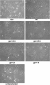
Morphology of ICAM-2 variants derived from SK-N-AS neuroblastoma cells. Cells plated at low density (~20% confluence) in CoStar tissue culture flasks were cultured at 37°C at 5% CO2 in humidified chambers, and allowed to proliferate to ~70% confluence. SK-N-AS cells transfected to express WT or gsv ICAM-2 have nascent neuronal morphologies. In comparison to SK-N-ASpIRES.ICAM-2 transfectants (WT), gsv transfectants were morphologically more pleomorphic and discohesive. All transfectants grew as attached monolayers. When detached and sized using a Coulter Counter, no differences in cell size were observed.
Figure 4
Figure 4
ICAM-2 WT and glycosylation variants inhibit NB cell migration in scratch assays. Expression of WT and gsv ICAM-2 inhibited SK-N-AS cell motility in vitro. A) Scratch assays to evaluate cell motility were performed using standard methods, and images acquired at 24 hour intervals. At each time point, the distance remaining across the scratch was calculated in pixels. Only wells in which initial scratches were of similar width were included in analyses. B) Analysis by two-way ANOVA followed by Bonferroni post test showed that at 72 hours post scratch, all variants except gsv4,5 migrated more slowly than transfectants that expressed no detectable ICAM-2 (neo) (P < 0.05* [gsv1-6] and P < 0.001*** [gsv1,4,5 and gsv1,2,5]). At 72 hours post scratch, all gsv transfectants migrated more rapidly than transfectants expressing ICAM-2 WT (P<0.01 – P<0.001, P values not indicated on graph).
Figure 5
Figure 5
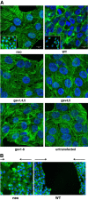
The cellular distribution of F-actin in control transfectants (neo) differs from that of transfectants expressing ICAM-2 WT. A) Control neo transfectants harbored transverse actin fibers. ICAM-2 WT induced juxtamembrane actin fiber distribution. ICAM-2 gsv expression did not induce a juxtamembrane localization of actin fibers. Each transfectant was plated at ~60% confluence and maintained under standard conditions at 37oC to allow the cells to adhere to chamber slides. Approximately 18 hours after plating, cells were fixed with 3.7% paraformaldehyde and incubated with FITC-conjugated phalloidin. Actin fibers were then visualized using confocal fluorescence microscopy by standard methods. Inset photomicrographs were acquired at 20x magnification and using a Zeiss Axio Observer Z.1 microscope platform in conjunction with Zen 2011 Blue imaging software (Carl Zeiss) (Photomicrographs of control cell lines (neo and WT) were published previously [13]). B) The distribution of actin fibers in cells at the leading edge of migration in scratch (wound healing) assays was similar to actin fiber distribution in stationary sub-confluent cultures of each cell line. Scratch assays were performed as described for experiments depicted in Figure 4. At 72 hours “post scratch”, cells were fixed and actin fibers visualized using FITC-phalloidin staining as described above, and fluorescence photomicrographs acquired using Nikon TE2000-U microscope with X-cite 120 mercury vapor short arc in conjunction with Q-capture ProV 5.1.1.14 software (Nikon Instruments, Inc.). Care was taken to acquire images of the control neo transfectants in areas where residual gaps from the original scratch were still visible, to confirm acquisition of the leading edge of migration.
Figure 6
Figure 6
WT and gsv ICAM-2 suppressed anchorage-independent growth in vitro. All four glycosylation site ICAM-2 variants suppressed anchorage-independent growth. Soft agar assays were performed using standard methods. Number of colonies of >20 cells were visualized 14-21 days after plating and results analyzed using a 2-tailed t test and GraphPad Prism software. P = 0.0124*. P < 0.0001***. Not shown in this Figure are control experiments demonstrating that the growth in soft agar of parental SK-N-AS cells was equivalent to control SK-N-ASpIRESneo transfectants.
Figure 7
Figure 7
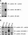
ICAM-2 WT and variants co-precipitated with α-actinin. A) IP/IB experiments with control cell lines demonstrate the expected association of ICAM-2, α-actinin and actin in lysates from SK-N-ASpIRES.ICAM-2 cells (labeled as WT), but not SK-N-ASpIRESneo cells (neo). Results for these control cell lines were published previously [13]. B) ICAM-2 glycosylation site variants associated simultaneously with α-actinin and actin. Immunoprecipitations (IP) were performed using whole cell lysates and a mouse monoclonal antibody to α-actinin (MAB1682, Millipore). Following protein separation by electrophoresis, immunoblots (IB) were performed with antibodies to α-actinin (sc-7454, Santa Cruz Biotech), ICAM-2 (AF244, R&D Systems), or actin (4968, Cell Signaling). The presence of ICAM-2 WT and variants in each preparation was confirmed by immunoblot analysis of input preparations (a representative blot is shown in Figure 2C) and also by immunoblot analysis of the proteins remaining in the supernatant following precipitation (not shown), to confirm the presence of sufficient/excess ICAM-2 protein or variant in each preparation used for immunoprecipitation experiments.
Figure 8
Figure 8
ICAM-2 variants significantly delayed but did not completely inhibit development of disseminated tumors in an in vivo model of metastatic neuroblastoma. Mice received intravenous injections of SK-N-AS cells expressing no detectable ICAM-2 (neo, N = 10), ICAM-2 WT (N = 10), or one of four hypo-glycosylated forms of ICAM-2 (N = 5/group). Kaplan-Meier survival plots were analyzed by log-rank (Mantel-Cox) test using GraphPad Prism 5 software (Version 5.02). Mice receiving cells expressing hypo-glycosylated ICAM-2 survived longer than mice receiving cells expressing no detectable ICAM-2, but not as long as mice receiving cells expressing ICAM-2 WT.

Similar articles

Cited by

References

    1. Kawauchi T. Cell adhesion and its endocytic regulation in cell migration during neural development and cancer metastasis. Int J Mol Sci. 2012;13(4):4564–4590. - PMC - PubMed
    1. Dallas MR, Liu G, Chen WC, Thomas SN, Wirtz D, Huso DL, Konstantopoulos K. Divergent roles of CD44 and carcinoembryonic antigen in colon cancer metastasis. FASEB J. 2012;26(6):2648–2656. doi: 10.1096/fj.12-203786. - DOI - PMC - PubMed
    1. Leda J, Yokoyama S, Tamura K, Takifuji K, Hotta T, Matsuda K, Oku Y, Nasu T, Kiriyama S, Yamamoto N, Nakamura Y, Shively JE, Yamaue H. Re-expression of CEACAM1 long cytoplasmic domain isoform is associated with invasion and migration of colorectal cancer. Int J Cancer. 2011;129(6):1351–1361. doi: 10.1002/ijc.26072. - DOI - PubMed
    1. Ngora H, Galli UM, Miyazaki K, Zoller M. Membrane-bound and exosomal metastasis-associated C4.4A promotes migration by associating with the α(6)β(4) integrin and MT1-MMP. Neoplasia. 2012;14(2):95–107. - PMC - PubMed
    1. Dalva MB, McClelland AC, Kayser MS. Cell adhesion molecules: signaling functions at the synapse. Nature Rev Neuroasience. 2007;8:206–220. - PMC - PubMed

Publication types

MeSH terms