. 2013 Sep;170(1):101-26.
doi: 10.1111/bph.12248.

A structural chemogenomics analysis of aminergic GPCRs: lessons for histamine receptor ligand design

Affiliations

A structural chemogenomics analysis of aminergic GPCRs: lessons for histamine receptor ligand design

A J Kooistra et al. Br J Pharmacol. 2013 Sep.

Abstract

Background and purpose: Chemogenomics focuses on the discovery of new connections between chemical and biological space leading to the discovery of new protein targets and biologically active molecules. G-protein coupled receptors (GPCRs) are a particularly interesting protein family for chemogenomics studies because there is an overwhelming amount of ligand binding affinity data available. The increasing number of aminergic GPCR crystal structures now for the first time allows the integration of chemogenomics studies with high-resolution structural analyses of GPCR-ligand complexes.

Experimental approach: In this study, we have combined ligand affinity data, receptor mutagenesis studies, and amino acid sequence analyses to high-resolution structural analyses of (hist)aminergic GPCR-ligand interactions. This integrated structural chemogenomics analysis is used to more accurately describe the molecular and structural determinants of ligand affinity and selectivity in different key binding regions of the crystallized aminergic GPCRs, and histamine receptors in particular.

Key results: Our investigations highlight interesting correlations and differences between ligand similarity and ligand binding site similarity of different aminergic receptors. Apparent discrepancies can be explained by combining detailed analysis of crystallized or predicted protein-ligand binding modes, receptor mutation studies, and ligand structure-selectivity relationships that identify local differences in essential pharmacophore features in the ligand binding sites of different receptors.

Conclusions and implications: We have performed structural chemogenomics studies that identify links between (hist)aminergic receptor ligands and their binding sites and binding modes. This knowledge can be used to identify structure-selectivity relationships that increase our understanding of ligand binding to (hist)aminergic receptors and hence can be used in future GPCR ligand discovery and design.

Keywords: GPCR; aminergic; chemical similarity; chemogenomics; crystal structures; histamine receptors; protein-ligand interactions; site-directed mutagenesis; structure-affinity relationship; transmembrane proteins.


Figures

Figure 1
Figure 1
(A) Conserved TM–fold of crystallized aminergic GPCRs (one crystal structure per receptor). The co-crystallized ligand doxepin (1) in H1R is depicted using black carbon atoms. (B) Top view of the H1R with doxepin (1, black carbon atoms). Magenta spheres depict C-alpha atoms from the binding pocket residues. The side chain of the key ionic anchor D3.32 is displayed in black. Both the major and minor binding pocket are highlighted. (C) Sequence alignment of putative binding site residues of the human H1R, human H2R, human H3R, human H4R, human M2R, rat and human M3R (the pocket residues for M3R rat and human are identical); human α1AR, turkey α1AR, human α2AR and human D3R. The lower case character preceding the receptor abbreviation indicates the species, h (human), r (rat) and m (turkey). Binding site residues are assigned based on the basis of 30 residues proposed by (Surgand et al., 2006) plus an additional 24 residues based on the six aminergic crystal structures and SDM studies (Table 1, Supporting Information Table S1) (Vroling et al., 2011). Residues in contact with the ligand in the crystal structure are coloured cyan. Magenta highlights residue W7.40, which is an aminergic family specific conserved residue. The conserved residue D3.32 is coloured red. Capital letters at the bottom indicate a partially conserved residue (>75%) and * indicates a fully conserved residue. All cysteines that form a disulphide bridge are highlighted in yellow.
Figure 2
Figure 2
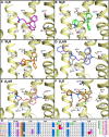
Binding mode of (A) doxepin (1, magenta carbon atoms) in human H1R (PDB code 3RZE (Shimamura et al., 2011)), (B) (R)-3-quinuclidinylbenzilate (2, green carbon atoms) in human M2R (PDB code 3UON (Haga et al., 2012)), (C) tiotropium (3, orange carbon atoms) in rat M3R (PDB code 4DAJ (Kruse et al., 2012)), (D) (S)-carvedilol (4, blue carbon atoms) in turkey β1AR (PDB code 4AMJ (Warne et al., 2012)), (E) (S)-carazolol (5, red carbon atoms) in human β2AR (PDB code 2RH1 (Cherezov et al., 2007)) and (F) (S)-eticlopride [(6, brown carbon atoms in D3R (PDB code 3PBL (Chien et al., 2010)]. The yellow ribbons represent parts of the backbone of transmembrane (TM) helices 2, 3, 5, 6 and 7. Selected binding site residues are depicted as ball-and-sticks with light grey carbon atoms. Oxygen, nitrogen, sulphur, hydrogen and chlorine atoms are coloured red, blue, yellow, cyan and green, respectively. Hydrogen bonds are depicted by black dashes. Polar hydrogen atoms of the ligand are shown, but are omitted for the pocket residues. The labels for W6.48 are omitted for all structures as well as F6.52 for H1R, β1AR, β2AR and D3R for clarity purposes. (G) Molecular interaction fingerprint (IFP) (Marcou and Rognan, 2007) bitstrings describing the binding poses of 1–6 (A–F), encoding different interaction types (negatively charged, positively charged, H-bond acceptor, H-bond donor, aromatic face-to-edge, aromatic-face-to-face and hydrophobic) for each residue in the binding site. For reasons of clarity, only the bit strings of residues D3.32, 5.42, 5.46, 6.52, 6.55, and 7.39 are shown. All binding modes are presented in a similar fashion throughout the manuscript. 2D structures of the molecules are presented in Figure 3A.
Figure 3
Figure 3
2D-structures of (A) co-crystallized ligands in the crystal structures (Figure 2) and (B) ligands shown in Table 1 or mentioned in the text. Most of the ligands in panel B are ordered according to their primary aminergic GPCR target: histamine receptors H1R (8, 11–20), H2R (8), H3R/H4R (810, 20–34), dopamine receptor D3R (3541), muscarinic receptors M2R/M3R (4249), beta-adrenergic receptors β1AR/β2AR (5060), but it should be noted that many of the ligands bind to multiple aminergic GPCR subfamilies (e.g. 48, 61, 62, 63).
Figure 4
Figure 4
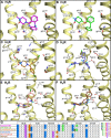
Proposed binding modes, based on SAR and SDM studies, of (A) clozapine (48) (magenta carbon atoms) in human H1R, (B) SBVS hit VUF13816 (13) (de Graaf et al., 2011) (green carbon atoms) in human H1R, (C) R-cetirizine (18) (green carbon atoms) and S-cetirizine (19) (orange carbon atoms) in human H1R, (D) VUF5228 (30) (blue carbon atoms) in a human H4R homology model (Istyastono et al., 2011b), (E) JNJ7777120 (32) (red carbon atoms) and (F) aminopyrimidine 33 (brown carbon atoms) (Schultes et al., 2012). Rendering and colour-coding are the same as in Figure 2. H4R homology models were build using the H1R (E, F) and β2AR (D) crystal structures as templates. (G) Molecular interaction fingerprint (IFP) bit strings describing the binding poses of 48, 13, 18, 19, 30, 32, 33 (A–F), encoding different interaction types with D3.32, 5.42, 5.46, 6.52, 6.55 and 7.39 (colour-coding as described in Figure 2). 2D structures of the molecules are presented in Figure 3B.
Figure 5
Figure 5
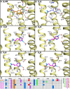
Binding mode of (A) isoproterenol (7, orange carbon atoms) in the turkey β1AR (PDB code 2Y03 (Warne et al., 2012)). Proposed binding modes of endogenous agonists, based on SDM studies, of (B) dopamine (35, blue carbon atoms) in the human D3R and the proposed binding modes of histamine (8, magenta carbon atoms) in the different human histamine receptors; (C) H1R, (D) H2R, (E) H3R and (F) H4R. Minor changes in the H1R crystal structure (i.e. rotation of K1915.39 and N1985.46) allow for accommodation of histamine. The H2R, H3R and H4R models are based on the H1R crystal structure. Rendering and colour-coding are the same as in Figure 2. (G) Molecular interaction fingerprint (IFP) bit strings describing the binding poses of 7–8 and 35 (A–F), encoding different interaction types with D3.32, 5.42, 5.46, 6.52, 6.55 and 7.39 (colour-coding as described in Figure 2). Two-dimensional structures of the molecules are presented in Figure 3A and B.
Figure 6
Figure 6
(A) The percentage (%) of the pairwise sequence identity between the ligand binding pockets (i.e. the selected 55 residues) of the histamine receptors as well as the aminergic receptors with a crystal structure available. The percentage (%) of the pairwise sequence identity for the TM helices is described in Supporting Information Table S3. (B) The average ligand similarity as calculated by EDprints (Kooistra et al., 2010) by comparing ligands from the ChEMBL database for each of the histamine receptors as well as the aminergic receptors with a crystal structure available (the scores are based on the average of the highest similarity scores). (C) The ligand overlap by comparing 9903 ligands from the ChEMBL database with annotated affinity for one or more of the discussed aminergic receptors (expressed as a percentage of the total number of ligands with experimentally determined binding affinity, i.e. if the experimentally determined radioligand displacement Ki or IC50 value is 10 μM or lower, for one or both receptors). (D) The structural distance between the ligand binding pockets as calculated by SiteAlign (Schalon et al., 2008) (i.e. the selected 54 residues using distance-3) for the crystallized aminergic GPCRs. The gradient from blue to white to red indicates a low to high similarity of sequences (A), similarity of ligands (B), ligand overlap (C) and similarity of structures (D) respectively. One has to be warned for the values with a grey colour (B, C) as this indicates a low number of ligands available for this analysis (n < 45) therefore they might be misleading.
Figure 7
Figure 7
Examples of ligand overlap within histamine receptor subtypes (H1R/H3R/H4R) as observed in the ChEMBL database (Gaulton et al., 2012) and the VU-MedChem fragment library (de Graaf et al., 2013). Compounds with annotated affinities (Ki/IC50) and a confidence factor of 8 or higher for H1R, H3R or H4R were retrieved from the ChEMBL database and considered as ligands for a receptor if the affinity for that receptor was ≤10 μM. The median affinity was used when multiple values were reported. Two-dimensional structures of some subtype selective as well as subtype unselective histamine ligands are presented.

Similar articles

Cited by

References

    1. Alberts GL, Pregenzer JF, Im WB. Contributions of cysteine 114 of the human D3 dopamine receptor to ligand binding and sensitivity to external oxidizing agents. Br J Pharmacol. 1998;125:705–710. - PMC - PubMed
    1. Alexander SP, Mathie A, Peters JA. Guide to receptors and channels (GRAC), 5th edition. Br J Pharmacol. 2011;164(Suppl. 1):S1–S324. - PMC - PubMed
    1. Ambrosio C, Molinari P, Cotecchia S, Costa T. Catechol-binding serines of beta(2)-adrenergic receptors control the equilibrium between active and inactive receptor states. Mol Pharmacol. 2000;57:198–210. - PubMed
    1. Attwood TK, Findlay JB. Fingerprinting G-protein-coupled receptors. Protein Eng. 1994;7:195–203. - PubMed
    1. Baker JG. The selectivity of beta-adrenoceptor agonists at human beta1-, beta2- and beta3-adrenoceptors. Br J Pharmacol. 2010;160:1048–1061. - PMC - PubMed

Publication types

LinkOut - more resources