. 2013 Jul 12;288(28):20280-92.
doi: 10.1074/jbc.M113.479337. Epub 2013 May 24.

The molecular basis for species-specific activation of human TRPA1 protein by protons involves poorly conserved residues within transmembrane domains 5 and 6

Affiliations

The molecular basis for species-specific activation of human TRPA1 protein by protons involves poorly conserved residues within transmembrane domains 5 and 6

Jeanne de la Roche et al. J Biol Chem. .

Abstract

The surveillance of acid-base homeostasis is concerted by diverse mechanisms, including an activation of sensory afferents. Proton-evoked activation of rodent sensory neurons is mainly mediated by the capsaicin receptor TRPV1 and acid-sensing ion channels. In this study, we demonstrate that extracellular acidosis activates and sensitizes the human irritant receptor TRPA1 (hTRPA1). Proton-evoked membrane currents and calcium influx through hTRPA1 occurred at physiological acidic pH values, were concentration-dependent, and were blocked by the selective TRPA1 antagonist HC030031. Both rodent and rhesus monkey TRPA1 failed to respond to extracellular acidosis, and protons even inhibited rodent TRPA1. Accordingly, mouse dorsal root ganglion neurons lacking TRPV1 only responded to protons when hTRPA1 was expressed heterologously. This species-specific activation of hTRPA1 by protons was reversed in both mouse and rhesus monkey TRPA1 by exchange of distinct residues within transmembrane domains 5 and 6. Furthermore, protons seem to interact with an extracellular interaction site to gate TRPA1 and not via a modification of intracellular N-terminal cysteines known as important interaction sites for electrophilic TRPA1 agonists. Our data suggest that hTRPA1 acts as a sensor for extracellular acidosis in human sensory neurons and should thus be taken into account as a yet unrecognized transduction molecule for proton-evoked pain and inflammation. The species specificity of this property is unique among known endogenous TRPA1 agonists, possibly indicating that evolutionary pressure enforced TRPA1 to inherit the role as an acid sensor in human sensory neurons.

Keywords: Acid-sensing Ion Channels (ASIC); Acidosis; Nociceptor; Pain; Patch Clamp; Species Specificity; TRP Channels.


Figures
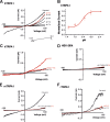
FIGURE 1.
FIGURE 1.
Protons activate hTRPA1. Currents were recorded during a 500-ms voltage ramp from −100 mV to 100 mV in cells held at −60 mV. A, membrane currents evoked by acidic solutions at pH 7.4, 7.0, 6.4, 6.0, and 5.4 and by 300 μm carvacrol on hTRPA1. B, concentration response curve for proton-evoked outward currents through hTRPA1. Peak current amplitudes at 100 mV were determined for all pH values and normalized to the amplitude evoked by pH 5.4. The line represents a fit of the data to the Hill equation. C, proton-evoked currents through hTRPA1 were blocked by the TRPA1 antagonist HC030031 (100 μm). Cells we challenged with pH 6.0 with and without HC030031. D, membrane currents in non-transfected HEK-293t cells challenged with acidic solutions. E and F, membrane currents evoked by acidic solutions and by 300 μm carvacrol in cells expressing mTRPA1 (E) or rTRPA1 (F).
FIGURE 2.
FIGURE 2.
Protons activate inward currents through hTRPA1. Currents were recorded in cells held at −60 mV. A, typical inward current evoked by pH 5.4 in a cell expressing hTRPA1. Note that coapplication of pH 5.4 with 100 μm HC-030031 (HC) resulted in a complete inhibition of the proton-evoked inward current. B, typical current trace displaying a proton-evoked current through hTRPA1 that is potentiated by 2 mm Ca2+. The column bars displays the average peak current amplitudes of currents evoked by pH 6.0 in the presence of 0 and 2 mm Ca2+. Data are expressed as mean ± S.E. C and D, typical responses evoked by application of pH 5.4 in cells expressing mTRPA1 (C) or in non-transfected HEK 293t cells (D). E, ASIC-like transient inward currents evoked by pH 5.4 in a non-transfected cell. Note that coapplication of pH 5.4 with 100 μm amiloride resulted in an almost complete inhibition of the ASIC-like inward current. F–I, current traces displaying the effects of pH 5.4 on acrolein- and carvacrol-evoked inward currents through hTRPA1 (F and H) and mTRPA1 (G and I). pH 5.4 was coapplied with acrolein or carvacrol when inward currents evoked by these agonists had reached a steady state.
FIGURE 3.
FIGURE 3.
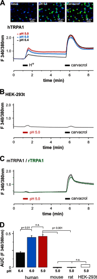
Protons evoke calcium influx through hTRPA1 but not through rodent TRPA1. A, upper panel, representative snapshots of hTRPA1-expressing cells loaded with FURA-2 during application of control solution (basal), pH 5.0, and 300 μm carvacrol. Lower panel, average effects of pH 6.4 (n = 148), 6.0 (n = 140), and 5.0 (n = 150) as well as 300 μm carvacrol on intracellular calcium in cells expressing hTRPA1 as determined by ratiometric imaging. Protons and carvacrol were both applied for 30 s. B and C, average effects of pH 5.0 on non-transfected HEK-293t cells (B) and on cells expressing mTRPA1 (C) or rTRPA1 (C) as determined by ratiometric imaging. Note that the minimal effect caused by application of protons and carvacrol in HEK-293t cells is due to an application artifact. D, areas under the curve for the average responses evoked by protons in hTRPA1, mTRPA1, rTRPA1, and non-transfected HEK-293t cells. All data are displayed as mean ± S.E. n.s., not significant.
FIGURE 4.
FIGURE 4.
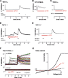
Proton-evoked calcium influx in mouse DRG neurons depends on TRPV1. A–C, ratiometric imaging of intracellular Ca2+ examined on DRG neurons derived from TRPV1−/− mice (A) (n = 181), wild-type C57Bl/6 mice (B), and TRPA1−/− mice (C). Protons were applied for 5 min. Although protons completely failed to evoke a calcium influx in neurons lacking TRPV1 (A), protons evoked a fast increase of intracellular calcium in wild-type neurons (B). This TRPV1-mediated calcium influx in neurons lacking TRPA1 could be blocked completely by the TRPV1 antagonist BCTC (10 μm) (C). D and E, protons inhibit calcium influx mediated by TRPA1 in TRPV1−/− DRG neurons. Coapplication of pH 5.0 and 100 μm AITC resulted in a suppression of calcium influx, as became obvious by a rebound effect following the washout of protons (D). Protons were applied for 5 min and AITC for 30 s. Coapplication of pH 5.0 and 300 μm carvacrol resulted in a suppression of calcium influx (E). Protons were applied for 5 min and carvacrol for 30 s. Data in A–E are displayed as mean ± S.E. F, original Ca2+ imaging recordings on DRG neurons derived from TRPV1/TRPA1−/− mice. Protons were applied for 60 s, AITC for 30 s, and capsaicin for 20 s. Upper panel, recordings on neurons transfected with hTRPA1. Note that a subpopulation of neurons responding to AITC also responded to pH 5.0. Lower panel, sham-transfected cells failed to respond to both protons and AITC. G, representative membrane currents evoked by pH 5.4 and 600 μm carvacrol in ND7/23 cells expressing hTRPA1. Currents were recorded as described in Fig. 1.
FIGURE 5.
FIGURE 5.
Transmembrane domains 5 and 6 dictate the species-specific effects of protons on TRPA1. A, alignment of TM domains 5/6 from hTRPA1 and mTRPA1. Non-identical amino acids are marked by colored letters. Amino acids involved in the multiple mutant hTRPA1-FGFATLIAM are underlined, and putative binding sites for protons are marked by bold letters. B and E, membrane currents of mTRPA1-hTM5/6 (B) and hTRPA1-mTM5/6 (E) were examined as described in Fig. 1. mTRPA1-hTM5/6 generated prominent proton-evoked inward currents at −60 mV (inset) and ramp currents upon application of pH 6.4 and 5.4. 100 μm HC030031 (HC) blocked outward currents evoked by pH 5.4. In contrast, hTRPA1-mTM5/6 failed to generate both inward currents (inset) and prominent outward currents when challenged by pH 6.4 and 5.4. C and F, typical acrolein-evoked inward currents through mTRPA1-hTM5/6 (C) and hTRPA1-mTM5/6 (F) (n = 8) modified by pH 5.4. C, coapplication of pH 5.4 with 100 μm acrolein resulted in a small potentiation of the acrolein-evoked current in mTRPA1-hTM5/6, and this effect was even smaller in hTRPA1-mTM5/6 (F). Cells were held at −60 mV, and protons were coapplied with acrolein when the inward current evoked by acrolein alone had reached a steady state. D and G, ratiometric calcium imaging on mTRPA1-hTM5/6 (D) and hTRPA1-mTM5/6 (G) expressed in HEK-293t cells. Protons and carvacrol were both applied for 30 s. Note that only a small fraction (14%) of all recorded carvacrol-sensitive cells also responded to pH 5.0 (average trace with the large peak following application of protons). When the mean response of all investigated cells was depicted, no significant proton-evoked effect could be observed (average trace lacking an effect following application of protons). hTRPA1-mTM5/6 completely failed to produce a calcium influx when pH 5.0 was applied but responded briskly upon application of 300 μm carvacrol.
FIGURE 6.
FIGURE 6.
Specific residues within TM domains 5/6 dictate the effects of protons on hTRPA1 and rhTRPA1. A, calcium imaging on cells expressing wild-type rhTRPA1 challenged with pH 5.0 and 300 μm carvacrol. B, typical ramp currents elicited in the presence of pH 7.4, pH 5.4, and 300 μm carvacrol in cells expressing rhTRPA1. Currents were examined as described in Fig. 1. C, current trace on a cell expressing rhTRPA1 examined at −60 mV. Note that rhTRPA1 completely failed to respond to pH 5.4, whereas 300 μm carvacrol elicited large inward currents. D, alignment of TM domain 6 from hTRPA1, rhTRPA1, and mTRPA1. Non-identical amino acids are marked by colored letters. E, average calcium imaging responses of cells expressing the hTRPA1 wild type, hTRPA1-V935F, hTRPA1-V942I, hTRPA1-S943A, and hTRPA1-T945A or rhTRPA1-wild type, rhTRPA1-I942V, rhTRPA1-C943S, and rhTRPA1–942V/C943S. Cells were challenged by pH 5.0 and 300 μm carvacrol. F, proton-evoked responses of hTRPA1 mutant constructs, normalized to the response obtained by carvacrol. G, original calcium imaging traces on cells expressing the rhTRPA1 wild type (black) or rhTRPA1–942V/C943S (red), again challenged by protons and carvacrol as described in E. I, representative membrane currents of the mutant rhTRPA1-I942V/C943S, examined as described in Fig. 1. Note that this mutant produces outwardly rectifying current evoked by protons. J, representative proton-evoked (pH 5.4) inward currents in cells expressing wild-type rhTRPA1-I942V/C943S, rhTRPA1-I942V, and rhTRPA1-C943S. Recordings were performed in an extracellular solution containing 2 mm Ca2+. The inward current was blocked by coapplication of 100 μm HC030031 (HC).
FIGURE 7.
FIGURE 7.
Activation of hTRPA1 by protons is not mediated by an interaction with intracellular cysteines. A and B, protons (pH 5.4) fail to activate and sensitize membrane currents through hTRPA1 when examined in cell-attached recordings. Ramp currents evoked by 500 ms long voltage ramps from −100 to 100 mV revealed outwardly rectifying currents evoked by 300 μm carvacrol but not by protons (A). Protons also failed to potentiate carvacrol-evoked currents recorded in the cell-attached mode (B). Protons were coapplied with carvacrol when the current evoked by carvacrol alone had reached a steady state. C, typical ramp current on hTRPA1 recorded in whole cell mode with an intracellular pH 5.4. Ramp currents were activated as described in Fig. 1. Note that pH 5.4 gates hTRPA1, although an intracellular acidosis is already established. D and E, current traces of hTRPA1-C621S/C641S/C665S (hTRP1–3C) evoked by pH 5.4 and 300 μm carvacrol. Ramp currents and inward currents (inset) through hTRPA1–3C could be evoked by pH 5.4. Note that 100 μm HC030031 completely blocked the membrane currents induced by pH 5.4 (D). E, average responses of hTRPA1–3C to application of pH 5.0 (30 s) and carvacrol (100 μm, 30 s) as examined by calcium imaging. Note that only a small fraction (6%) of all recorded carvacrol-sensitive cells also responded to pH 5.0 (average trace with the large peak following application of protons). When the mean response of investigated cells was depicted, no significant proton-evoked effect could be observed (average trace lacking an effect following application of protons). F, proton-evoked inward current that is not reduced by 5 mm DTT. DTT was applied together with pH 6.0 when inward currents evoked by pH 6.0 alone had reached a steady state.

Similar articles

Cited by

References

    1. Basbaum A. I., Bautista D. M., Scherrer G., Julius D. (2009) Cellular and molecular mechanisms of pain. Cell 139, 267–284 - PMC - PubMed
    1. Reeh P. W., Kress M. (2001) Molecular physiology of proton transduction in nociceptors. Curr. Opin. Pharmacol 1, 45–51 - PubMed
    1. Krishtal O. A., Pidoplichko V. I. (1981) Receptor for protons in the membrane of sensory neurons. Brain Res. 214, 150–154 - PubMed
    1. Holzer P. (2009) Acid-sensitive ion channels and receptors. Handb. Exp. Pharmacol. 194, 283–332 - PMC - PubMed
    1. Deval E., Gasull X., Noël J., Salinas M., Baron A., Diochot S., Lingueglia E. (2010) Acid-sensing ion channels (ASICs). Pharmacology and implication in pain. Pharmacol. Ther. 128, 549–558 - PubMed

Publication types

MeSH terms

LinkOut - more resources