. 2013 Aug;87(15):8372-87.
doi: 10.1128/JVI.00392-13. Epub 2013 May 22.

Unique N-linked glycosylation of CasBrE Env influences its stability, processing, and viral infectivity but not its neurotoxicity

Affiliations

Unique N-linked glycosylation of CasBrE Env influences its stability, processing, and viral infectivity but not its neurotoxicity

Krystal M Renszel et al. J Virol. 2013 Aug.

Abstract

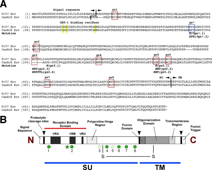
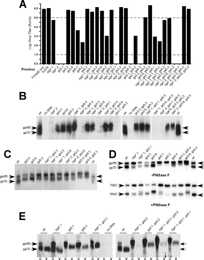
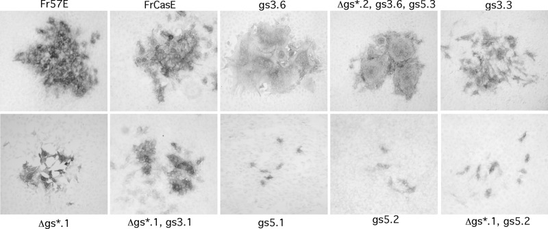
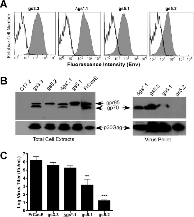
The envelope protein (Env) from the CasBrE murine leukemia virus (MLV) can cause acute spongiform neurodegeneration analogous to that induced by prions. Upon central nervous system (CNS) infection, Env is expressed as multiple isoforms owing to differential asparagine (N)-linked glycosylation. Because N-glycosylation can affect protein folding, stability, and quality control, we explored whether unique CasBrE Env glycosylation features could influence neurovirulence. CasBrE Env possesses 6/8 consensus MLV glycosylation sites (gs) but is missing gs3 and gs5 and contains a putative site (gs*). Twenty-nine mutants were generated by modifying these three sites, individually or in combination, to mimic the amino acid sequence in the nonneurovirulent Friend 57 MLV. Three basic viral phenotypes were observed: replication defective (dead; titer < 1 focus-forming unit [FFU]/ml), replication compromised (RC) (titer = 10(2) to 10(5) FFU/ml); and wild-type-like (WTL) (titer > 10(5) FFU/ml). Env protein was undetectable in dead mutants, while RC and WTL mutants showed variations in Env expression, processing, virus incorporation, virus entry, and virus spread. The newly introduced gs3 and gs5 sites were glycosylated, whereas gs* was not. Six WTL mutants tested in mice showed no clear attenuation in disease onset or severity versus controls. Furthermore, three RC viruses tested by neural stem cell (NSC)-mediated brainstem dissemination also induced acute spongiosis. Thus, while unique N-glycosylation affected structural features of Env involved in protein stability, proteolytic processing, and virus assembly and entry, these changes had minimal impact on CasBrE Env neurotoxicity. These findings suggest that the Env protein domains responsible for spongiogenesis represent highly stable elements upon which the more variable viral functional domains have evolved.


Figures

Fig 1
Fig 1
Mutagenesis strategy to assess the contribution of Env glycosylation to neuropathogenesis. (A) Amino acid sequence comparison between the nonneurovirulent Friend Env (clone 57) (Fr57) and the neurovirulent CasBrE Env (clone 15-1). Consensus glycosylation sites (based on Kayman et al. [23]) are boxed in red, with their number designations, and a putative glycosylation sequence (NXT) boxed in blue and designated gs*. Shown below the sequences are the specific mutations introduced into CasBrE Env to mimic the Friend 57 Env. The mutant designations are in parentheses and refer to the specific consensus sites, with the number of amino acid changes after the decimal point. These mutations were introduced singly or in combination to generate 29 different mutant Env genes (listed in Table 1) that were cloned into the FrCasE provirus (7). Highlighted in yellow are amino acid residues involved in ecotropic receptor interactions (56). (B) Schematic diagram of MLV Env depicting the known functional domains, along with the relative locations of the glycosylation sites (green circles) within the surface-expressed domain (SU) and the proteolytic processing sites (arrowheads). The light-green circles indicate missing MLV consensus glycosylation sites in CasBrE Env that are present in Friend Env. The star indicates the putative glycosylation site gs*, unique to CasBrE Env. VRA, VRB, and VRC are the variable regions of the receptor binding domain that confer receptor specificity.
Fig 2
Fig 2
CasBrE Env glycosylation mutations alter Env expression, processing, and incorporation into virions. (A) Virus titers arising after transfection of NIH 3T3 cells with the various gs mutant proviral DNAs tested on M. dunni fibroblasts. Mutants with titers of >105 FFU/ml were classified as WTL (above the upper dashed line), those with titers of 101 to 105 FFU/ml were classified as RC, and those with titers of <101 FFU/ml were classified as dead viruses (below the lower dashed line). (B to D) Immunoblots for CasBrE Env protein after protein separation on 9% SDS-PAGE. (B) Env expression in total cell extracts from transfected NIH 3T3 cells showing both precursor (gpr85) and processed (gp70) isoforms for various gs mutants. Note the different isoform expression levels (from equivalent protein loads), as well as the absence of detectable expression with certain mutants, such as Δgs*.2; Δgs*.2, gs5.3; Δgs*.2, gs3.6; gs3.1, gs5.3; Δgs*.1, gs3.6, gs5.3; and Δgs*.2, gs3.1, gs5.3 (gs3.1 not shown), which correlates with dead virus. (C) Examples of SDS-PAGE mobility changes for Env mutants with added gs3 and/or gs5 consensus sites, suggesting an increase in apparent Mr due to more extensive glycosylation. No relative mobility changes were apparent between mutants with or without gs* (cf. gs5.3 versus Δgs*.1, gs5.3), suggesting that this NXT/S site was not utilized; however, the gs*, gs5.1, and gs5.2 mutations showed an effect on precursor-processing efficiency and protein stability (B to E). (D) Env immunoblotting for select mutants after treating whole 3T3 cell extracts without (−) and with (+) PNGase F to remove N-linked glycosylation. (E) Semiquantitative comparison of select RC mutant Env proteins in equivalent whole-cell extracts (C) and pellets from cell culture supernatants subjected to centrifugation (V).
Fig 3
Fig 3
CasBrE Env gs mutations alter virus spread. To assess the effects of gs mutations on Env infectivity, foci on M. dunni fibroblasts were examined after exposure to limiting dilutions of control (Fr57E and FrCasE) and gs mutant viruses. Four basic focus morphologies were observed and are illustrated: (i) wild-type contiguous foci (Fr57E, FrCasE, and gs3.3), (ii) small wild type (Δgs*.1 and Δgs*.1, gs3.1), (iii) fusogenic (gs3.6 and Δgs*.2, gs3.6, gs5.3), and (iv) defective (gs5.1; gs5.2; and Δgs*.1, gs5.2). Some viruses, such as Δgs*.2, gs3.6, gs5.3 (not shown), exhibited both small wild-type and defective foci. The results for all the gs mutants are summarized in Table 1.
Fig 4
Fig 4
WTL gs mutant viruses induce CNS spongiform neurodegeneration after intraperitoneal inoculation. (A) Serum viremia titers associated with inoculation of mutant viruses indicating that in vivo titers for some viruses could be 1 to 2 log units lower than for wild-type FrCasE. Significant differences from FrCasE are indicated by asterisks (*, P < 0.05; **, P < 0.01; ***, P < 0.001). The error bars indicate standard deviations. (B) Examples of H&E-stained sections from the brainstems of mice infected with wt and select WTL mutant viruses at the time of sacrifice (19 to 21 days p.i.). Note the presence of abundant large and small vacuoles associated with the neuropil (arrows) and cells showing cytoplasmic effacement (arrowheads). (C) Summary of the brainstem pathology scores for each group of mutant-virus-inoculated mice, with the scatter plots illustrating the animal-to-animal variability of pathological severity and the mean pathology score represented by the solid lines. In individual mice showing brainstem pathology scores of <1, spinal cord histology was also examined and revealed spongiform changes of >2.0, consistent with the appearance of clinical neurological signs (Table 2). (D) Pathology comparison in individual brain regions in mice infected with either FrCasE or the gs5.3 mutant at 16 days p.i. for both viruses. The mean pathology score for each group is indicated by the horizontal line, and a difference of at least one full point would be considered a meaningful result. DCN, deep cerebellar nuclei.
Fig 5
Fig 5
WTL and RC glycosylation mutant Envs are variably expressed in C17.2 NSCs and incorporated into infectious virions. (A) Live-cell FACS analysis comparison of uninfected NSCs (black open traces) and NSCs infected with the WTL virus gs3.3 or RC virus Δgs*.1, gs5.1, or gs5.2 (gray filled traces). Fluorescence intensity represents cell surface Env expression detected with the CasBrE Env-specific monoclonal antibody 697. The peak shifts compared with the control suggest that ostensibly all NSCs expressed mutant Env on their plasma membranes. (B) Immunoblot assessment of the Env (top) and Gag (bottom) expression in NSC total cell extracts and in virus pellets after centrifugation of culture supernatants. The small spots on either side of the gs5.1 sample are artifacts associated with human cytokeratin contamination (skin) on the gel comb, which cross-reacts with the anti-CasBrE Env 697 monoclonal antibody. (C) Graph of a virus titration assay performed on cell culture supernatants taken from C17.2 NSCs infected with wild-type or the indicated gs mutant virus, indicating the same general replication trends as were observed for NIH 3T3 cells. Significant difference from FrCasE is indicated by asterisks (**, P < 0.01; ***, P < 0.001).
Fig 6
Fig 6
NSC-mediated CNS delivery of RC gs mutant viruses induces significant spongiform neuropathology despite restricted peripheral replication. (A) Representative H&E-stained paraffin-embedded sections from the brainstems of mice transplanted with C17.2 NSCs infected with wt (FrCasE) or WTL (gs3.3) or RC (Δgs*.1, gs5.1, or gs5.2) mutant viruses at 21 days posttransplantation. Note that pathology is focal and includes both neuropil vacuolation and cells with cytoplasmic effacement for all the different virus-NSC combinations. Pathology roughly colocalized with engrafted NSCs visualized in adjacent sections via immunostaining for β-galactosidase, a genetic marker engineered into C17.2 cells (not shown). (B) Scatter plot of the pathology scores observed for all the animals examined in each group, indicating the animal-to-animal variability. The horizontal lines indicate the mean pathology scores for the groups. (C) Peripheral viremia titers that develop in mice transplanted with infected NSCs. Note that the gs5.1/2 RC viruses remain replication restricted in vivo. The error bars indicate standard deviations.
Fig 7
Fig 7
Neurodegeneration is associated with expression of RC mutant Envs in NSC-transplanted mice. (A) Representative 5-μm paraffin-embedded brainstem sections immunostained for CasBrE Env (brown; left). These sections were taken adjacent to or near sections showing spongiosis by H&E staining. Env staining was detected in cells with highly ramified morphologies (brackets), consistent with virus spread to host glia, as well as in cells with more amoeboid morphology and unbranched processes, consistent with transplanted NSCs (arrows) and, as shown on the right for gs5.1, with immunostaining for β-galactosidase (β-Gal) (9). Highly localized Env expression was noted with the gs5.2 and gs5.1 transplants and roughly correlated with β-galactosidase expression. More widespread Env expression was observed with the gs3.3 and gs*.1 transplants. (B) (Left) Immunoblot detection of Env isoforms arising within the brainstems of mice transplanted with virus-infected NSCs. (Right) Shorter exposure of a portion of the blot, illustrating the multiple Env isoforms observed for Δgs*.1 and gs3.3 mutants. Extracts were made from 10 10-μm frozen brain sections taken adjacent to a section that was determined to be Env positive by immunofluorescence staining. Each lane represents a sample taken from a different mouse. Equivalent protein was loaded in each lane of the gel, except for the FrCasE sample, which was 20% of that loaded for the other lanes. Note the differences apparent in SDS-PAGE mobility for both precursor and processed Envs with the different glycosylation mutations.

Similar articles

Cited by

References

    1. Gardner MB. 1991. Retroviral leukemia and lower motor neuron disease in wild mice: natural history, pathogenesis, and genetic resistance. Adv. Neurol. 56:473–479 - PubMed
    1. Lynch WP, Czub S, McAtee FJ, Hayes SF, Portis JL. 1991. Murine retrovirus-induced spongiform encephalopathy: productive infection of microglia and cerebellar neurons in accelerated CNS disease. Neuron 7:365–379 - PubMed
    1. Nagra RM, Masliah E, Wiley CA. 1993. Synaptic and dendritic pathology in murine retroviral encephalitis. Exp. Neurol. 124:283–288 - PubMed
    1. Czub S, Lynch WP, Czub M, Portis JL. 1994. Kinetic analysis of spongiform neurodegenerative disease induced by a highly virulent murine retrovirus. Lab. Invest. 70:711–723 - PubMed
    1. Czub M, Czub S, McAtee FJ, Portis JL. 1991. Age-dependent resistance to murine retrovirus-induced spongiform neurodegeneration results from central nervous system-specific restriction of virus replication. J. Virol. 65:2539–2544 - PMC - PubMed

Publication types

Substances

LinkOut - more resources