. 2013 May 16;8(5):e63162.
doi: 10.1371/journal.pone.0063162. Print 2013.

Intravenous immunglobulin binds beta amyloid and modifies its aggregation, neurotoxicity and microglial phagocytosis in vitro

Affiliations

Intravenous immunglobulin binds beta amyloid and modifies its aggregation, neurotoxicity and microglial phagocytosis in vitro

Susann Cattepoel et al. PLoS One. .

Abstract

Intravenous Immunoglobulin (IVIG) has been proposed as a potential therapeutic for Alzheimer's disease (AD) and its efficacy is currently being tested in mild-to-moderate AD. Earlier studies reported the presence of anti-amyloid beta (Aβ) antibodies in IVIG. These observations led to clinical studies investigating the potential role of IVIG as a therapeutic agent in AD. Also, IVIG is known to mediate beneficial effects in chronic inflammatory and autoimmune conditions by interfering with various pathological processes. Therefore, we investigated the effects of IVIG and purified polyclonal Aβ-specific antibodies (pAbs-Aβ) on aggregation, toxicity and phagocytosis of Aβ in vitro, thus elucidating some of the potential mechanisms of action of IVIG in AD patients. We report that both IVIG and pAbs-Aβ specifically bound to Aβ and inhibited its aggregation in a dose-dependent manner as measured by Thioflavin T assay. Additionally, IVIG and the purified pAbs-Aβ inhibited Aβ-induced neurotoxicity in the SH-SY5Y human neuroblastoma cell line and prevented Aβ binding to rat primary cortical neurons. Interestingly, IVIG and pAbs-Aβ also increased the number of phagocytosing cells as well as the amount of phagocytosed fibrillar Aβ by BV-2 microglia. Phagocytosis of Aβ depended on receptor-mediated endocytosis and was accompanied by upregulation of CD11b expression. Importantly, we could also show that Privigen dose-dependently reversed Aβ-mediated LTP inhibition in mouse hippocampal slices. Therefore, our in vitro results suggest that IVIG may have an impact on different processes involved in AD pathogenesis, thereby promoting further understanding of the effects of IVIG observed in clinical studies.


Conflict of interest statement

Competing Interests: Susann Cattepoel, Alexander Schaub, Miriam Ender, Annette Gaida, Alain Kropf, Ursula Guggisberg, Reinhard Bolli and Sylvia M. Miescher are employees of CSL Behring AG, except Marc W. Nolte, who is employee of CSL Behring GmbH, and Louis Fabri, who is employee of CSL Limited. This does not alter the authors' adherence to all the PLOS ONE policies on sharing data and materials. Paul A. Adlard and David Finkelstein declare no conflict of interest.

Figures

Figure 1
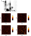
Figure 1. Characterization of Aβ preparations.
The Aβ preparations that were used in subsequent assays were characterized by SDS-PAGE (A) and AFM (B–E). A Silver Staining of Aβ42 monomers (m), oligomers (o), oligomers cross-linked with Peroxynitrite (o*), fibrils (f) and fibrils cross-linked with peroxynitrite (f*) separated on a 4–12% Bis-tris gel. B–E AFM of blank (B), Aβ42 monomers in NaOH 10 mM (C), oligomers in PBS (D), fibrils in HCl 10 mM (E). WB and AFM revealed cross-linked Aβ oligomers ranging in size from dimers to hexamers. Every batch of oligomers was tested accordingly to assure consistent quality for comparable results. M Marker.
Figure 2
Figure 2. Specific binding of Privigen and pAbs-Aβ to Aβ oligomers.
Determination of specific binding to Aβ42 oligomers by ELISA (A and B) and real-time binding by BioLayer Interferometry (C). A Privigen, pAbs-Aβ and 6E10 showed different binding activities to the Aβ42 oligomer-coated plate, with 6E10 mAb exhibiting the strongest binding followed by pAbs-Aβ and Privigen. Privigen Fc fragment showed no binding to Aβ42 oligomers. B A competition ELISA showed that the binding of Privigen, pAbs-Aβ and 6E10 could be competed by pre-incubation with Aβ42, thereby confirming specificity to Aβ42 oligomers. C BioLayer Interferometry (Octet System). Binding curves of pAbs-Aβ, 6E10 and Privigen Fc fragment on monomeric biotinylated Aβ. measured at 133 nM. In an independent experiment, the apparent KD value for the pAbs-Aβ was calculated to be 1.5*10−7±1.8*10−8 M and 2.1*10−8±1.3*10−9 M for 6E10. Privigen Fc showed no binding to the Biosensor tip.
Figure 3
Figure 3. Inhibition of Aβ fibril formation by incubation with Privigen and pAbs-Aβ.
Thioflavin T assay showing aggregation kinetics of recombinant Aβ42 (5 µM) alone or co-incubated with either A Privigen, Privigen F(ab′)2 fragment, and Privigen Fc fragment at 100 µM or with B Privigen, pAbs-Aβ and 6E10 at 1 µM. Recombinant Aβ42 was incubated with various compounds at 100 µM C or 1 µM D and Thioflavin T fluorescence was measured at t = 12 h. Aβ aggregation was significantly decreased by incubation with Privigen and Privigen F(ab)2 at 100 µM and pAbs-Aβ and 6E10 at 1 µM but not with Privigen Fc at 100 µM (* p<0.05; ** p<0.005).
Figure 4
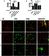
Figure 4. Privigen and pAbs-Aβ prevented Aβ-mediated cytotoxicity in SH-SY5Y cells and inhibited Aβ-binding to primary cortical neurons.
A and B SH-SY5Y cells were treated with 10 µM monomeric Aβ42 alone or co-incubated with A Privigen, Privigen Fc fragment and Privigen F(ab′)2 fragment at 100 µM, or with B Privigen, pAbs-Aβ and 6E10 at 1 µM. Cell death was measured by LDH release, normalized to Aβ and expressed in percent. Aβ-mediated neurotoxicity was significantly reduced by Privigen and Privigen F(ab′)2 fragment at 100 µM (A) and by pAbs-Aβ and 6E10 at 1 µM (B) but not by Privigen Fc fragment at 100 µM (A) and Privigen at 1 µM (B) (** p<0.005). C to E Staining of rat primary cortical neurons (DIV5) after treatment with C 10 µM monomeric Aβ alone or co-incubated with D Privigen (45 µM) or E pAbs-Aβ (0.5 µM). Confocal images showed that Privigen at 45 µM and pAbs-Aβ at 0.5 µM apparently disrupted the binding of Aβ to the neuronal cell body and dendrites, which resulted in the distribution of Aβ in the medium as immune-complexes. Microtubule-associated protein 2 (green), Aβ42 (red), antibody test substances (blue), immune-complexes (violet). Bar 100 µm.
Figure 5
Figure 5. Privigen and pAbs-Aβ increased the phagocytosis of fibrillar Aβ.
BV-2 cells were treated for 30 min with 2 µM FITC-labelled Aβ fibrils alone or co-incubated with Privigen and Privigen Fc fragment at 45 µM, A and B or with Privigen, pAbs-Aβ and 6E10 at 0.5 µM, C and D. Cytochalasin D was added as an inhibitor for receptor-mediated endocytosis and fucoidan as an inhibitor of scavenger receptor A/B. At 45 µM only Privigen significantly increased the number of phagocytosing cells (A) and the amount of phagocytosed fibrillar Aβ in the FITC-positive cell population (B). At 0.5 µM Privigen slightly whereas pAbs-Aβ and 6E10 significantly increased the number of FITC-positive cells (C). Only with Privigen and pAbs-Aβ the amount of phagocytosed Aβ was significantly increased, whereas 6E10 resulted only in a slightly not statistically significant increased phagocytosis of fibrillar Aβ (D) (** p<0.005).
Figure 6
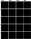
Figure 6. Privigen and pAbs-Aβ increased CD11b expression on BV-2 cells and internalized Aβ co-localized with lysosomes.
BV-2 cells were treated for 4 h with A 2 µM FITC-labelled Aβ fibrils alone or co-incubated with B Privigen (45 µM) and C pAbs-Aβ (0.5 µM), D Privigen (45 µM) and Fucoidan or E Privigen (45 µM)and Blebbistatin. Incubation with Privigen and pAbs-Aβ resulted in increased uptake of FITC-Aβ fibrils and upregulation of CD11b expression Co-incubation with Privigen and Fucoidan did not abolish Aβ phagocytosis and upregulation of CD11b was still detectable. Incubation of Aβ alone or with Fucoidan showed weak uptake of FITC-Aβ into microglia but without upregulation of CD11b expression. The intracellular FITC-Aβ signal was localized in punctate structures that co-localized with CD11b immunoreactivity and LAMP1 immunoreactivity in cells incubated with Privigen and pAbs-Aβ. CD11b (white), LAMP1 (red), FITC-Aβ (green). Bar 50 µm.
Figure 7
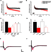
Figure 7. Privigen reversed Aβ-mediated LTP inhibition.
LTP assay using mouse brain sections showed that A oligomeric Aβ42 caused an impairment of LTP, untreated slices exhibited normal LTP. B Privigen protected against Aβ42 when applied at a concentration of 0.5 µM. Privigen at 0.25 µM only slightly improved LTP inhibition. Applied alone at 0.5 µM Privigen had no effect on LTP. C Summary of the LTP data 90 min after TBS. ***P≤0.001; compared to Untreated; ##P≤0.01 compared to Aβ42. D Summary of the LTP data 90 min after TBS. Proline did not impair LTP and did not modify the effect of Aβ42 on LTP. ***P≤0.001; compared to Untreated; ##P≤0.01 compared to Aβ42. E and F Representative fEPSP traces showing evoked responses before (black) and 90 minutes after TBS stimulation (red) for E Aβ42 treated slices with fEPSP 90 min after TBS (red) almost as large as before TBS (black) and F Privigen (0.5 µM)+Aβ42 treated slices clearly showing that Privigen rescued the Aβ-mediated LTP impairment, since the evoked response after TBS (red) is nearly twice as large as the fEPSP before TBS (black). Calibration bars are 0.5 mV and 15 ms. The number of slices was n = 6 for each group.

Similar articles

Cited by

References

    1. Hardy J, Selkoe DJ (2002) The amyloid hypothesis of Alzheimer's disease: progress and problems on the road to therapeutics. Science 297: 353–356 297/5580/353 [pii]. doi:10.1126/science.1072994. - DOI - PubMed
    1. Weiner HL, Frenkel D (2006) Immunology and immunotherapy of Alzheimer's disease. Nat Rev Immunol 6: 404–416 nri1843 [pii]; doi:10.1038/nri1843. - DOI - PubMed
    1. Bard F, Cannon C, Barbour R, Burke RL, Games D, et al. (2000) Peripherally administered antibodies against amyloid beta-peptide enter the central nervous system and reduce pathology in a mouse model of Alzheimer disease. Nat Med 6: 916–919 doi:10.1038/78682. - DOI - PubMed
    1. DeMattos RB, Bales KR, Cummins DJ, Dodart JC, Paul SM, et al. (2001) Peripheral anti-A beta antibody alters CNS and plasma A beta clearance and decreases brain A beta burden in a mouse model of Alzheimer's disease. Proc Natl Acad Sci U S A 98: 8850–8855 151261398 [pii]. doi:10.1073/pnas.151261398. - DOI - PMC - PubMed
    1. Morgan D (2011) Immunotherapy for Alzheimer's disease. J Intern Med 269: 54–63 doi:10.1111/j.1365-2796.2010.02315.x. - DOI - PMC - PubMed

Publication types

Grants and funding

The study was fully funded by CSL Behring AG. The funders had no role in study design, data collection and analysis, decision to publish, or preparation of the manuscript.