

Increased TRPV4 expression in urinary bladder and lumbosacral dorsal root ganglia in mice with chronic overexpression of NGF in urothelium
- PMID: 23690258
- PMCID: PMC3779511
- DOI: 10.1007/s12031-013-0033-5
Increased TRPV4 expression in urinary bladder and lumbosacral dorsal root ganglia in mice with chronic overexpression of NGF in urothelium
Abstract
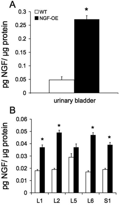
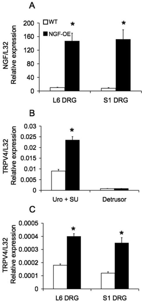
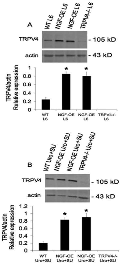
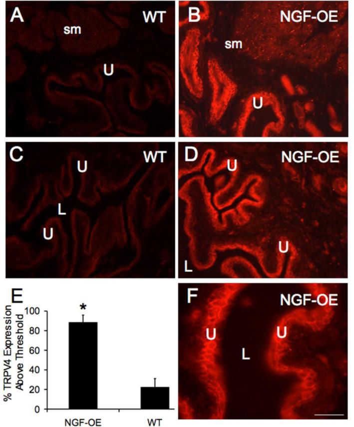
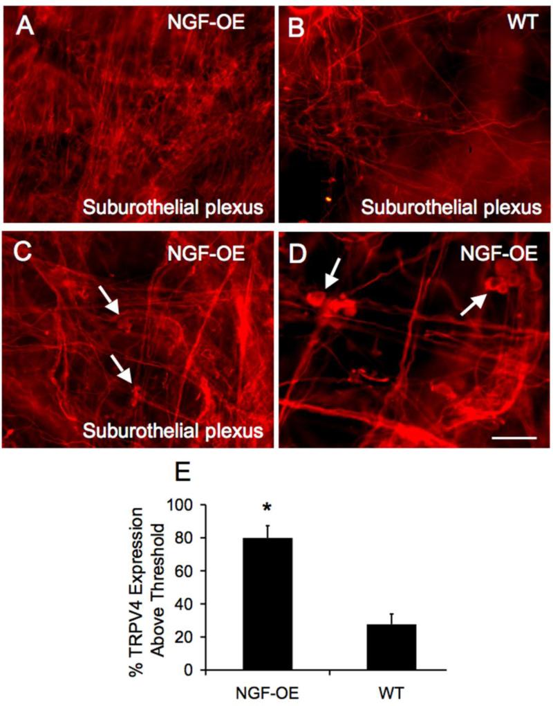
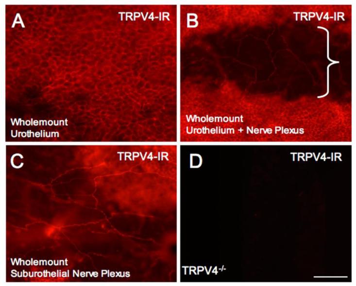
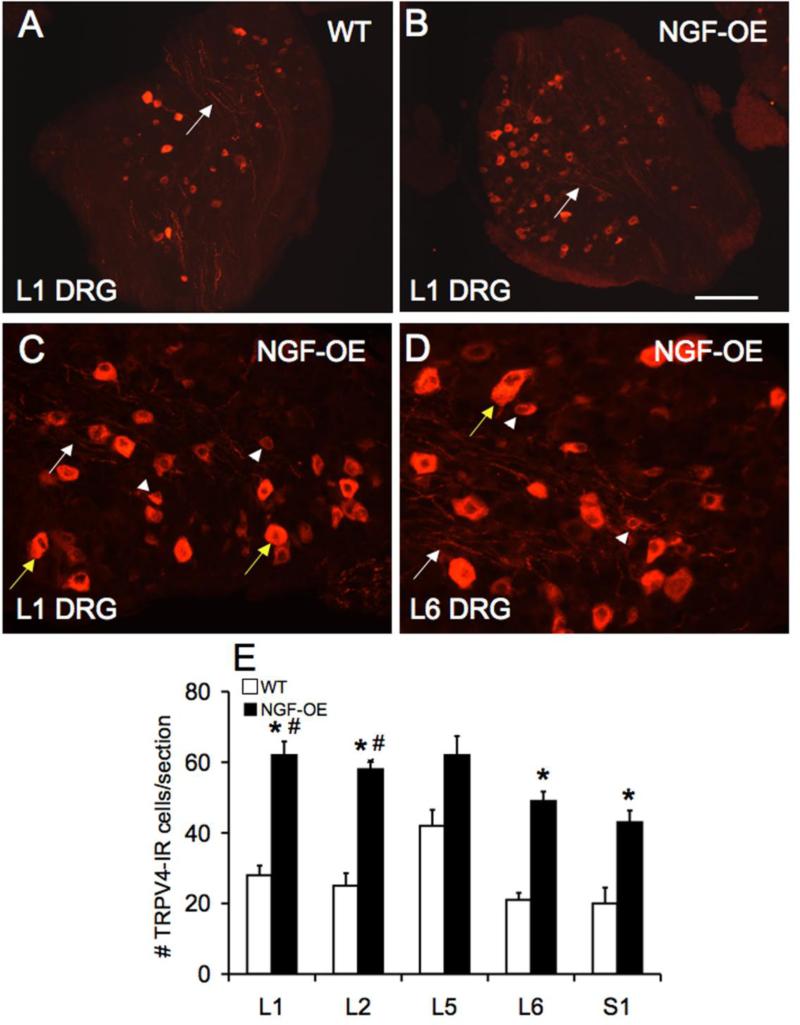
Transient receptor potential vanilloid (TRPV) family member 4 (TRPV4) expression has been demonstrated in urothelial cells and dorsal root ganglion (DRG) neurons, and roles in normal micturition reflexes as well as micturition dysfunction have been suggested. TRP channel expression and function is dependent upon target tissue expression of growth factors. These studies expand upon the target tissue dependence of TRPV4 expression in the urinary bladder and lumbosacral DRG using a recently characterized transgenic mouse model with chronic overexpression of nerve growth factor (NGF-OE) in the urothelium. Immunohistochemistry with image analyses, real-time quantitative polymerase chain reaction, and Western blotting were used to determine TRPV4 protein and transcript expression in the urinary bladder (urothelium + suburothelium, detrusor) and lumbosacral DRG from littermate wild-type (WT) and NGF-OE mice. Antibody specificity controls were performed in TRPV4(-/-) mice. TRPV4 transcript and protein expression was significantly (p ≤ 0.001) increased in the urothelium + suburothelium and suburothelial nerve plexus of the urinary bladder and in small- and medium-sized lumbosacral (L1, L2, L6-S1) DRG cells from NGF-OE mice compared to littermate WT mice. NGF-OE mice exhibit significant (p ≤ 0.001) increases in NGF transcript and protein in the urothelium + suburothelium and lumbosacral DRG. These studies demonstrate regulation of TRPV4 expression by NGF in lower urinary tract tissues. Ongoing studies are characterizing the functional roles of TRPV4 expression in the sensory limb (DRG, urothelium) of the micturition reflex.
Figures
Similar articles
-
Effects of CYP-Induced Cystitis on Growth Factors and Associated Receptor Expression in Micturition Pathways in Mice with Chronic Overexpression of NGF in Urothelium.J Mol Neurosci. 2016 Aug;59(4):531-43. doi: 10.1007/s12031-016-0774-z. Epub 2016 Jun 3. J Mol Neurosci. 2016. PMID: 27259880 Free PMC article.
-
Accelerated onset of the vesicovesical reflex in postnatal NGF-OE mice and the role of neuropeptides.Exp Neurol. 2016 Nov;285(Pt B):110-125. doi: 10.1016/j.expneurol.2016.06.021. Epub 2016 Jun 21. Exp Neurol. 2016. PMID: 27342083 Free PMC article.
-
TRPV4 blockade reduces voiding frequency, ATP release, and pelvic sensitivity in mice with chronic urothelial overexpression of NGF.Am J Physiol Renal Physiol. 2019 Dec 1;317(6):F1695-F1706. doi: 10.1152/ajprenal.00147.2019. Epub 2019 Oct 21. Am J Physiol Renal Physiol. 2019. PMID: 31630542 Free PMC article.
-
Transient receptor potential vanilloid type 4 (TRPV4) in urinary bladder structure and function.Curr Top Membr. 2022;89:95-138. doi: 10.1016/bs.ctm.2022.06.002. Epub 2022 Jul 18. Curr Top Membr. 2022. PMID: 36210154 Free PMC article. Review.
-
Emerging roles of the TRPV4 channel in bladder physiology and dysfunction.J Physiol. 2021 Jan;599(1):39-47. doi: 10.1113/JP279776. Epub 2020 Oct 27. J Physiol. 2021. PMID: 33052604 Review.
Cited by
-
NGF-Induced Nav1.7 Upregulation Contributes to Chronic Post-surgical Pain by Activating SGK1-Dependent Nedd4-2 Phosphorylation.Mol Neurobiol. 2021 Mar;58(3):964-982. doi: 10.1007/s12035-020-02156-1. Epub 2020 Oct 16. Mol Neurobiol. 2021. PMID: 33063281
-
Effects of CYP-Induced Cystitis on Growth Factors and Associated Receptor Expression in Micturition Pathways in Mice with Chronic Overexpression of NGF in Urothelium.J Mol Neurosci. 2016 Aug;59(4):531-43. doi: 10.1007/s12031-016-0774-z. Epub 2016 Jun 3. J Mol Neurosci. 2016. PMID: 27259880 Free PMC article.
-
TRPV1 and mast cell involvement in repeated variate stress-induced urinary bladder dysfunction in adult female mice.Am J Physiol Renal Physiol. 2024 Sep 1;327(3):F476-F488. doi: 10.1152/ajprenal.00125.2024. Epub 2024 Jul 11. Am J Physiol Renal Physiol. 2024. PMID: 38991005
-
Accelerated onset of the vesicovesical reflex in postnatal NGF-OE mice and the role of neuropeptides.Exp Neurol. 2016 Nov;285(Pt B):110-125. doi: 10.1016/j.expneurol.2016.06.021. Epub 2016 Jun 21. Exp Neurol. 2016. PMID: 27342083 Free PMC article.
-
Non-surgical treatments for the management of early osteoarthritis.Knee Surg Sports Traumatol Arthrosc. 2016 Jun;24(6):1775-85. doi: 10.1007/s00167-016-4089-y. Epub 2016 Apr 4. Knee Surg Sports Traumatol Arthrosc. 2016. PMID: 27043347 Review.
References
-
- Andersson KE, Gratzke C, Hedlund P. The role of the transient receptor potential (TRP) superfamily of cation-selective channels in the management of the overactive bladder. B.J.U. Int. 2010;106:1114–1127. - PubMed
-
- Araki I. TRP channels in urinary bladder mechanosensation. Adv. Exp. Med. Biol. 2011;704:861–879. - PubMed
Publication types
MeSH terms
Substances
Grants and funding
- 5 P30 RR 032135/RR/NCRR NIH HHS/United States
- R56 DK060481/DK/NIDDK NIH HHS/United States
- P30 GM103498/GM/NIGMS NIH HHS/United States
- R29 DK051369/DK/NIDDK NIH HHS/United States
- DK065989/DK/NIDDK NIH HHS/United States
- R01 DK060481/DK/NIDDK NIH HHS/United States
- DK060481/DK/NIDDK NIH HHS/United States
- DK051369/DK/NIDDK NIH HHS/United States
- R01 DK051369/DK/NIDDK NIH HHS/United States
- P30 RR032135/RR/NCRR NIH HHS/United States
- R01 DK065989/DK/NIDDK NIH HHS/United States
- 8 P30 GM 103498/GM/NIGMS NIH HHS/United States
LinkOut - more resources
Full Text Sources
Other Literature Sources
Miscellaneous
