. 2013 May 10;2(3):e000201.
doi: 10.1161/JAHA.113.000201.

Angiopoietin-like 2 promotes atherogenesis in mice

Affiliations

Angiopoietin-like 2 promotes atherogenesis in mice

Nada Farhat et al. J Am Heart Assoc. .

Abstract

Background: Angiopoietin like-2 (angptl2), a proinflammatory protein, is overexpressed in endothelial cells (ECs) from patients with coronary artery disease (CAD). Whether angptl2 contributes to atherogenesis is unknown. We tested the hypothesis that angptl2 promotes inflammation and leukocyte adhesion onto ECs, thereby accelerating atherogenesis in preatherosclerotic dyslipidemic mice.

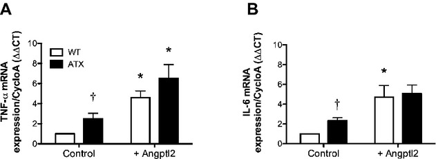
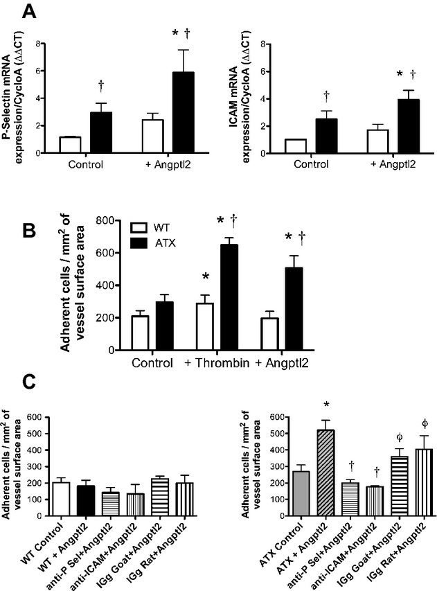
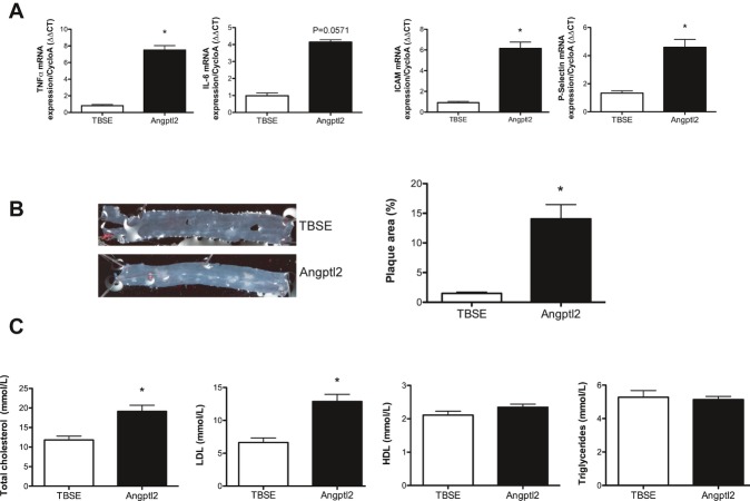
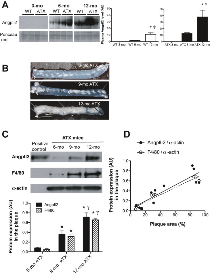
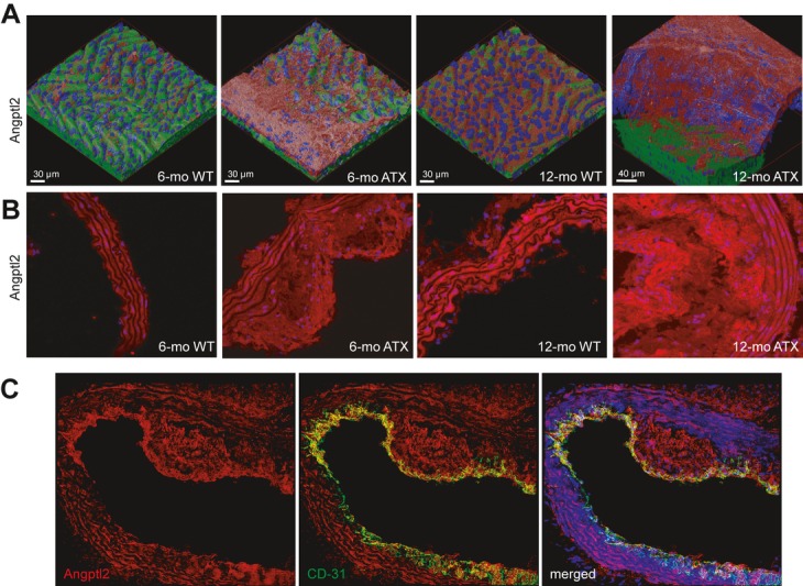
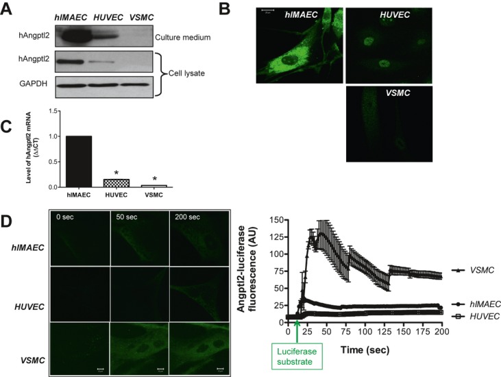
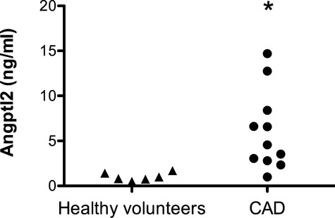
Methods and results: In ECs freshly isolated from the aorta, basal expression of TNF-α and IL-6 mRNA was higher in 3-month-old severely dyslipidemic mice (LDLr(-/-); hApoB100(+/+) [ATX]) than in control healthy wild-type (WT) mice (P<0.05) and was increased in both groups by exogenous angptl2 (100 nmol/L). Angptl2 stimulated the adhesion of leukocytes ex vivo on the native aortic endothelium of ATX, but not WT mice, in association with higher expression of ICAM-1 and P-selectin in ECs (P<0.05). Antibodies against these endothelial adhesion molecules prevented leukocyte adhesion. Intravenous administration of angptl2 for 1 month in preatherosclerotic 3-month-old ATX mice increased (P<0.05) total cholesterol and LDL-cholesterol levels, strongly induced (P<0.05) the expression of endothelial proinflammatory cytokines and adhesion molecules while accelerating atherosclerotic lesion formation by 10-fold (P<0.05). Plasma and aortic tissue levels of angptl2 increased (P<0.05) with age and were higher in 6- and 12-month-old ATX mice than in age-matched WT mice. Angptl2 accumulated to high levels in the atherosclerotic lesions (P<0.05). Finally, angptl2 was greatly expressed (P<0.05) in ECs cultured from CAD patients, and circulating angptl2 levels were 6-fold higher in CAD patients compared with age-matched healthy volunteers.

Conclusions: Angptl2 contributes to the pathogenesis of atherosclerosis.

Keywords: CAD; adhesion molecules; aging; freshly isolated mouse endothelial cells; inflammation; mouse model of atherosclerosis.


Figures

Figure 1.
Figure 1.
Angptl2 stimulates the expression of inflammatory markers in ECs. Quantitative RT‐PCR analysis of expression of TNF‐α mRNA (a) and IL‐6 mRNA (b) was performed in ECs freshly extracted from aortas of 3‐month‐old WT and LDLr−/−; hApoB+/+(ATX) mice stimulated and unstimulated (control) with purified recombinant angptl2 (100 nmol/L). Results were normalized to cyclophilin A (CycloA) expression, and average gene expression level in WT control cells was arbitrarily set at 1. Data are mean±SEM of n=4 WT and n=4 ATX mice; each experiment was performed in triplicate. *P<0.05 vs control; †P<0.05 vs WT mice. Angptl2 indicates angiopoietin like‐2; EC, endothelial cell; RT‐PCR, real‐time polymerase chain reaction; TNF‐α, tumor necrosis factor α; IL, interleukin; WT, wild type; LDL, low‐density lipoprotein; ΔΔCT, target gene threshold cycle quantification.
Figure 2.
Figure 2.
Angptl2 stimulates leukocyte adhesion to the native endothelium in ATX but not WT mice. A, Expression of P‐selectin and ICAM‐1 mRNA in freshly isolated aortic ECs from WT (n=7) and LDLr−/−; hApoB+/+ (ATX; n=6) mice was quantified by quantitative RT‐PCR and normalized to cyclophilin A after stimulation or not with purified recombinant angptl2 (100 nmol/L). Data are mean±SEM of n mice. *P<0.05 vs control; †P<0.05 vs condition‐matched in WT mice. B, Leukocytes from n=12 WT and n=6 ATX mice were labeled with 51Cr and incubated with aortic segments stimulated or not (control) with thrombin (10 U/mL) or purified recombinant angptl2 (100 nmol/L). Adherent 51Cr‐leukocytes were counted, and results are expressed as number of adherent cells/mm2 of endothelium surface area. Data are mean±SEM of n mice. *P<0.05 vs control; †P<0.05 vs condition‐matched in WT mice. C, Aortic segments from n=16 WT mice (left) and n=9 ATX mice (right) were preincubated with anti‐P‐selectin or anti‐ICAM‐1 antibodies or the corresponding isotype‐matched IgG prior to stimulation with purified recombinant angptl2 (100 nmol/L). Adhesion of 51Cr‐leukocytes was then quantified. Data are mean±SEM of n mice. *P<0.05 vs control; †P<0.05 vs +angptl2 condition; φP<0.05 vs antibody+angptl2 condition. Angptl2 indicates angiopoietin like‐2; WT, wild type; EC, endothelial cell; LDL, low‐density lipoprotein; RT‐PCR, real‐time polymerase chain reaction; IgG, immunoglobulin G; ICAM‐1, intercellular adhesion molecule‐1.
Figure 3.
Figure 3.
Angplt2 mRNA abundance and cell surface expression of cell adhesion molecules in leukocytes from both WT and ATX mice. Basal and angptl2‐induced mRNA expression of (A) CD18, (B) CD62L, and (C) CD162 in leukocytes from 3‐month‐old WT (n=6) and ATX (n=6) mice, were quantified by quantitative RT‐PCR and normalized by cyclophilin A. Cell surface protein expression of (D) CD18, (E) CD62L, and (F) CD162 was quantified in control and angptl2‐treated WT (n=6) and ATX (n=6) leukocytes by flow cytometry. Leukocytes were labeled using monoclonal anti‐CD18, anti‐CD62, and anti‐CD162L antibodies or with corresponding isotype‐matched IgG (data not shown). Data are mean±SEM of n mice. *P<0.05 vs control condition. Angptl2 indicates angiopoietin like‐2; WT, wild type; RT‐PCR, real‐time polymerase chain reaction.
Figure 4.
Figure 4.
Chronic angptl2 infusion for 1 month ATX mice accelerates atherogenesis in 3‐month‐old ATX mice. A, Infusion with angptl2 promotes the expression of inflammatory cytokines and adhesion molecules in freshly scraped ECs from the aorta evaluated by quantitative RT‐PCR and normalized by cyclophilin A. B, angptl2 accelerates the formation of atherosclerotic plaque. C, angptl2 increases total cholesterol and LDL plasma levels. Data are mean±SEM of n=5 angptl2‐infused mice and n=5 TBSE‐infused mice. *P<0.05 vs TBSE condition. Angptl2 indicates angiopoietin like‐2; EC, endothelial cell; WT, wild type; RT‐PCR, real‐time polymerase chain reaction; LDL, low‐density lipoprotein; TBSE, Tris‐base, Sodium chloride, EDTA.
Figure 5.
Figure 5.
Angptl2 plasma levels and angptl2 expression in atherosclerotic plaque increase with age and atherosclerosis. A, Samples corresponding to equal amounts of total protein were collected from plasma in wild‐type (WT; n=4) and LDLr−/−; hApoB+/+ (ATX; n=4) mice at 3, 6, and 12 months of age. Low abundant proteins in the plasma samples were enriched using Bio‐Rad ProteoMiner protein enrichment kits. Following enrichment, the samples were resolved on SDS‐PAGE, transferred to nitrocellulose membranes, and probed with an antibody against angptl2. Uniform protein loading and transfer were verified by staining membranes with Ponceau red. Results (arbitrary units, AU) are presented as the mean±SEM of 4 mice in each group. *P<0.05 vs 3‐month‐old mice; ϕP<0.05 vs 6‐month‐old mice. B, Photographs of atherosclerotic lesions in abdominal aortas from 6‐, 9‐, and 12‐month‐old LDLr−/−; hApoB+/+ (ATX) mice. C, Proteins were specifically extracted from the lesion, and Western blot analysis was performed for angptl2 (n=4 of 6‐, 9‐, and 12‐month‐old ATX mice) and F4/80 (n=3 of 6‐, 9‐, and 12‐month‐old ATX mice), a marker of mature macrophages. D, Correlation between angptl2 level (Pearson r=0.8959, P<0.0001, r2=0.8026, n=12), F4/80 (Pearson r=0.9682, P<0.0001, r2=0.9373, n=9), and the surface area of the lesion. Data are mean±SEM of n mice. *P<0.05 vs 6‐month‐old ATX mice; γP<0.05 vs 9‐month‐old ATX mice. Angptl2 indicates angiopoietin like‐2; LDL, low‐density lipoprotein; SDS‐PAGE, sodium dodecyl sulfate polyacrylamide gel electrophoresis.
Figure 6.
Figure 6.
Aortic angptl2 immunofluorescence increases with age and the progression of atherosclerosis, with marked accumulation within atherosclerotic plaque. Immunofluorescence was used to visualize angptl2 in (A) fresh longitudinally opened aortas and (B) frozen aortic sections of WT and LDLr−/−; hApoB+/+ (ATX) mice at 6 and 12 months of age. A, z‐stacks were acquired, deconvolved, and 3D images rendered. Angptl2 accumulates in plaque and in media. Angptl2 levels are shown in red and basal membrane in green. Nuclei are shown in blue. C, Double‐immunostaining of angptl2 (red) and CD‐31 (green) in frozen aortic sections from 6‐month‐old ATX mice, showing that angptl2 colocalizes with the endothelial marker CD‐31. Angptl2 indicates angiopoietin like‐2; WT, wild type; LDL, low‐density lipoprotein.
Figure 7.
Figure 7.
Expression and secretion of angptl2 is greater in ECs than in VSMCs, but angptl2 binding is higher in VSMCs than in ECs. A, Western blot analysis of endogenous angptl2 secreted into the culture medium overnight (16 h) by hIMAECs, HUVECs, and VSMCs. Angptl2 protein expression was also quantified in the cell lysate. B, Endogenous angptl2 expression in the cells was detected by immunofluorescence using a confocal microscope. Scale bar represents 20 μm. C, Quantitative RT‐PCR analysis of angptl2 mRNA expression was performed in the cell lysate from cultured cells (n=3 hIMAECs, n=3 HUVECs, n=3 VSMCs). Results were normalized to GAPDH expression, and average gene expression in hIMAECs was arbitrarily set at 1. Data are mean±SEM of n=3; each experiment was performed in duplicate. *P<0.05 vs hIMAECs. D, To assess angptl2 binding, hIMAECs, HUVECs, and VSMCs were incubated for 10 minutes with human angptl2‐luciferase (100 nmol/L) in phenol red‐free medium. Cells were then washed, and the binding of angptl2‐luciferase to cell surface was revealed by adding the luciferase substrate. A selection of confocal time‐series images acquired in a single living cell at 0, 50, and 200 seconds is shown (left). The average angptl2‐luciferase fluorescent signals (AU) recorded in VSCMs (n=6 cells), hIMAECs (n=6 cells), and HUVECs (n=4 cells) were derived from the time‐series images (right). Data are mean±SEM, and the assay was performed 3 to 4 times. EC indicates endothelial cell; VSMC, vascular smooth muscle cell; hangptl2, human angiopoietin like‐2; hIMAEC, human internal mammary artery endothelial cell; HUVEC, human umbilical vein endothelial cell; RT‐PCR, real‐time polymerase chain reaction; AU, arbitrary unit.
Figure 8.
Figure 8.
Plasma angptl2 levels are higher in CAD patients than in healthy volunteers. Circulating angptl2 levels were quantified in CAD patients (n=11) and in age‐matched healthy volunteers (n=6) by ELISA. Data are presented as a dot plot. *P<0.05 vs healthy volunteers. Angptl2 indicates angiopoietin like‐2; CAD, coronary artery disease; ELISA, enzyme‐linked immunosorbent assay.

Similar articles

Cited by

References

    1. Hato T, Tabata M, Oike Y. The role of angiopoietin‐like proteins in angiogenesis and metabolism. Trends Cardiovasc Med. 2008; 18:6-14 - PubMed
    1. Morisada T, Kubota Y, Urano T, Suda T, Oike Y. Angiopoietins and angiopoietin‐like proteins in angiogenesis. Endothelium. 2006; 13:71-79 - PubMed
    1. Quagliarini F, Wang Y, Kozlitina J, Grishin NV, Hyde R, Boerwinkle E, Valenzuela DM, Murphy AJ, Cohen JC, Hobbs HH. Atypical angiopoietin‐like protein that regulates angptl3. Proc Natl Acad Sci USA. 2012; 109:19751-19756 - PMC - PubMed
    1. Ando Y, Shimizugawa T, Takeshita S, Ono M, Shimamura M, Koishi R, Furukawa H. A decreased expression of angiopoietin‐like 3 is protective against atherosclerosis in ApoE‐deficient mice. J Lipid Res. 2003; 44:1216-1223 - PubMed
    1. Kathiresan S, Melander O, Guiducci C, Surti A, Burtt NP, Rieder MJ, Cooper GM, Roos C, Voight BF, Havulinna AS, Wahlstrand B, Hedner T, Corella D, Tai ES, Ordovas JM, Berglund G, Vartiainen E, Jousilahti P, Hedblad B, Taskinen MR, Newton‐Cheh C, Salomaa V, Peltonen L, Groop L, Altshuler DM, Orho‐Melander M. Six new loci associated with blood low‐density lipoprotein cholesterol, high‐density lipoprotein cholesterol or triglycerides in humans. Nat Genet. 2008; 40:189-197 - PMC - PubMed

Publication types