. 2013 Jul;33(14):2671-82.
doi: 10.1128/MCB.00154-13. Epub 2013 May 6.

LKB1 controls human bronchial epithelial morphogenesis through p114RhoGEF-dependent RhoA activation

Affiliations

LKB1 controls human bronchial epithelial morphogenesis through p114RhoGEF-dependent RhoA activation

Xiaojian Xu et al. Mol Cell Biol. 2013 Jul.

Abstract

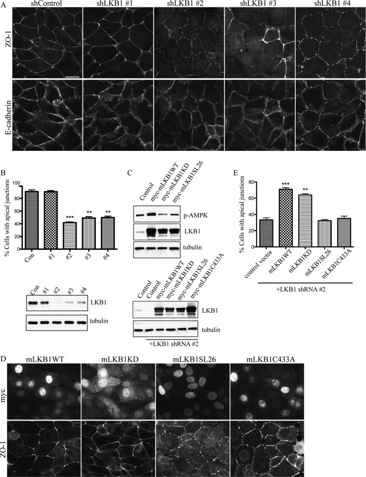
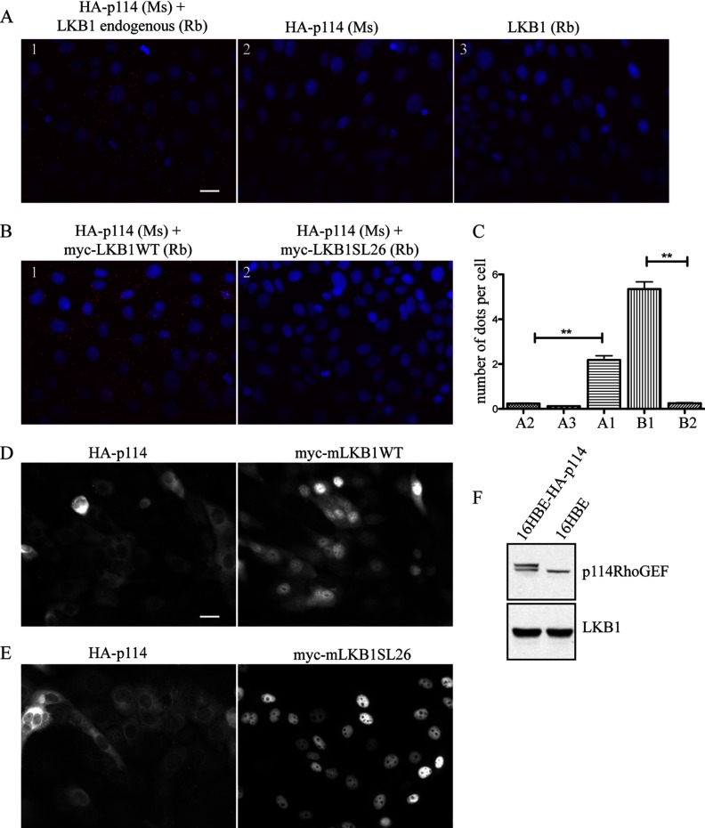
LKB1 is a Ser/Thr kinase that plays an important role in controlling both energy metabolism and cell polarity in metazoan organisms. LKB1 is also a tumor suppressor, and homozygous, inactivating mutations are found in a wide range of human cancers. In lung cancer, inactivating mutations are found in 10 to 50% of cases, but the consequences of functional loss in this context are poorly understood. We report here that LKB1 is required for the maturation of apical junctions in the human bronchial epithelial cell line 16HBE14o- (16HBE). This activity is dependent on an interaction with the Rho guanine nucleotide exchange factor p114RhoGEF but is independent of LKB1 kinase activity. Together, LKB1 and p114RhoGEF control RhoA activity in these cells to promote apical junction assembly.


Figures

Fig 1
Fig 1
LKB1 controls apical junction formation in 16HBE cells. (A) At 2 days after infection with lentiviral vectors harboring control or 4 distinct shRNAs targeting LKB1 (shLKB1 to shLKB4), 16HBE cells were seeded on coverslips. At 2 days after plating, confluent cells were fixed and stained with anti-ZO-1 (top) and anti-E-cadherin (bottom). Bar, 20 μm. (B) (Top) Quantification of apical junction formation from three independent experiments. Error bar, SEMs. **, P < 0.01; ***, P < 0.001. (Bottom) At 5 days after infection, cells were lysed and analyzed by Western blotting with the indicated antibodies. Con, control. (C) (Top) Myc-tagged LKB1WT, LKB1KD (K78M), or LKB1SL26 was transiently transfected into 16HBE cells stably expressing HA-STRAD. One day later, cells were lysed and analyzed by Western blotting with the indicated antibodies. (Bottom) 16HBE cells stably expressing mLKB1WT, mLKB1KD (K78M), mLKB1SL26, or mLKB1C433A were infected with shRNA targeting LKB1 lentiviral vector, and 2 and 5 days later cells were lysed and analyzed by Western blotting with the indicated antibodies. (D) Cells described for panel C were replated, grown to confluence, and fixed and stained with anti-ZO-1 (bottom) and anti-myc (top). (E) Quantification of apical junction formation as described for panel D from three independent experiments. Error bars, SEMs. **, P < 0.01; ***, P < 0.001.
Fig 2
Fig 2
LKB1 and STRAD activate RhoA and enhance phospho-PRK recruitment to apical junctions. (A) 16HBE cells stably expressing HA-tagged STRAD were infected with pLL3.7 lentiviral vectors encoding GFP-tagged LKB1 constructs. Twenty-four hours later, cells were harvested and Rho activity (Rho.GTP) was determined using a standard pulldown assay. LKB1WT, LKB1KD (K78M), and LKB1P38A induced 6- to 8-fold increases in the levels of active RhoA.GTP relative to the level for the control, LKB1SL26, or LKB1SL26/P38A. Total phospho-PRK was determined in the input cell lysates. Error bars, SEMs. **, P < 0.01; *, P < 0.05. WB, Western blotting. (B) 16HBE cells expressing GFP-tagged hLKB1P38A and hLKB1SL26/P38A were fixed and stained with DAPI (4′,6-diamidino-2-phenylindole). GFP signals were visualized directly. Bar, 20 μm. (C) At 1 day after infection, cells were stained for anti-phospho-PRK. GFP signals were visualized directly. GFP indicated infected cells. Bar, 20 μm.
Fig 3
Fig 3
p114RhoGEF controls apical junction formation in 16HBE cells and interacts with LKB1. (A) At 2 days after infection with lentiviral vectors harboring shRNAs targeting p114RhoGEF, 16HBE cells were seeded on coverslips. Two days later, confluent cells were fixed and stained with anti-ZO-1 (top) and anti-E-cadherin (bottom). Bar, 20 μm. (B) (Top) Quantification of apical junction formation from three independent experiments. Error bars, SEMs. *, P < 0.05; **, P < 0.01. (Bottom) At 5 days after infection, cells were lysed and analyzed by Western blotting with the indicated antibodies. (C) Schematic organization of hLKB1 and mouse p114RhoGEF (numbers represent amino acids). hLKB1 possesses a farnesylation site at aa 430. SL26 represents a 9-bp, in-frame deletion in the kinase domain. p114RhoGEF has a potential PBM at its C terminus. NRD, N-terminal regulatory domain; DH, Dbl homology domain. (D to F) HEK293T cells cotransfected with the indicated combinations of constructs. Proteins were immunoprecipitated (IP) from cell lysates using anti-mouse IgG or anti-Flag antibody. Input and immunoprecipitated lysates were analyzed by Western blotting.
Fig 4
Fig 4
LKB1 and p114RhoGEF C-terminal domains have a dominant negative effect on apical junction formation in 16HBE cells. (A) 16HBE cells stably expressing GFP-tagged LKB1WT, LKB1KD, LKB1SL26, LKB1CRD (aa 309 to 433), or LKB1CRDΔCAAX (aa 309 to 429) were stained with anti-ZO-1 (bottom) and visualized directly with GFP signaling (top). (B) 16HBE cells stably expressing HA-tagged p114RhoGEF-FL (p114-FL), p114RhoGEF-N (p114-N), p114RhoGEF-M (p114-M), p114RhoGEF-C (p114-C), or p114RhoGEF-CΔPBM (p114-CΔPBM) were stained with anti-ZO-1 and anti-HA antibodies. Bars, 20 μm.
Fig 5
Fig 5
PLA to visualize the interaction between LKB1 with p114RhoGEF in 16HBE cells. (A) 16HBE cells stably expressing HA-tagged p114RhoGEF were fixed and stained with a mixture of mouse (Ms) anti-HA and rabbit (Rb) anti-LKB1 (A1), with anti-HA alone (A2), or with anti-LKB1 alone (A3). PLA signals were visualized by addition of a mixture of anti-mouse and anti-rabbit antibody reagents provided by the company (see Materials and Methods). Bar, 20 μm. (B) 16HBE cells stably expressing HA-tagged p114RhoGEF and either myc-LKB1WT (B1) or myc-LKB1SL26 (B2) were fixed and stained with a mixture of mouse anti-HA and rabbit anti-myc antibodies (left). PLA signals were visualized by addition of a mixture of anti-mouse and anti-rabbit antibody reagents provided by the company (see Materials and Methods). (C) Quantification of PLA signals. Data represent the number of positive dots per cell. Six random nonoverlapping images were taken, and signals were quantified using Volocity image analysis software. Error bars, SEMs. **, P < 0.01. (D) Fluorescent images of stable cells expressing HA-p114RhoGEF and myc-mLKB1WT stained with anti-HA antibody (left) and anti-myc antibody (right). Bar, 20 μm. (E) Fluorescent images of stable cells expressing HA-p114RhoGEF and myc-mLKB1SL26 stained with anti-HA antibody (left) and anti-myc antibody (right). (F) Lysates from 16HBE cells stably expressing HA-tagged p114RhoGEF and control cells were examined on Western blots with anti-p114RhoGEF or anti-LKB1 antibodies.
Fig 6
Fig 6
p114RhoGEF is required for LKB1-dependent RhoA activation. (Left) 16HBE cells stably expressing HA-tagged STRAD cells were transfected with siRNA control (siCon) or siRNA oligonucleotide targeting p114RhoGEF. Three days later, cells were infected with lentiviral vectors encoding LKB1WT, LKB1KD, and LKB1SL26. Twenty-four hours later, cells were harvested and the levels of activated Rho (Rho.GTP) were determined using a pulldown assay, followed by Western blotting. (Right) Quantification of the data from three experiments is shown. Error bars, SEMs. **, P < 0.01.
Fig 7
Fig 7
LKB1 and p114RhoGEF regulate the maturation of primordial junctions to apical junctions. 16HBE cells infected with lentiviral vectors harboring control shRNA (A), shRNA2 targeting LKB1 (B), and shRNA3 targeting p114RhoGEF (C) were tested. At 5 days after infection, confluent monolayers were subjected to a calcium-switch assay. After calcium readdition, cells were fixed at 1 h and 6 h and stained with anti-ZO-1 (top) and anti-E-cadherin (bottom). Bar, 20 μm.

Similar articles

Cited by

References

    1. Hearle N, Schumacher V, Menko FH, Olschwang S, Boardman LA, Gille JJ, Keller JJ, Westerman AM, Scott RJ, Lim W, Trimbath JD, Giardiello FM, Gruber SB, Offerhaus GJ, de Rooij FW, Wilson JH, Hansmann A, Moslein G, Royer-Pokora B, Vogel T, Phillips RK, Spigelman AD, Houlston RS. 2006. Frequency and spectrum of cancers in the Peutz-Jeghers syndrome. Clin. Cancer Res. 12:3209–3215 - PubMed
    1. Wingo SN, Gallardo TD, Akbay EA, Liang MC, Contreras CM, Boren T, Shimamura T, Miller DS, Sharpless NE, Bardeesy N, Kwiatkowski DJ, Schorge JO, Wong KK, Castrillon DH. 2009. Somatic LKB1 mutations promote cervical cancer progression. PLoS One 4:e5137.10.1371/journal.pone.0005137 - DOI - PMC - PubMed
    1. Makowski L, Hayes DN. 2008. Role of LKB1 in lung cancer development. Br. J. Cancer 99:683–688 - PMC - PubMed
    1. Tiainen M, Ylikorkala A, Makela TP. 1999. Growth suppression by Lkb1 is mediated by a G(1) cell cycle arrest. Proc. Natl. Acad. Sci. U. S. A. 96:9248–9251 - PMC - PubMed
    1. Xu XJ, Omelchenko T, Hall A. 2010. LKB1 tumor suppressor protein regulates actin filament assembly through Rho and its exchange factor Dbl independently of kinase activity. BMC Cell Biol. 11:77.10.1186/1471-2121-11-77 - DOI - PMC - PubMed

Publication types

MeSH terms

Substances

LinkOut - more resources