. 2013 Sep 1;22(17):3460-76.
doi: 10.1093/hmg/ddt201. Epub 2013 May 6.

APP/PS1 mice overexpressing SREBP-2 exhibit combined Aβ accumulation and tau pathology underlying Alzheimer's disease

Affiliations

APP/PS1 mice overexpressing SREBP-2 exhibit combined Aβ accumulation and tau pathology underlying Alzheimer's disease

Elisabet Barbero-Camps et al. Hum Mol Genet. .

Abstract

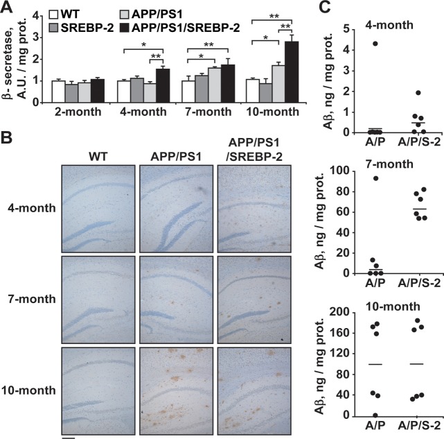
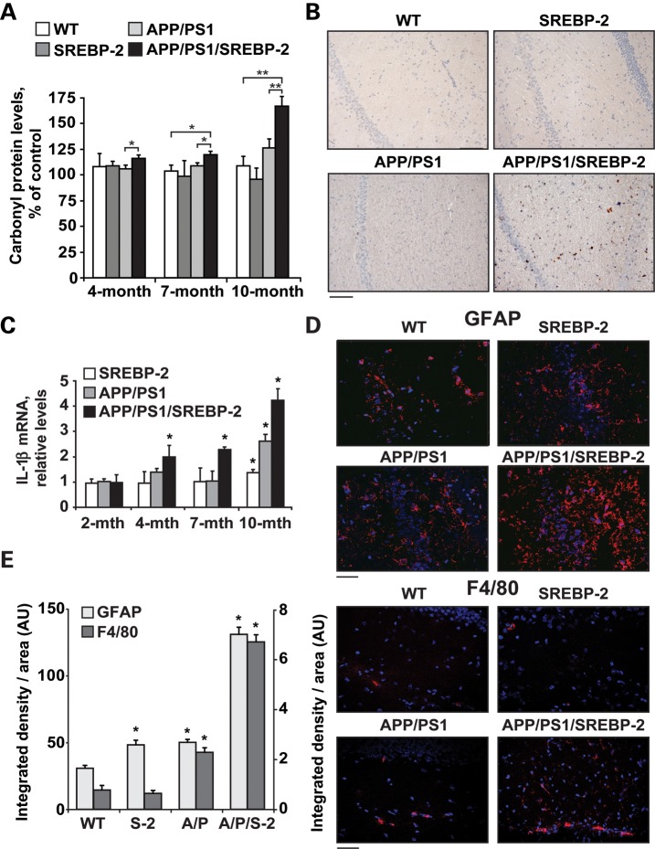
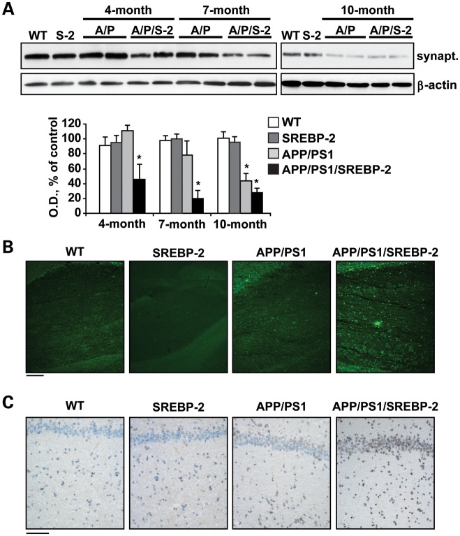
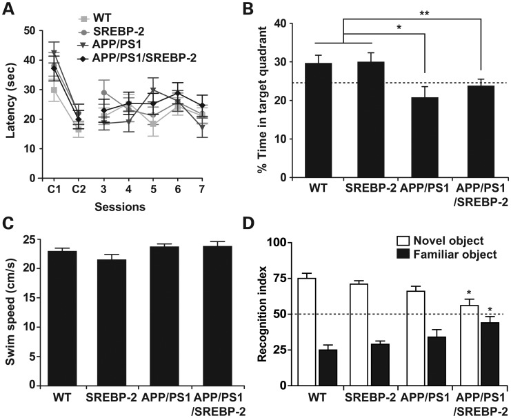
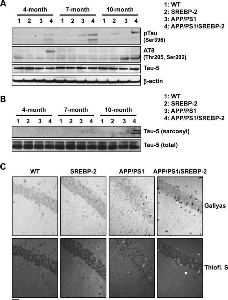
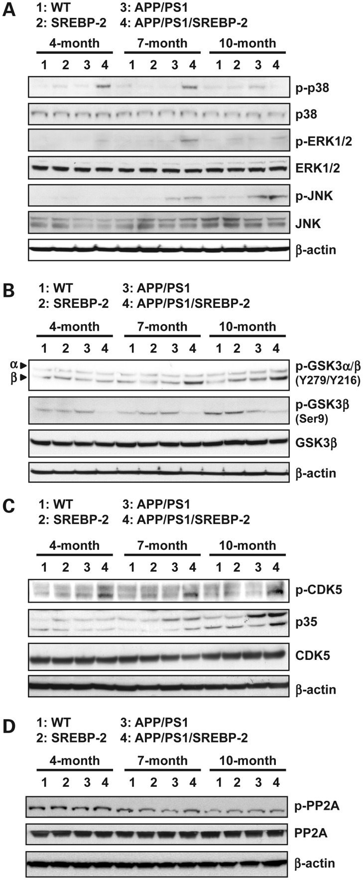
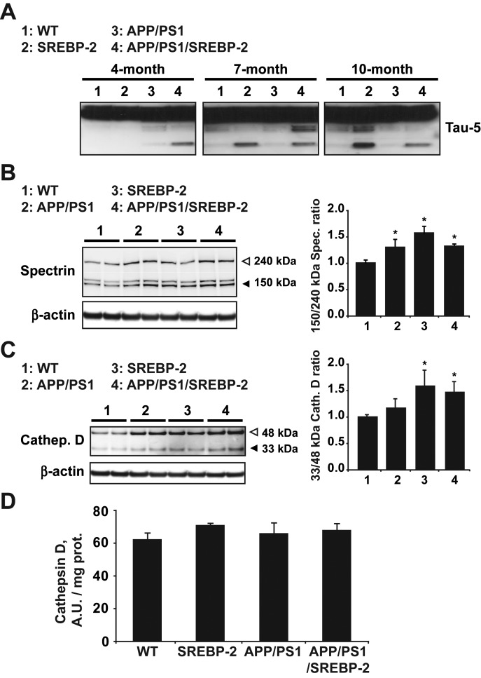
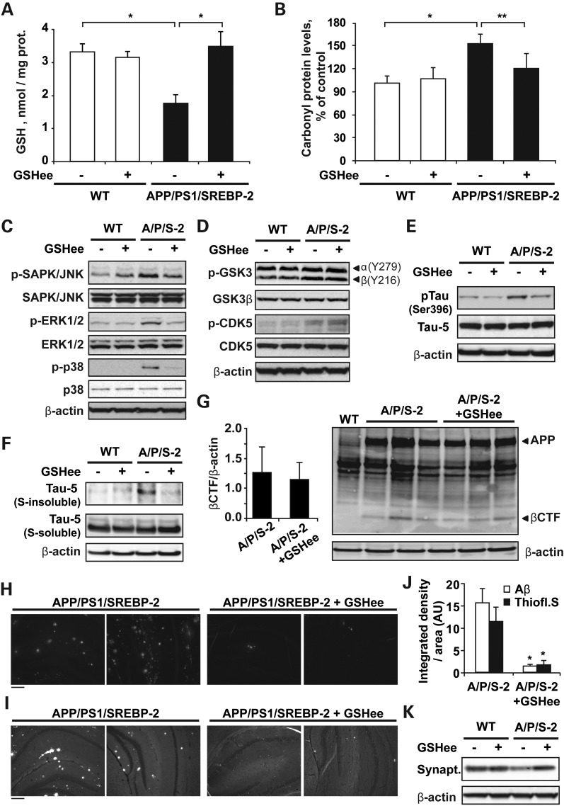
Current evidence indicates that excess brain cholesterol regulates amyloid-β (Aβ) deposition, which in turn can regulate cholesterol homeostasis. Moreover, Aβ neurotoxicity is potentiated, in part, by mitochondrial glutathione (mGSH) depletion. To better understand the relationship between alterations in cholesterol homeostasis and Alzheimer's disease (AD), we generated a triple transgenic mice featuring sterol regulatory element-binding protein-2 (SREBP-2) overexpression in combination with APPswe/PS1ΔE9 mutations (APP/PS1) to examine key biochemical and functional characteristics of AD. Unlike APP/PS1 mice, APP/PS1/SREBP-2 mice exhibited early mitochondrial cholesterol loading and mGSH depletion. Moreover, β-secretase activation and Aβ accumulation, correlating with oxidative damage and neuroinflammation, were accelerated in APP/PS1/SREBP-2 mice compared with APP/PS1 mice. Triple transgenic mice displayed increased synaptotoxicity reflected by loss of synaptophysin and neuronal death, resulting in early object-recognition memory impairment associated with deficits in spatial memory. Interestingly, tau pathology was present in APP/PS1/SREBP-2 mice, manifested by increased tau hyperphosphorylation and cleavage, activation of tau kinases and neurofibrillary tangle (NFT) formation without expression of mutated tau. Importantly, in vivo treatment with the cell permeable GSH ethyl ester, which restored mGSH levels in APP/PS1/SREBP-2 mice, partially prevented the activation of tau kinases, reduced abnormal tau aggregation and Aβ deposition, resulting in attenuated synaptic degeneration. Taken together, these results show that cholesterol-mediated mGSH depletion is a key event in AD progression, accelerating the onset of key neuropathological hallmarks of the disease. Thus, therapeutic approaches to recover mGSH may represent a relevant strategy in the treatment of AD.


Figures

Figure 1.
Figure 1.
Increased cholesterol promotes Aβ synthesis and loading. (A) β-Secretase activity analyzed in brain extracts from WT and transgenic mice (AU: arbitrary units). *P < 0.05, **P < 0.01 (n = 3). Values are expressed as mean ± SD (B) Representative photomicrographs of hippocampus labeled with human anti-Aβ(1-42) and counterstained with hematoxylin showing early Aβ depositions in APP/PS1/SREBP-2 mice. Scale bar: 100 μm. (C) Brain from WT and indicated mutant mice (A/P: APP/PS1 mice; A/P/S-2: APP/PS1/SREBP-2 mice) were homogenized with guanidine HCl extraction buffer, and the homogenates were analyzed by ELISAs for quantitative assessment of the human Aβ(1-42) content at the indicated ages (n = 6 per genotype).
Figure 2.
Figure 2.
SREBP-2 overexpression in APP/PS1 mice exacerbates oxidative stress and neuroinflammation. (A) Protein carbonyl content from brain of the indicated genotype analyzed spectrophotometrically. *P < 0.05, **P < 0.01 (n = 6). (B) Immunohistochemical staining of 8-hydroxyguanosine (8-OHG). Representative photomicrographs of hippocampus showing nuclear presence of 8-OHG in 7-month-old APP/PS1-SREBP-2 mice, which is indicative of oxidative DNA damage. Scale bar: 100 μm. (C) mRNA levels of IL-1β in brain from the indicated genotype analyzed by a quantitative PCR. Absolute mRNA values were determined, normalized to 18S and reported as relative levels referred to the expression in WT mice. *P < 0.05 versus WT values (n = 6). (D and E) Activation of astrocytes and microglia analyzed by GFAP and F4/80 immunostaining, respectively. (D) Representative confocal images of GFAP and F4/80 immunofluorescence (red) of hippocampal sections from 7- and 4-month-old mice, respectively. Nuclei were stained with Hoechst 33258 (blue). Scale bar: 50 μm. (E) Quantification of GFAP (left axis) and F4/80 (right axis) immunoreactivity by integrated density analysis. *P < 0.01 (n = 6). Values are expressed as mean ± SD.
Figure 3.
Figure 3.
APP/PS1/SREBP-2 mice exhibit enhanced neuronal damage. (A) Representative immunoblotting showing synaptophysin (synapt.) protein levels from WT and indicated mutant mice (S-2: SREBP-2 mice; A/P: APP/PS1 mice; A/P/S-2: APP/PS1/SREBP-2 mice). Densitometric values of the bands representing synaptophysin immunoreactivity were normalized with the values of the corresponding β-actin bands (OD: normalized optical density). *P < 0.05 versus WT values (n = 3). Values are expressed as mean ± SD. (B) Representative images of degenerated neurons in hippocampal regions from 10-month-old mice by Fluoro-Jade B staining. Scale bar: 100 μm. (C) Representative images of apoptotic cells in hippocampus from 10-month-old mice by a terminal deoxynucleotidyl transferase mediated nick-end labeling assay (TUNEL assay). Scale bar: 50 μm.
Figure 4.
Figure 4.
SREBP-2 overexpression in APP/PS1 mice does not worsen the spatial memory deficits but impairs short-term recognition memory. MWM was used to evaluate the spatial memory deficits in 7-month-old mice (n = 10–15 per genotype). (A) Cognitive performance in cued tasks (C1 and C2) and acquisition sessions (3–7) measured as mean time to locate the escape platform. All mouse strains showed similar latencies to reach the platform in the nonspatial variant of the MWM (C1 and C2). Learning rates during spatial memory acquisition were similar between groups. (B) Performance in the probe trial measured as mean percentage time spent in the target quadrant. APP/PS1 and APP/PS1/SREBP-2 mice showed no preference for the target quadrant relative to the other quadrants, whereas mice without APP/PS1 expression preferred the target quadrant. *P < 0.01, **P < 0.05. (C) Swimming speed exhibiting no significant differences among groups. (D) A novel object recognition test was used to assess deficits in short-term recognition memory in 7-month-old mice (n = 7–12 per genotype). Shown is the recognition index, which is the amount of time exploring the novel or familiar object versus the overall exploration time multiplied by 100. APP/PS1 mice that overexpress SREBP-2 exhibited a similar preference between novel and familiar objects. *P < 0.05 versus WT values. Values are expressed as mean ± SD.
Figure 5.
Figure 5.
SREBP-2 overexpression in APP/PS1 mice enhances the development of neurofibrillary pathology. (A) Protein extracted from brain homogenates of WT and mutant mice at the indicated ages analyzed by western blot to assess phosphorylation levels of tau at different epitopes recognized by the indicated phosphorylation-dependent and site-specific anti-tau antibodies. Total tau levels were detected by the phosphorylation-independent antibody Tau-5. (B) Protein immunoblots showing total (non-phosphorylated and phosphorylated) tau (Tau-5) in crude and sarcosyl-insoluble fractions from brain homogenates of WT and mutant mice at the indicated ages. (C) Representative sections of hippocampus from 10-month-old mice showing increased Gallyas-stained NFTs and thioflavin-S-positive cells in APP/PS1/SREBP-2 mice. Scale bar: 50 μm.
Figure 6.
Figure 6.
Activation profiles of different kinases associated with tau phosphorylation. (A) Representative immunoblots of cerebral homogenates from WT and indicated mutant mice using antibodies against the non-phosphorylated and active phosphorylated forms of ERK 1/2, JNK and p38 MAPK. As shown, all three MAPK pathways were activated in APP/PS1/SREBP-2 mice. (B) Western blot analysis of GSK-3β activation with antibodies against Y279 and Y216 (activating sites) and Ser9 (inhibitory site) indicating enhanced GSK-3β activity in APP/PS1 mice that overexpress SREBP-2. (C) Representative immunoblots showing increased levels of phosphorylated CDK5 and enhanced cleavage of p35 (CDK5 activator protein) at 10 months of age in brain extracts from APP/PS1/SREBP-2 mice. (D) Western blot analysis of PP2A activation using the PP2A C subunit antibody and the phospho-specific antibody that recognize the inhibitory phosphorylation at Tyr307 in subunit C.
Figure 7.
Figure 7.
Increased proteolytic cleavage of tau protein in mice harboring the SREBP-2 transgene. (A) Representative immunoblot of total tau showing cleaved tau presence in brain extracts from SREBP-2 and APP/PS1/SREBP-2 mice at the indicated ages. (B) Western blot analysis of calpain activity by means of increase in the 145/150 kDa specific spectrin cleaved products. Quantification of the 145/150:240 kDa spectrin ratio exhibited a significant calpain activity increase in brain extracts from 7-month-old mutant mice compared with WT mice. (C) Representative protein immunoblot of cathepsin D (33 and 48 kDa, mature and immature forms, respectively) from brain extracts of 7-month-old WT and indicated mutant mice. Quantification of the 33:48 kDa ratio indicates an increased processing of cathepsin D in cerebral extracts from SREBP-2 and APP/PS1/SREBP-2 mice. (D) Cathepsin D activity analyzed in brain extracts from WT and transgenic mice (AUs). *P < 0.05 versus WT values (n = 3). Values are expressed as mean ± S.D.
Figure 8.
Figure 8.
In vivo GSH ethyl ester treatment recovers mGSH and prevents tau pathology and Aβ deposition in APP/PS1/SREBP-2 mice. 7-month-old WT and APP/PS1/SREBP-2 mice were treated with GSH ethyl ester (GSHee; i.p. 1.25 mmol/kg/day) for 2 weeks. (A and B) Mitochondrial GSH levels and the protein carbonyl content after GSHee therapy. *P < 0.01, **P < 0.05 (n = 6). Values are expressed as mean ± SD. (C) Representative immunoblots (non-phosphorylated and active phosphorylated forms) of JNK/SAPK, ERK 1/2, and p38 MAP kinases showing reduced activation in brain extracts of APP/PS1/SREBP-2 mice after GSHee treatment. (D) Western blot analysis of GSK-3β activation with antibodies against Y279 and Y216 (activating sites) and CDK5 phosphorylation showing an enhanced activity of both kinases in APP/PS1/SREBP-2 mice independently of GSHee treatment. (E) Representative immunoblots showing reduced tau phosphorylation at Ser 396 in brain extracts of APP/PS1/SREBP-2 mice after GSHee treatment. Total tau levels were detected by the phosphorylation-independent antibody Tau-5. (F) Protein immunoblots of total tau (Tau-5) in crude (s-soluble) and sarcosyl-insoluble (s-insoluble) fractions showing decreased presence of insoluble tau in APP/PS1/SREBP-2 mice after GSHee treatment. (G) Western blot analysis of β-CTFs using Aβ antibody (clone 6E10). The levels of β-CFTs were normalized to β-actin (left graph). (H and I) Representative immunofluorescent images of amyloid plaques stained with an Aβ antibody (clone 6E10) (H) and thioflavin S (I). (J) Quantification of Aβ immunoreactivity and thioflavin staining by integrated density analysis showing decreased amyloid burden in hippocampus from APP/PS1/SREBP-2 mice after GSHee treatment. (AUs). *P < 0.001 (n = 4). Values are expressed as mean ± SD. (K) Representative immunoblot of synaptophysin (synapt.) protein levels.

Similar articles

Cited by

References

    1. Cutler R.G., Kelly J., Storie K., Pedersen W.A., Tammara A., Hatanpaa K., Troncoso J.C., Mattson M.P. Involvement of oxidative stress-induced abnormalities in ceramide and cholesterol metabolism in brain aging and Alzheimer's disease. Proc. Natl Acad. Sci. USA. 2004;101:2070–2075. - PMC - PubMed
    1. Bandaru V.V., Troncoso J., Wheeler D., Pletnikova O., Wang J., Conant K., Haughey N.J. ApoE4 disrupts sterol and sphingolipid metabolism in Alzheimer's but not normal brain. Neurobiol. Aging. 2009;30:591–599. - PMC - PubMed
    1. Xiong H., Callaghan D., Jones A., Walker D.G., Lue L.F., Beach T.G., Sue L.I., Woulfe J., Xu H., Stanimirovic D.B., et al. Cholesterol retention in Alzheimer's brain is responsible for high beta- and gamma-secretase activities and Abeta production. Neurobiol. Dis. 2008;29:422–437. - PMC - PubMed
    1. Panchal M., Loeper J., Cossec J.C., Perruchini C., Lazar A., Pompon D., Duyckaerts C. Enrichment of cholesterol in microdissected Alzheimer's disease senile plaques as assessed by mass spectrometry. J. Lipid Res. 2010;51:598–605. - PMC - PubMed
    1. Fernandez A., Llacuna L., Fernandez-Checa J.C., Colell A. Mitochondrial cholesterol loading exacerbates amyloid beta peptide-induced inflammation and neurotoxicity. J. Neurosci. 2009;29:6394–6405. - PMC - PubMed

Publication types

MeSH terms