

Imatinib mesylate inhibits cell growth of malignant peripheral nerve sheath tumors in vitro and in vivo through suppression of PDGFR-β
- PMID: 23642185
- PMCID: PMC3654969
- DOI: 10.1186/1471-2407-13-224
Imatinib mesylate inhibits cell growth of malignant peripheral nerve sheath tumors in vitro and in vivo through suppression of PDGFR-β
Abstract
Background: Malignant peripheral nerve sheath tumors (MPNSTs) are highly aggressive and associated with poor prognosis. Basic research to develop new treatment regimens is critically needed.
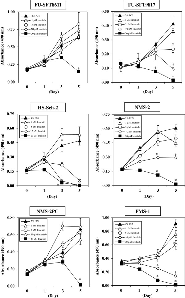
Methods: The effects of imatinib mesylate on MPNSTs were examined in six human MPNST cell lines and in a xenograft mouse model.
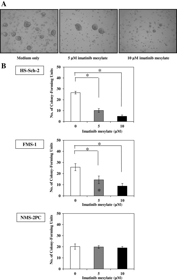
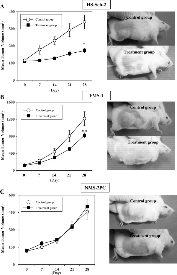
Results: The results showed expression of platelet-derived growth factor receptor-β and suppression of its phosphorylation by imatinib mesylate in all six cell lines. Imatinib mesylate effectively suppressed MPNST cell growth in vitro at concentrations similar to those used clinically (1.46 - 4.6 μM) in three of six cell lines. Knockdown of PDGFR-β by transfection with a specific siRNA also caused significant reduction in cell proliferation in the sensitive cell lines, but not in the resistant cell lines. Furthermore, imatinib mesylate also significantly suppressed colony formation within soft agar and tumor growth in xenograft models using two of the three sensitive MPNST cell lines. There was excellent agreement between in vitro and in vivo sensitivity to imatinib mesylate, suggesting possible selection of imatinib-sensitive tumors by in vitro analysis.
Conclusions: The results suggest that imatinib mesylate may be useful in the treatment of MPNST patients and in vitro studies may help select cells that are sensitive to imatinib mesylate in vivo.
Figures


Similar articles
-
Imatinib mesylate inhibits cell invasion of malignant peripheral nerve sheath tumor induced by platelet-derived growth factor-BB.Lab Invest. 2007 Aug;87(8):767-79. doi: 10.1038/labinvest.3700591. Epub 2007 Jun 11. Lab Invest. 2007. PMID: 17558420
-
Imatinib mesylate inhibits platelet-derived growth factor receptor phosphorylation of melanoma cells but does not affect tumorigenicity in vivo.J Invest Dermatol. 2004 Feb;122(2):400-5. doi: 10.1046/j.0022-202X.2004.22231.x. J Invest Dermatol. 2004. PMID: 15009722
-
Imatinib mesylate inhibits tumorigenicity of malignant fibrous histiocytoma cells in vivo.Anticancer Res. 2007 Jan-Feb;27(1A):423-9. Anticancer Res. 2007. PMID: 17352263
-
The role of imatinib in the treatment of pulmonary hypertension.Drugs Today (Barc). 2013 Mar;49(3):203-11. doi: 10.1358/dot.2013.49.3.1937430. Drugs Today (Barc). 2013. PMID: 23527324 Review.
-
PDGFRA mutations in gastrointestinal stromal tumors: frequency, spectrum and in vitro sensitivity to imatinib.J Clin Oncol. 2005 Aug 10;23(23):5357-64. doi: 10.1200/JCO.2005.14.068. Epub 2005 May 31. J Clin Oncol. 2005. PMID: 15928335 Review.
Cited by
-
Activation of Receptor Tyrosine Kinases Mediates Acquired Resistance to MEK Inhibition in Malignant Peripheral Nerve Sheath Tumors.Cancer Res. 2021 Feb 1;81(3):747-762. doi: 10.1158/0008-5472.CAN-20-1992. Epub 2020 Nov 17. Cancer Res. 2021. PMID: 33203698 Free PMC article.
-
Comprehensive establishment and characterization of orthoxenograft mouse models of malignant peripheral nerve sheath tumors for personalized medicine.EMBO Mol Med. 2015 May;7(5):608-27. doi: 10.15252/emmm.201404430. EMBO Mol Med. 2015. PMID: 25810463 Free PMC article.
-
Immunohistochemical Expression of Platelet-Derived Growth Factor Receptor β (PDGFR-β) in Canine Cutaneous Peripheral Nerve Sheath Tumors: A Preliminary Study.Vet Sci. 2022 Jul 9;9(7):345. doi: 10.3390/vetsci9070345. Vet Sci. 2022. PMID: 35878362 Free PMC article.
-
Programming of Schwann Cells by Lats1/2-TAZ/YAP Signaling Drives Malignant Peripheral Nerve Sheath Tumorigenesis.Cancer Cell. 2018 Feb 12;33(2):292-308.e7. doi: 10.1016/j.ccell.2018.01.005. Cancer Cell. 2018. PMID: 29438698 Free PMC article.
-
Platelet-derived growth factor (PDGF) signalling in cancer: rapidly emerging signalling landscape.Cell Biochem Funct. 2015 Jul;33(5):257-65. doi: 10.1002/cbf.3120. Epub 2015 Jul 7. Cell Biochem Funct. 2015. PMID: 26153649 Free PMC article. Review.
References
-
- Lewis JJ, Brennan MF. Soft tissue sarcomas. Curr Probl Surg. 1996;33(10):817–872. - PubMed
-
- Woodruff JM, Kourea HP, Louis DN, Scheithauer BW. World health organization classification of tumors pathology and genetics of tumor of the nervous system. Lyon, France: IARC Press; 2000. Malignant peripheral nerve sheath tumor (MPNST) pp. 172–174.
-
- Ferner RE, Gutmann DH. International consensus statement on malignant peripheral nerve sheath tumors in neurofibromatosis. Cancer Res. 2002;62(5):1573–1577. - PubMed
MeSH terms
Substances
LinkOut - more resources
Full Text Sources
Other Literature Sources
Miscellaneous
