. 2013;9(4):e1003323.
doi: 10.1371/journal.ppat.1003323. Epub 2013 Apr 18.

Factor H binds to the hypervariable region of many Streptococcus pyogenes M proteins but does not promote phagocytosis resistance or acute virulence

Affiliations

Factor H binds to the hypervariable region of many Streptococcus pyogenes M proteins but does not promote phagocytosis resistance or acute virulence

Mattias C U Gustafsson et al. PLoS Pathog. 2013.

Abstract

Many pathogens express a surface protein that binds the human complement regulator factor H (FH), as first described for Streptococcus pyogenes and the antiphagocytic M6 protein. It is commonly assumed that FH recruited to an M protein enhances virulence by protecting the bacteria against complement deposition and phagocytosis, but the role of FH-binding in S. pyogenes pathogenesis has remained unclear and controversial. Here, we studied seven purified M proteins for ability to bind FH and found that FH binds to the M5, M6 and M18 proteins but not the M1, M3, M4 and M22 proteins. Extensive immunochemical analysis indicated that FH binds solely to the hypervariable region (HVR) of an M protein, suggesting that selection has favored the ability of certain HVRs to bind FH. These FH-binding HVRs could be studied as isolated polypeptides that retain ability to bind FH, implying that an FH-binding HVR represents a distinct ligand-binding domain. The isolated HVRs specifically interacted with FH among all human serum proteins, interacted with the same region in FH and showed species specificity, but exhibited little or no antigenic cross-reactivity. Although these findings suggested that FH recruited to an M protein promotes virulence, studies in transgenic mice did not demonstrate a role for bound FH during acute infection. Moreover, phagocytosis tests indicated that ability to bind FH is neither sufficient nor necessary for S. pyogenes to resist killing in whole human blood. While these data shed new light on the HVR of M proteins, they suggest that FH-binding may affect S. pyogenes virulence by mechanisms not assessed in currently used model systems.


Conflict of interest statement

The authors have declared that no competing interests exist.

Figures

Figure 1
Figure 1. Streptococcal M proteins vary in ability to bind human FH and C4BP.
(A) Schematic representation of the human complement regulators FH and C4BP and of an M protein. For FH and C4BP, each circle represents an SCR domain. While FH is a single chain with 20 SCR domains, C4BP typically contains 7 α-chains with 8 SCR domains and one β-chain with 3 SCRs. An M protein has an N-terminal HVR and a more conserved C-terminal region that includes C repeats and the wall-anchoring region. The location of M protein binding sites in FH and C4BP are indicated. (B) SDS-PAGE analysis of the purified recombinant M proteins studied, five of which were of class I, and two of class II. The presence of doublet or triplet bands is typical for M proteins expressed in E. coli or S. pyogenes , . (C) Binding of human FH, C4BP or fibrinogen (Fg) to pure M proteins immobilized in microtiter wells. The wells were coated with 0.1 µg M protein and human ligands (50 µl) were added at the following concentrations: FH 2 µg/ml, C4BP 1 µg/ml, Fg 0.28 µg/ml. Bound ligands were detected with specific antibodies. Binding is given in percent of maximal binding for each ligand. (D) Binding of human FH analyzed for the M-positive M5, M6 and M18 S. pyogenes strains and their M-negative mutants (ΔM5, ΔM6, and ΔM18, respectively). Bacterial suspensions were incubated with pure FH (50 µg/ml). After two washes bound protein was eluted and analyzed by western blot, employing anti-FH for detection. Pure FH was included as a control in the blot (right).
Figure 2
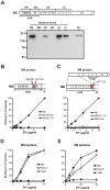
Figure 2. Human FH binds to the HVR of the M5 and M6 proteins.
(A) Binding of human FH to chromosomal mutants of the M5 strain. The upper part of the panel shows the location of deletions (ΔN1, ΔN2, ΔB and ΔC) causing the surface expression of truncated M5 proteins, and the positions of A, B and C repeats in M5. The lower part of the panel shows results of binding tests with pure FH and the mutants expressing truncated M5 proteins. The wild type M5 strain and a strain (ΔM5) lacking the entire M5 protein were also included. In the binding tests, pure FH was incubated with a suspension of the strain indicated. After washes, bound protein was eluted from the bacteria and analyzed by western blot, employing anti-FH for detection. Pure FH was included as a control in the blot (right). (B and C) Binding of FH to purified derivatives of the M5 protein (B) and the M6 protein (C). The proteins indicated were used to coat microtiter wells, using 0.1 µg per well, and analyzed for ability to bind pure FH, added at the concentration indicated. In B, wild type M5 protein was compared with two M5 derivatives (M5Δ80-86 and M5Δ87-93) with short deletions in the HVR, as indicated. In C, wild type M6 protein was compared with a deletion derivative having a short deletion in the HVR (M6Δ97-110), and with a dimerized construct derived from the C repeat region of M6 (M6-Crep), as indicated. (D and E) Binding of FH to whole S. pyogenes M5 bacteria (D) or M6 bacteria (E), and to bacterial mutants of these strains lacking C repeats (M5ΔC and M6ΔC) or no M protein (ΔM5 and ΔM6).
Figure 3
Figure 3. Isolated HVRs derived from M5, M6 and M18 specifically bind FH.
(A) Analysis by non-reducing SDS-PAGE of HVRs dimerized via a C-terminal cysteine residue. As previously observed , dimerized HVRs move more slowly than expected in gels. (B) Binding of FH to isolated HVRs immobilized in microtiter wells. The wells were coated with 0.1 µg of the HVRs, as indicated, and tested for ability to bind added FH. (C) Immobilized HVRs, derived from FH-binding M proteins, specifically bind FH among all proteins in human serum. Whole human serum was applied to columns in which the HVRs indicated had been immobilized. After washings, bound protein was eluted and analyzed by SDS-PAGE. A column without HVR was used as control. Pure FH was included as a reference in the gel analysis (right). (D) Sequence alignment of the three FH-binding HVRs that were studied in isolated form. This alignment does not include a C-terminal Cys residue included to allow dimerization. The lengths of these HVR vary slightly from those previously reported , because the position of the C-terminal end was chosen to allow optimal dimerization and FH-binding. Asterisks indicate residues identical in all three sequences. Pair-wise identities (based on regions present in both sequences) are indicated to the right.
Figure 4
Figure 4. FH-binding M proteins show species specificity and bind to the same region in FH.
(A) The M5-HVR binds FH present in normal human serum (NHS) but not FH in normal mouse serum (NMS) or normal rabbit serum (NRS). In three separate tests, the sera were applied to a column containing the M5-HVR. After washes, bound protein was eluted and analyzed by SDS-PAGE. (B) Binding of a chimeric FH, expressed by Tg mice, to M proteins. The chimeric FH includes the SCR6-8 region of human FH (top). Serum from the Tg mice, or from wild type (wt) C57Bl/6 mice, was analyzed for presence of FH able to bind to the M protein indicated, immobilized in microtiter wells. (C) Different FH-binding HVRs bind to the same or overlapping site(s) in FH. A biotinylated form of the M5, M6 or M18 protein was used to detect FH immobilized in microtiter wells, and binding was inhibited with the four HVRs indicated, added at a concentration of 10 µM.
Figure 5
Figure 5. Antigenic properties of FH-binding HVRs.
(A) Isolated HVRs show limited or no antigenic cross-reactivity. Antiserum to one HVR (specified at the top of a panel) was analyzed for reactivity with four immobilized HVRs, as indicated. The HVRs of M5 and M18 exhibited limited cross-reactivity, while the HVR of M6 did not cross-react with the other two HVRs. The non-FH-binding M1-HVR was included as a control. Pre-immune sera were used to obtain background values, which were subtracted from the values obtained with immune sera. (B) Binding of FH to the HVR of an M protein is inhibited by antiserum to the corresponding HVR. The biotinylated M protein indicated above each panel was used to detect FH immobilized in microtiter wells. The binding was inhibited with rabbit anti-HVR serum, diluted as indicated. Preimmune serum was used as a control.
Figure 6
Figure 6. Phagocytosis tests with M1 and M3 strains.
(A) Suspensions of the bacterial strains indicated were analyzed for ability to bind human FH. After incubation of a bacterial suspension with pure FH (50 µg/ml), the bacteria were washed twice and bound protein was eluted and analyzed by western blot, employing anti-FH for detection. The analysis included wild type M5, M1 and M3 strains and their M-negative mutants (ΔM5, ΔM1 and ΔM3, respectively). Pure FH was included as a control in the blot (right). (B) Phagocytosis assay in whole human blood with M1 and M3 strains, and M-negative mutants, as indicated. MF, multiplication factor.
Figure 7
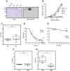
Figure 7. Characterization of a chimeric FH expressed by Tg mice, and infection experiments with Tg and normal mice.
(A) SDS-PAGE and western blot analysis of purified chimeric FH (chim. FH), derived from mouse and human FH. This FH was compared with pure human Y402 FH (hFH) and pure mouse FH (mFH). Streptococcal protein Rib was included as negative control. The blot was probed with antiserum to human FH. Bound antibodies were detected with radiolabeled protein G followed by autoradiography. (B) Binding of the M5 protein to immobilized FH proteins. The proteins used were those shown in panel A. The wells of microtiter plates were coated with the protein indicated, using 0.1 µg protein per well, with protein Rib as negative control. The immobilized proteins were analyzed for ability to bind biotinylated pure M5 protein, added at the concentrations indicated, and bound M protein was detected by the addition of radiolabeled streptavidin. (C) Comparison of bacterial growth in wild type mice and Tg mice. The mice (n = 8 for wt and n = 8 for Tg) were subjected to invasive infection with a sublethal dose of the S. pyogenes M5 strain and sacrificed after 18 h, followed by determination of bacterial counts in the spleen. (D) Survival of wild type (wt) and Tg mice after infection with M5 bacteria. The mice (n = 7 for wt and n = 10 for Tg) were subjected to invasive infection with an ∼LD90 dose of the S. pyogenes M5 strain and survival was recorded regularly, as indicated. (E) Survival of wild type (wt) and Tg mice after infection with M5 bacteria. The mice (n = 12 for wt and n = 12 for Tg) were subjected to invasive infection, as in D, but with a lower bacterial dose. (F) Growth of the S. pyogenes M5 strain in wild type mice injected with pure human Y402 FH (2×100 µg) or with PBS. The mice (n = 6 for FH and n = 6 for PBS) received FH or PBS 4 h before the infection and mixed with the infecting bacteria. Bacterial counts in spleens were analyzed, as in (C). (G) Growth of the S. pyogenes M5 strain in wild type mice injected with normal human serum (NHS) or normal mouse serum (NMS). The mice (n = 8 for NHS and n = 7 for NMS) were injected with two 200 µl doses of serum, one dose given 4 h before the infection and one mixed with the infecting bacteria. Analysis as in (C).
Figure 8
Figure 8. Binding of human proteins to M proteins, with focus on the HVR.
The figure summarizes current knowledge in the field, with emphasis on the M proteins studied here. Fg, fibrinogen; HSA, human serum albumin; IgA-Fc, Fc-part of IgA. See text for details.

Similar articles

Cited by

References

    1. Ricklin D, Hajishengallis G, Yang K, Lambris JD (2010) Complement: a key system for immune surveillance and homeostasis. Nat Immunol 11: 785–797. - PMC - PubMed
    1. Carroll MC, Isenman DE (2012) Regulation of humoral immunity by complement. Immunity 37: 199–207. - PMC - PubMed
    1. Lachmann PJ (2009) The amplification loop of the complement pathways. Adv Immunol 104: 115–149. - PubMed
    1. Schmidt CQ, Herbert AP, Hocking HG, Uhrín D, Barlow PN (2008) Translational mini-review series on complement factor H: structural and functional correlations for factor H. Clin Exp Immunol 151: 14–24. - PMC - PubMed
    1. Zipfel PF, Skerka C (2009) Complement regulators and inhibitory proteins. Nat Rev Immunol 9: 729–740. - PubMed

Publication types

MeSH terms

LinkOut - more resources