Regulation of VH replacement by B cell receptor-mediated signaling in human immature B cells
- PMID: 23630348
- PMCID: PMC3660396
- DOI: 10.4049/jimmunol.1102503
Regulation of VH replacement by B cell receptor-mediated signaling in human immature B cells
Abstract
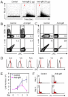
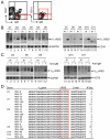
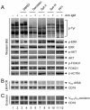
VH replacement provides a unique RAG-mediated recombination mechanism to edit nonfunctional IgH genes or IgH genes encoding self-reactive BCRs and contributes to the diversification of Ab repertoire in the mouse and human. Currently, it is not clear how VH replacement is regulated during early B lineage cell development. In this article, we show that cross-linking BCRs induces VH replacement in human EU12 μHC(+) cells and in the newly emigrated immature B cells purified from peripheral blood of healthy donors or tonsillar samples. BCR signaling-induced VH replacement is dependent on the activation of Syk and Src kinases but is inhibited by CD19 costimulation, presumably through activation of the PI3K pathway. These results show that VH replacement is regulated by BCR-mediated signaling in human immature B cells, which can be modulated by physiological and pharmacological treatments.
Figures


Similar articles
-
Internalization of B cell receptors in human EU12 μHC⁺ immature B cells specifically alters downstream signaling events.Biomed Res Int. 2013;2013:807240. doi: 10.1155/2013/807240. Epub 2013 Oct 9. Biomed Res Int. 2013. PMID: 24222917 Free PMC article.
-
CD19 amplifies B lymphocyte signal transduction by regulating Src-family protein tyrosine kinase activation.J Immunol. 1999 Jun 15;162(12):7088-94. J Immunol. 1999. PMID: 10358152
-
Tyrosine kinase activation in the decision between growth, differentiation, and death responses initiated from the B cell antigen receptor.Adv Immunol. 2000;75:283-316. doi: 10.1016/s0065-2776(00)75007-3. Adv Immunol. 2000. PMID: 10879287 Review.
-
The molecular basis and biological significance of VH replacement.Immunol Rev. 2004 Feb;197:231-42. doi: 10.1111/j.0105-2896.2004.0107.x. Immunol Rev. 2004. PMID: 14962199 Review.
-
Molecular mechanism of serial VH gene replacement.Ann N Y Acad Sci. 2003 Apr;987:270-3. doi: 10.1111/j.1749-6632.2003.tb06060.x. Ann N Y Acad Sci. 2003. PMID: 12727651 Review.
Cited by
-
Antibody repertoire diversification through VH gene replacement in mice cloned from an IgA plasma cell.Proc Natl Acad Sci U S A. 2015 Feb 3;112(5):E450-7. doi: 10.1073/pnas.1417988112. Epub 2015 Jan 21. Proc Natl Acad Sci U S A. 2015. PMID: 25609671 Free PMC article.
-
Role of Sex Hormone Levels and Psychological Stress in the Pathogenesis of Autoimmune Diseases.Cureus. 2017 Jun 5;9(6):e1315. doi: 10.7759/cureus.1315. Cureus. 2017. PMID: 28690949 Free PMC article. Review.
-
Internalization of B cell receptors in human EU12 μHC⁺ immature B cells specifically alters downstream signaling events.Biomed Res Int. 2013;2013:807240. doi: 10.1155/2013/807240. Epub 2013 Oct 9. Biomed Res Int. 2013. PMID: 24222917 Free PMC article.
-
Heavy-chain receptor editing unbound.Proc Natl Acad Sci U S A. 2015 Feb 24;112(8):2297-8. doi: 10.1073/pnas.1501480112. Epub 2015 Feb 17. Proc Natl Acad Sci U S A. 2015. PMID: 25691748 Free PMC article. No abstract available.
-
VH replacement in primary immunoglobulin repertoire diversification.Proc Natl Acad Sci U S A. 2015 Feb 3;112(5):E458-66. doi: 10.1073/pnas.1418001112. Epub 2015 Jan 21. Proc Natl Acad Sci U S A. 2015. PMID: 25609670 Free PMC article.
References
-
- Schatz DG, Oettinger MA, Baltimore D. The V(D)J recombination activating gene, RAG-1. Cell. 1989;59:1035. - PubMed
-
- Oettinger MA. Activation of V(D)J recombination by RAG1 and RAG2. Trends Genet. 1992;8:413. - PubMed
-
- Lewis SM. The mechanism of V(D)J joining: lessons from molecular, immunological, and comparative analyses. Adv. Immunol. 1994;56:27. - PubMed
-
- Jung D, Alt FW. Unraveling V(D)J recombination: insights into gene regulation. Cell. 2004;116:299. - PubMed
-
- Rajewsky K. Clonal selection and learning in the antibody system. Nature. 1996;381:751. - PubMed
Publication types
MeSH terms
Substances
Grants and funding
LinkOut - more resources
Full Text Sources
Other Literature Sources
Miscellaneous

