

Human infections with the emerging avian influenza A H7N9 virus from wet market poultry: clinical analysis and characterisation of viral genome
- PMID: 23623390
- PMCID: PMC7134567
- DOI: 10.1016/S0140-6736(13)60903-4
Human infections with the emerging avian influenza A H7N9 virus from wet market poultry: clinical analysis and characterisation of viral genome
Abstract
Background: Human infection with avian influenza A H7N9 virus emerged in eastern China in February, 2013, and has been associated with exposure to poultry. We report the clinical and microbiological features of patients infected with influenza A H7N9 virus and compare genomic features of the human virus with those of the virus in market poultry in Zhejiang, China.
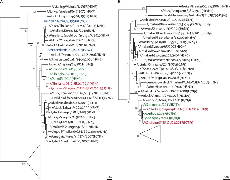
Methods: Between March 7 and April 8, 2013, we included hospital inpatients if they had new-onset respiratory symptoms, unexplained radiographic infiltrate, and laboratory-confirmed H7N9 virus infection. We recorded histories and results of haematological, biochemical, radiological, and microbiological investigations. We took throat and sputum samples, used RT-PCR to detect M, H7, and N9 genes, and cultured samples in Madin-Darby canine kidney cells. We tested for co-infections and monitored serum concentrations of six cytokines and chemokines. We collected cloacal swabs from 86 birds from epidemiologically linked wet markets and inoculated embryonated chicken eggs with the samples. We identified and subtyped isolates by RT-PCR sequencing. RNA extraction, complementary DNA synthesis, and PCR sequencing were done for one human and one chicken isolate. We characterised and phylogenetically analysed the eight gene segments of the viruses in the patient's and the chicken's isolates, and constructed phylogenetic trees of H, N, PB2, and NS genes.
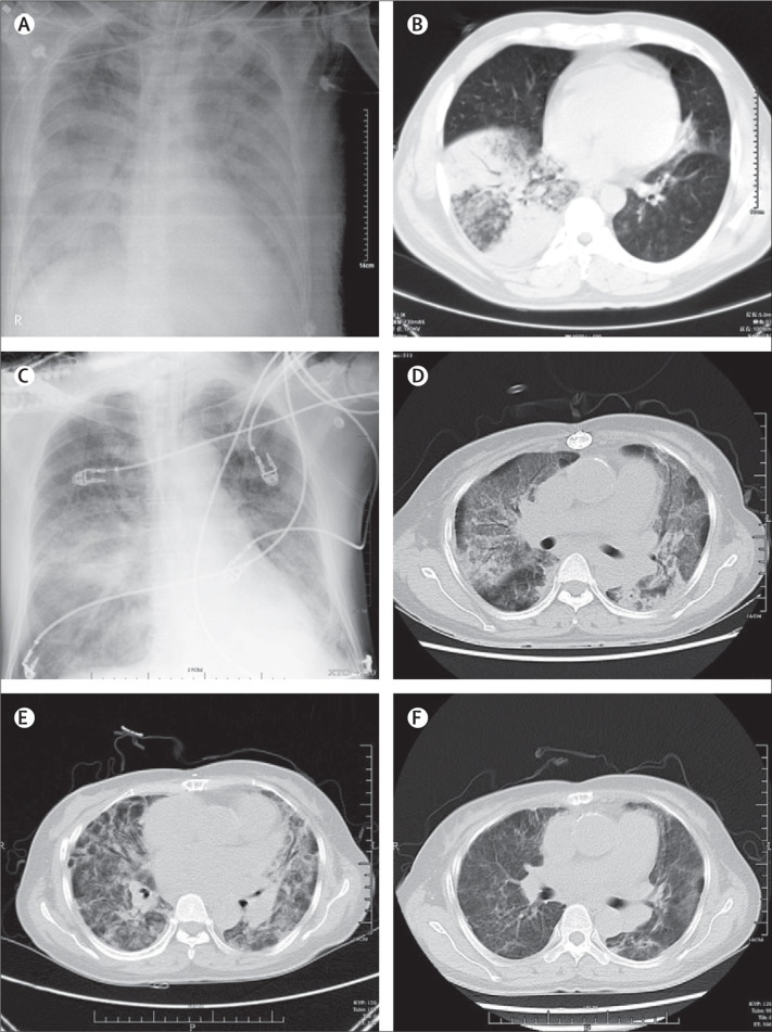
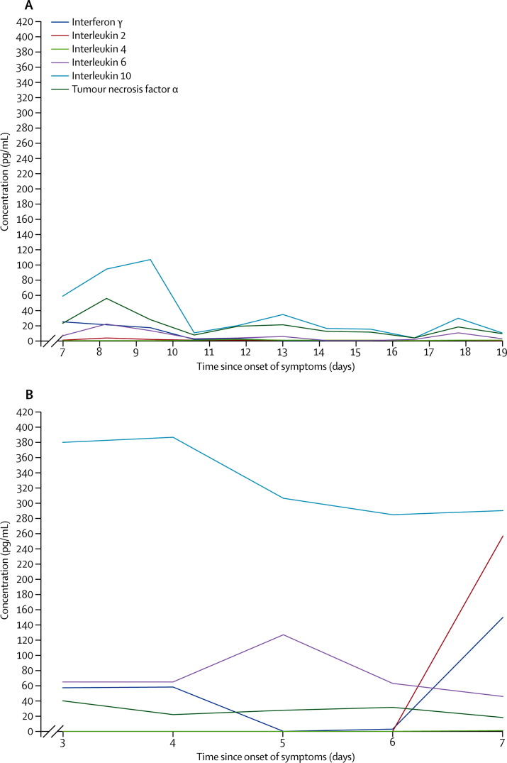
Findings: We identified four patients (mean age 56 years), all of whom had contact with poultry 3-8 days before disease onset. They presented with fever and rapidly progressive pneumonia that did not respond to antibiotics. Patients were leucopenic and lymphopenic, and had impaired liver or renal function, substantially increased serum cytokine or chemokine concentrations, and disseminated intravascular coagulation with disease progression. Two patients died. Sputum specimens were more likely to test positive for the H7N9 virus than were samples from throat swabs. The viral isolate from the patient was closely similar to that from an epidemiologically linked market chicken. All viral gene segments were of avian origin. The H7 of the isolated viruses was closest to that of the H7N3 virus from domestic ducks in Zhejiang, whereas the N9 was closest to that of the wild bird H7N9 virus in South Korea. We noted Gln226Leu and Gly186Val substitutions in human virus H7 (associated with increased affinity for α-2,6-linked sialic acid receptors) and the PB2 Asp701Asn mutation (associated with mammalian adaptation). Ser31Asn mutation, which is associated with adamantane resistance, was noted in viral M2.
Interpretation: Cross species poultry-to-person transmission of this new reassortant H7N9 virus is associated with severe pneumonia and multiorgan dysfunction in human beings. Monitoring of the viral evolution and further study of disease pathogenesis will improve disease management, epidemic control, and pandemic preparedness.
Funding: Larry Chi-Kin Yung, National Key Program for Infectious Diseases of China.
Copyright © 2013 Elsevier Ltd. All rights reserved.
Figures

Comment in
-
Avian influenza A H7N9 in Zhejiang, China.Lancet. 2013 Jun 1;381(9881):1882-3. doi: 10.1016/S0140-6736(13)60936-8. Epub 2013 Apr 27. Lancet. 2013. PMID: 23628442 No abstract available.
Similar articles
-
Influenza H7N9 and H9N2 viruses: coexistence in poultry linked to human H7N9 infection and genome characteristics.J Virol. 2014 Mar;88(6):3423-31. doi: 10.1128/JVI.02059-13. Epub 2014 Jan 8. J Virol. 2014. PMID: 24403589 Free PMC article.
-
Human infection with a novel avian-origin influenza A (H7N9) virus.N Engl J Med. 2013 May 16;368(20):1888-97. doi: 10.1056/NEJMoa1304459. Epub 2013 Apr 11. N Engl J Med. 2013. PMID: 23577628
-
Clinical and epidemiological characteristics of a fatal case of avian influenza A H10N8 virus infection: a descriptive study.Lancet. 2014 Feb 22;383(9918):714-21. doi: 10.1016/S0140-6736(14)60111-2. Epub 2014 Feb 5. Lancet. 2014. PMID: 24507376
-
Viral lung infections: epidemiology, virology, clinical features, and management of avian influenza A(H7N9).Curr Opin Pulm Med. 2014 May;20(3):225-32. doi: 10.1097/MCP.0000000000000047. Curr Opin Pulm Med. 2014. PMID: 24637225 Review.
-
[The effects of closure to live poultry markets on Avian influenza A (H7N9) epidemics in China].Zhonghua Liu Xing Bing Xue Za Zhi. 2017 Dec 10;38(12):1716-1718. doi: 10.3760/cma.j.issn.0254-6450.2017.12.027. Zhonghua Liu Xing Bing Xue Za Zhi. 2017. PMID: 29294594 Review. Chinese.
Cited by
-
Anemia and H7N9 Bird Flu: A Forgotten Problem.Indian J Hematol Blood Transfus. 2015 Jun;31(2):320. doi: 10.1007/s12288-013-0306-8. Epub 2013 Nov 8. Indian J Hematol Blood Transfus. 2015. PMID: 25825585 Free PMC article. No abstract available.
-
Genetically Related Avian Influenza H7N9 Viruses Exhibit Different Pathogenicity in Mice.Animals (Basel). 2023 Nov 28;13(23):3680. doi: 10.3390/ani13233680. Animals (Basel). 2023. PMID: 38067031 Free PMC article.
-
Resistance to neuraminidase inhibitors conferred by an R292K mutation in a human influenza virus H7N9 isolate can be masked by a mixed R/K viral population.mBio. 2013 Jul 16;4(4):e00396-13. doi: 10.1128/mBio.00396-13. mBio. 2013. PMID: 23860768 Free PMC article.
-
Vital Members in the More Dysbiotic Oropharyngeal Microbiotas in H7N9-Infected Patients.Front Med (Lausanne). 2020 Aug 11;7:396. doi: 10.3389/fmed.2020.00396. eCollection 2020. Front Med (Lausanne). 2020. PMID: 32850904 Free PMC article.
-
Interspecies transmission and emergence of novel viruses: lessons from bats and birds.Trends Microbiol. 2013 Oct;21(10):544-55. doi: 10.1016/j.tim.2013.05.005. Epub 2013 Jun 14. Trends Microbiol. 2013. PMID: 23770275 Free PMC article. Review.
References
-
- Yuen KY, Chan PKS, Peiris M. Clinical features and rapid viral diagnosis of human disease associated with avian influenza A H5N1 virus. Lancet. 1998;351:467–471. - PubMed
-
- Koopmans M, Wilbrink B, Conyn M. Transmission of H7N7 avian influenza A virus to human beings during a large outbreak in commercial poultry farms in the Netherlands. Lancet. 2004;363:587–593. - PubMed
Publication types
MeSH terms
LinkOut - more resources
Full Text Sources
Other Literature Sources
Medical
Miscellaneous
