. 2013 Jun;41(11):5769-83.
doi: 10.1093/nar/gkt314. Epub 2013 Apr 24.

Reconstitution of hemisomes on budding yeast centromeric DNA

Affiliations

Reconstitution of hemisomes on budding yeast centromeric DNA

Takehito Furuyama et al. Nucleic Acids Res. 2013 Jun.

Abstract

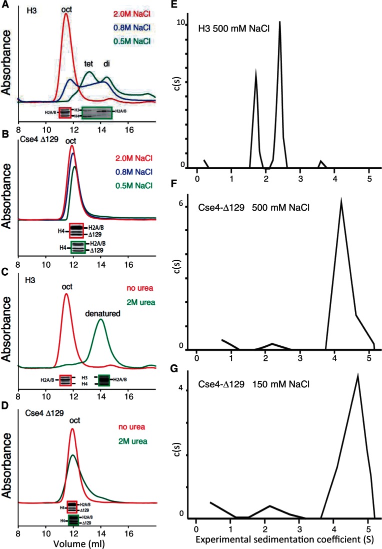
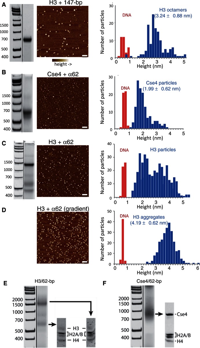
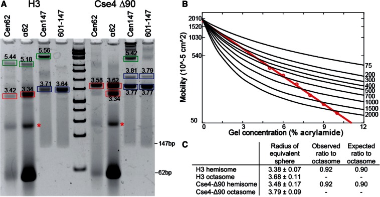
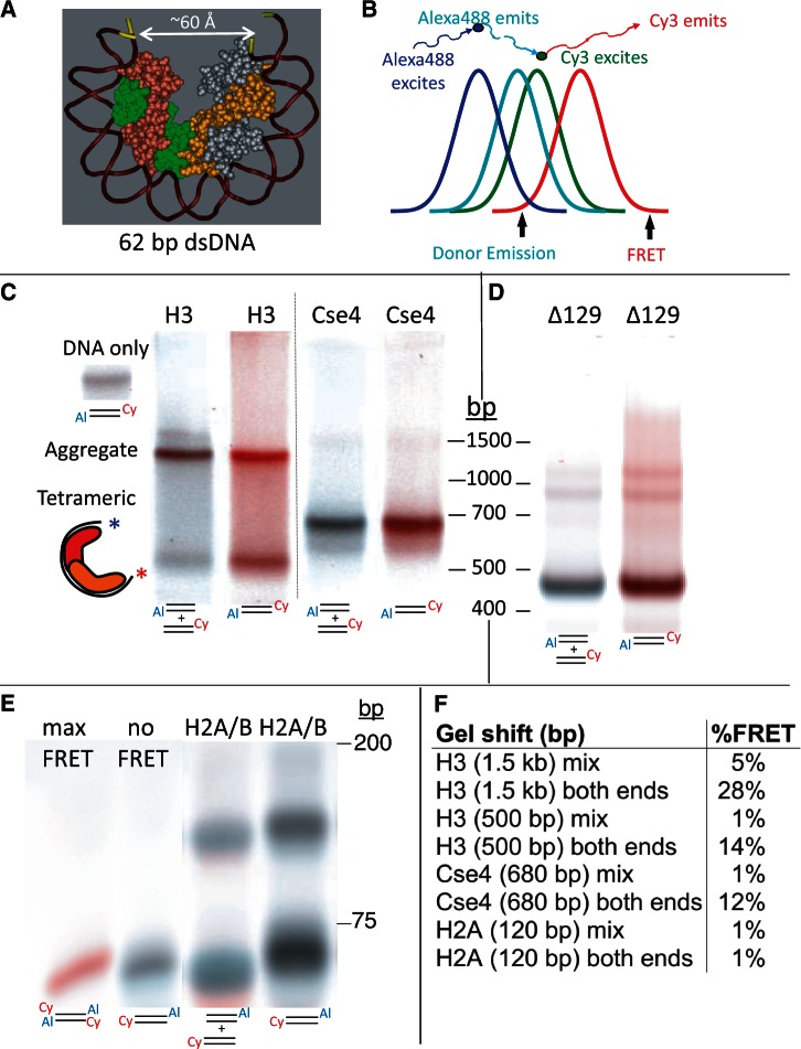
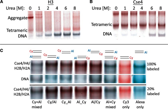
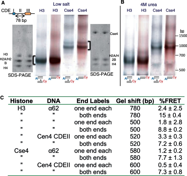
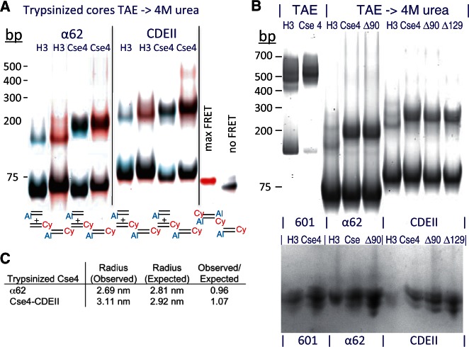
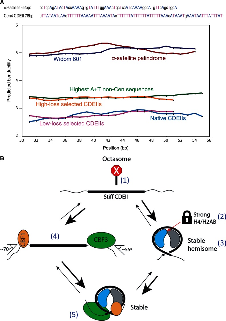
The structure of nucleosomes that contain the cenH3 histone variant has been controversial. In budding yeast, a single right-handed cenH3/H4/H2A/H2B tetramer wraps the ∼80-bp Centromere DNA Element II (CDE II) sequence of each centromere into a 'hemisome'. However, attempts to reconstitute cenH3 particles in vitro have yielded exclusively 'octasomes', which are observed in vivo on chromosome arms only when Cse4 (yeast cenH3) is overproduced. Here, we show that Cse4 octamers remain intact under conditions of low salt and urea that dissociate H3 octamers. However, particles consisting of two DNA duplexes wrapped around a Cse4 octamer and separated by a gap efficiently split into hemisomes. Hemisome dimensions were confirmed using a calibrated gel-shift assay and atomic force microscopy, and their identity as tightly wrapped particles was demonstrated by gelFRET. Surprisingly, Cse4 hemisomes were stable in 4 M urea. Stable Cse4 hemisomes could be reconstituted using either full-length or tailless histones and with a 78-bp CDEII segment, which is predicted to be exceptionally stiff. We propose that CDEII DNA stiffness evolved to favor Cse4 hemisome over octasome formation. The precise correspondence between Cse4 hemisomes resident on CDEII in vivo and reconstituted on CDEII in vitro without any other factors implies that CDEII is sufficient for hemisome assembly.


Figures

Figure 1.
Figure 1.
Cse4 octamers resist dissociation in low-salt and denaturing conditions. (A and B) Superdex 200 gel-filtration chromatography of octamers containing (A) H3 or (B) (tail-deleted) Cse4-Δ129 following equilibration of the column with 2.0 (red), 0.8 (blue) or 0.5 M (green) NaCl. (C and D) Same as (A and B) except that the column was equilibrated with either 2 M NaCl (red) or 2 M NaCl + 2 M urea (green). Red- and green-bordered insets show SDS–PAGE Coomassie-stained images of corresponding peak fractions. In each panel, exactly the same sample was injected into the column; however, non-specific absorption to the column increases with lower salt concentrations, resulting in slightly lower recovery of total proteins. (E–G) Sedimentation velocity analyses of octamers containing H3 in 500 mM NaCl (E), Cse4-Δ129 in 500 mM NaCl (F) or Cse4-Δ129 in 150 mM NaCl (G).
Figure 2.
Figure 2.
Pseudo-octasomes split in half during low-salt dialysis. (A) Gel-shift image (native 7% PAGE), AFM image and height distribution of control H3 octamers assembled with 147-bp Widom 601 duplex DNA. (B) Same as (A) except using Cse4/α62 particles (native 6% PAGE). (C) Same as (A) except using H3/α62 particles (native 6% PAGE). In (A–C), DNA standards are included only as a rough guide, as migration of nucleosomal particles varies depending on gel concentration and running conditions. (D) AFM image and height distribution of H3 particles produced by gradient dialysis from 2 to 0.25 M, followed by step dialysis to 0.25 mM, conditions that increase aggregation. Bar in each image = 100 nm. DNA duplexes (147-bp 601) added to each sample provided an internal height standard. (E and F) Gel-shifted bands contain all four histones. Bands from native 6% gels were excised as indicated and loaded onto an SDS–PAGE gel to determine the histone composition of the particles. Particles were assembled using (E) H3 or (F) Cse4.
Figure 3.
Figure 3.
Ferguson plot analysis of gel-shifted bands. (A) Example of a 7% native gel used for Ferguson plot analysis of gel-shifted species. Calculated particle radii bands are boxed: hemisomes (red), octasomes (blue) and aggregates (green). Asterisks indicate an unidentified α62-derived species that lacks protein based on SDS–PAGE analysis (data not shown). (B) Example of a Ferguson analysis for a Cse4-Δ90/α62 particle (red), where black lines indicate DNA standard mobilities for the corresponding 5–10% series. (C) Calculated dimensions based on three independent experiments, along with observed and expected hemisome:octasome ratios (see text). We compared the distribution of measurements between hemisomes and octasomes by t-test, and the P-value was determined to be 0.0003 for both H3 and Cse4-Δ90.
Figure 4.
Figure 4.
A gelFRET assay directly confirms hemisome formation. (A) Structural model based on PDB 1KX5, showing that a 62-bp duplex is sufficient to wrap a hemisome. (B) FRET strategy using Alexa488 as donor and Cy3 as acceptor, where colors illustrate wavelength and curves represent approximate spectral overlap. (C) GelFRET analysis of a representative gel-shift experiment in which H3 or Cse4 octamers and mixed singly or doubly end-labeled duplexes (as indicated below each lane with 5′ Cy3 on α62 R and 3′ Alexa488 on α62 F) were combined in 2 M NaCl, subjected to dialysis versus 10 mM HEPES (pH 7.5) + 0.25 mM EDTA and electrophoresed on a native 6% polyacrylamide gel, followed by scanning for gelFRET. A doubly end-labeled DNA band showing no FRET signal served as a negative control. Al = Alexa 488. Cy = Cy3 (D) Deletion of the Cse4 N-terminal tail increases gel-shift mobility. Octamers containing tail-deleted Cse4 were assembled with α62 DNA duplexes labeled for gelFRET. DNA standards are included only as a rough guide. (E) As a negative control, H2A/H2B dimers were used for assembly (Supplementary Figure S5). When DNA fragments are in large molar excess, single H2A/H2B dimers bind and cause a gel shift (34). Rapidly migrating dye-coupled fragments resolve from one another with Alexa488 (which is coupled via an amino-C7 linker) causing slower migration than Cy3 (which is directly coupled) of otherwise identical fragments, and resulting in misalignment of mixed Cy3 and Alexa488 fragments. (F) Quantification of gelFRET emissions for the bands shown in the figure.
Figure 5.
Figure 5.
Cse4 hemisomes are stable in high concentrations of urea. (A and B) Native 6% gelFRET showing gel-shifted particles after dialysis versus 10 mM HEPES (pH7.5) + 0.25 mM EDTA containing the indicated concentrations of urea, using H3 octamers (A) and Cse4 octamers (B). The slowly migrating Cse4 band varies between experiments (see Supplementary Figure S7) and might represent an intact pseudo-octasome, a ‘stack’ of hemisomes or a stable aggregate. (C) Cse4 octamers and α62 duplexes were dialyzed versus 4 M urea + 10 mM HEPES (pH 7.5) + 0.25 mM EDTA using different combinations of end-labeled fluorophores as indicated, where Cy or Al on left indicates a 5′ label on α62 F oligonucleotide, and on right for α62 R. For each lane, a DNA band corresponding to the band marked by asterisks in Figure 3A is shown in the middle image and served as a negative control. The lower image shows gel shifts for labeled α62 duplexes that had been diluted 5-fold with unlabeled α62 duplexes.
Figure 6.
Figure 6.
Stable Cse4 and H3 hemisomes form over CDEII DNA. (A) Both H3 and Cse4 split into hemisomes with 78-bp Cen4 CDEII DNA duplexes after dialysis versus 0.25 mM HEPES (pH 7.5) and native 6% PAGE. Loading and electrophoresis of gel slices excised from these gels (brackets) onto SDS–PAGE gels confirms that all four histones were present in the gel-shifted particles. (B) Same as (A) except that dialysis was versus 4 M urea + 0.25 mM HEPES (pH 7.5). (C) Assembly mixtures containing a 4:1 ratio of unlabeled:labeled duplex in 2 M NaCl were dialyzed versus low salt and electrophoresed. %FRET values from four experiments were calculated as in Figure 5E. The free DNA band in each lane served as a no-FRET standard (see Supplementary Figure S6), and a doubly labeled duplex with both fluorophores at each end run in a separate lane served as a 100% standard. Mean and standard deviations for three experiments are shown.
Figure 7.
Figure 7.
Hemisomes form on tailless histone cores. (A) Gel-shifts and gelFRET of particles were assembled in TAE (Supplementary Figure S10), then transferred to 4 M urea using trypsinized H3 or Cse4 cores and α62 or Cen4 CDEII duplexes labeled as indicated, together with maximum-FRET and no-FRET controls. (B) Gel-shifts of trypsinized cores from H3, Cse4, Cse4-Δ90 (Δ90) and Cse4-Δ129 (Δ129) mixed with unlabeled duplexes of 601-145, α62 and Cen4 CDEII duplexes. Top panel: Ethidium bromide-stained gel. Bottom panel: Gel-shifted bands were excised, loaded onto an 18% SDS–PAGE gel in the same order, electrophoresed and silver stained. DNA standards are included only as a rough guide. (C) Ferguson plot analysis of trypsinized Cse4/α62 and Cse4/CDEII shows close correspondences between observed and expected radii of equivalent spheres, where the radius refers to the radius of the geometric mean. Unfortunately, assembly reactions with trypsinized cores and 147-bp DNA resulted in aggregation (data not shown). Averages are shown for samples dialyzed versus low-salt and low-salt followed by 4 M urea, which were similar. AFM was not performed on trypsinized particles because histone tails are required to immobilize nucleosomes on the mica surface (28).
Figure 8.
Figure 8.
CDEII DNA is exceptionally stiff. (A) Bendability predictions for DNA segments using the Bend-it server (http://hydra.icgeb.trieste.it/dna/bend_it.html) (38). Predictions for the 147-bp α-satellite palindrome (brown) and Widom 601 (dark blue) duplexes used in this and previous studies for assembling cenH3 octasomes show that these sequences are predicted to be bendable relative to the 24 most AT-rich non-centromeric (green) and high-loss selected centromeric sequences (orange). The latter sequences are predicted to be more bendable than native CDEII (light blue) and low-loss selected CDEII (magenta) sequences. Sequences used to split octamers in this study are shown with As and Ts highlighted to illustrate that CDEIIs are especially rich in runs of As and Ts. Default parameters were used, except that the window size was lengthened for 147-bp sequences to compensate for their much longer lengths and to simplify the display, although similar averages were obtained using the default window size of 31 bp. (B) Five factors contribute to stable occupancy of Cse4 hemisomes at CDEII: (1) The extreme stiffness of CDEII DNA (thick line) makes wrapping more than once around an octasome difficult relative to the incomplete wrapping around a horseshoe-shaped hemisome. As a result, hemisomes are stable on CDEII DNA, but octasomes are not. (2) The H4/H2B junction at the center of the Cse4 hemisome is strong, unlike the H4/H2B junction in H3 nucleosomes, which is weak relative to the H3/H3 junction and therefore causes octamers to dissociate into an (H3/H4)2 tetramer and two H2A/H2B dimers. (3) The Cse4 hemisome is stable even in the absence of Cbf1 and CBF3. (4) Cbf1 and CBF3 bind avidly to and sharply bend the DNA immediately flanking CDEII and so expose only ∼80 bp of DNA, which can accommodate heterotypic tetramers, but not octamers. (5) Although highly stable H3 hemisomes can form on CDEII in vitro, they are strongly disfavored in vivo because the CBF3 complex is known to specifically recruit Cse4, which would displace H3-containing nucleosomes. Cse4/H4 heterodimers are depicted in blue and H2A/H2B in gray.

Similar articles

Cited by

References

    1. Black BE, Cleveland DW. Epigenetic centromere propagation and the nature of CENP-A nucleosomes. Cell. 2011;144:471–479. - PMC - PubMed
    1. Henikoff S, Furuyama T. The unconventional structure of centromeric nucleosomes. Chromosoma. 2012;121:341–352. - PMC - PubMed
    1. Malik HS, Henikoff S. Major evolutionary transitions in centromere complexity. Cell. 2009;138:1067–1082. - PubMed
    1. Camahort R, Shivaraju M, Mattingly M, Li B, Nakanishi S, Zhu D, Shilatifard A, Workman JL, Gerton JL. Cse4 is part of an octameric nucleosome in budding yeast. Mol. Cell. 2009;35:794–805. - PMC - PubMed
    1. Dalal Y, Furuyama T, Vermaak D, Henikoff S. Structure, dynamics, and evolution of centromeric nucleosomes. Proc. Natl Acad. Sci. USA. 2007;104:15974–15981. - PMC - PubMed

Publication types

MeSH terms