. 2013 Jul;33(13):2574-85.
doi: 10.1128/MCB.01677-12. Epub 2013 Apr 22.

p90 RSK2 mediates antianoikis signals by both transcription-dependent and -independent mechanisms

Affiliations

p90 RSK2 mediates antianoikis signals by both transcription-dependent and -independent mechanisms

Lingtao Jin et al. Mol Cell Biol. 2013 Jul.

Abstract

How invasive and metastatic tumor cells evade anoikis induction remains unclear. We found that knockdown of RSK2 sensitizes diverse cancer cells to anoikis induction, which is mediated through phosphorylation targets including apoptosis signal-regulating kinase 1 (ASK1) and cyclic AMP (cAMP) response element-binding protein (CREB). We provide evidence to show that RSK2 inhibits ASK1 by phosphorylating S83, T1109, and T1326 through a novel mechanism in which phospho-T1109/T1326 inhibits ATP binding to ASK1, while phospho-S83 attenuates ASK1 substrate MKK6 binding. Moreover, the RSK2→CREB signaling pathway provides antianoikis protection by regulating gene expression of protein effectors that are involved in cell death regulation, including the antiapoptotic factor protein tyrosine kinase 6 (PTK6) and the proapoptotic factor inhibitor-of-growth protein 3 (ING3). PTK6 overexpression or ING3 knockdown in addition to ASK1 knockdown further rescued the increased sensitivity to anoikis induction in RSK2 knockdown cells. These data together suggest that RSK2 functions as a signal integrator to provide antianoikis protection to cancer cells in both transcription-independent and -dependent manners, in part by signaling through ASK1 and CREB, and contributes to cancer cell invasion and tumor metastasis.


Figures
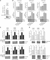
Fig 1
Fig 1
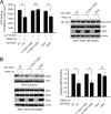
Loss of RSK2 sensitizes diverse cancer cells to anoikis induction. (A) Stable knockdown of RSK2 by shRNA does not affect apoptosis induced by the control agent cycloheximide (CHX) compared to control cells harboring an empty vector. (B) Stable knockdown of RSK2 by two different shRNA clones sensitizes metastatic HNSCC 212LN (left), breast cancer SKBR3 (middle), and lung cancer A549 (right) cells to detachment-induced anoikis compared to control cells harboring an empty vector or a vector harboring shRNA targeting eGFP. Cells were cultured on a 1%-agar-treated dish to achieve detachment, and control cells were treated with the anticancer agent cycloheximide, an inhibitor of protein biosynthesis, to induce apoptosis. Apoptosis was assessed by FACS analysis in cells stained with FITC-conjugated annexin V and propidium iodide (top) or by caspase 3/7 activity using a Caspase-Glo 3/7 assay (bottom). (C) Overexpression of murine RSK2 (top) or shRNA-resistant human RSK2 (bottom) rescued anoikis induced by RSK2 knockdown and conferred resistance to cells from undergoing anoikis. Stable RSK2 knockdown cells were transiently transfected with murine RSK2 Y707A cDNA or shRNA-resistant human RSK2 Y707A cDNA prior to anoikis induction. (D) Treatment with small-molecule RSK inhibitors, fmk (10 μM) (top), or SL0101 (100 μM) (bottom) effectively decreases RSK2 kinase activity and sensitizes 212LN, SKBR3, and A549 cells to induction of anoikis. For fmk treatment, RSK2 activity was assessed by RSK2 immunoprecipitation (IP) and Western blotting (WB), using a specific phospho-RSK antibody that recognizes phospho-S380 (S386 for RSK2 numbering). Phosphorylation of the RSK2 substrate GSK3α/β was assessed for SL0101. P values were determined by Student's t test. All the error bars shown in the figures represent mean values ± standard deviations from three independent experiments (∗, 0.01 < P < 0.05; ∗∗, P < 0.01; ns, not significant).
Fig 2
Fig 2
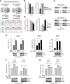
RSK2 inhibits ASK1 to protect metastatic cancer cells from detachment-induced anoikis. (A) Phosphoantibody microarray analysis identified novel phosphorylation targets of RSK2, whose phosphorylation states decreased in HNSCC cells when RSK2 was stably knocked down by shRNA. The signal intensities of phosphorylated proteins and the total protein levels were determined. The ratio of each protein was determined as the ratio between the percentages of phosphorylated proteins in total proteins in HNSCC-pLKO.1-RSK2 shRNA and HNSCC-pLKO.1 cells. Antiapoptotic or proapoptotic protein factors with phosphorylation status decreased or increased more than 15% in metastatic HNSCCs with RSK2 knockdown are shown. S83 phosphorylation, which inhibits ASK1, was decreased 31% in 886LN cells with stable knockdown of RSK2 compared to control cells. (B and C) Knockdown or overexpression of proapoptotic ASK1 results in reduced (B) or increased (C) sensitivity to anoikis induction in 212LN cells. (D) RNAi-mediated knockdown of ASK1 attenuates the increased sensitivity to anoikis induction in 212LN (left) and SKBR3 (right) cells with stable knockdown of RSK2. (E) Inhibition of RSK2 by a specific RSK inhibitor, fmk (6 μM), results in increased sensitivity to anoikis induction in 212LN cells, while knockdown of ASK1 rescues the phenotype of cells treated with fmk. (F) Overexpression of a constitutively active form of RSK2, the Y707A mutant, results in decreased ASK1 kinase activity. GST-tagged ASK1 was coexpressed with or without the RSK2 Y707A mutant in 293T cells. GST-ASK1 was pulled down by GST beads and used for an in vitro ASK1 kinase assay using recombinant, inactive human MKK6 protein as an exogenous substrate. ASK1 activity was assessed by the phosphorylation levels of MKK6 detected by a specific phospho-Ser/Thr antibody. (G) Western blot results show that expression of a constitutively active form of RSK2, the Y707A mutant, results in decreased activation levels of p38 (left) and JNK (right) in 212LN cells. Activation of p38 and JNK was assessed by specific antibodies recognizing phospho-p38 and phospho-SAPK/JNK, respectively. (H) Stable knockdown of RSK2 results in increased phosphorylation and activation levels of p38 and JNK in 212LN cells.
Fig 3
Fig 3
RSK2 inhibits ASK1 by phosphorylating S83. (A) Knockdown of RSK2 results in decreased phospho-S83 levels of ASK1 in 212LN cells. Phospho-S83 levels were assessed by a specific phospho-ASK1 antibody (p-S83). (B) Enforced RSK2 expression results in increased phosphorylation levels of S83 but not of S967 or T845 of ASK1. 293T cells were transiently transfected with the HA-tagged constitutively active RSK2 Y707A mutant. S83, S967, and T845 phosphorylation levels of endogenous ASK1 were determined by using specific phospho-ASK1 antibodies recognizing individual phosphorylation sites. (C) RSK2 directly phosphorylates ASK1 at S83 but not at S967 or T845. An in vitro RSK2 kinase assay was performed by using recombinant active RSK2 (rRSK2) incubated with either WT recombinant ASK1 (rASK1) or a kinase-dead mutant form, K709M, purified from E. coli. (D) RSK2 directly phosphorylates WT ASK1 at S83 but is unable to phosphorylate the S83A mutant. Purified recombinant WT ASK1 and S83A mutant proteins were used in an in vitro RSK2 kinase assay. (E) An in vitro RSK2 kinase assay was performed by using the GST-tagged ASK1 K709M mutant enriched from 293T cells by GST pulldown.
Fig 4
Fig 4
T1109 and T1326 are identified as new RSK2 phosphorylation sites that, when phosphorylated, inhibit ASK1. (A) Mass spectrometry spectra of phosphothreonine peptide fragments of ASK1 containing T1109 and T1326. GST-ASK1 K709M protein purified from 293T cells was incubated with recombinant active RSK2. Bands from SDS-PAGE gels were excised and applied for LC-MS/MS. (B and C) Substitution of either T1109, T1326, or S83 did not affect RSK2-dependent attenuation of ASK1 kinase activity (B), whereas the T1109A/T1326A double mutant showed resistance to RSK2-dependent inhibition (C). Distinct GST-tagged ASK1 variants were coexpressed in the presence or absence of a constitutively active form of RSK2, the Y707A mutant, in 293T cells. At 24 h posttransfection, cells were harvested, and GST-ASK1 proteins were pulled down by GST beads. An in vitro ASK1 kinase assay was performed by using myelin basic protein as a substrate. (D) Expression of the ASK1 T1109D/T1326D mutant in a dominant negative form results in less proapoptotic activity that with WT ASK1 in cells with RSK2 knockdown. RSK2 knockdown cells were transiently transfected with ASK1 variants. An anoikis assay was performed at 48 h posttransfection.
Fig 5
Fig 5
Phosphorylation at T1109/T1326 and S83 inhibits ASK1 by attenuating ATP and substrate binding, respectively. (A, left) Substitution of T1109/T1326, but not S83, reversed the decreased ATP binding to ASK1 in the presence of RSK2 CA (constitutively active Y707A mutant form). GST-ASK1 variants expressed in the presence or absence of RSK2 CA were enriched from 293T cell lysates by GST pulldown, followed by incubation with [α-32P]ATP. Unbound [α-32P]ATP was washed away, and bound [α-32P]ATP on ASK1 was measured with a scintillation counter. (Right) Western blot results show expression levels of GST-ASK1 and RSK2 in 293T cells. (B) Mutation at S83, but not at T1109/T1326, results in an increase of MKK6 binding to ASK1 in the presence of RSK2 CA. GST-ASK1 variants were pulled down from cells coexpressed with or without RSK2 CA. Bound endogenous MKK6 was detected by immunoblotting. (Left) Representative immunoblotting result. (Right) Relative intensity of the MKK6 bands from three different experiments, which are normalized to values for the control samples without RSK2 expression.
Fig 6
Fig 6
RSK2 protects cells from anoikis induction, in part by signaling through CREB to upregulate the antiapoptotic factor PTK6 and downregulate the proapoptotic factor ING3. (A) DNA microarray analysis identifies novel transcription targets of the RSK2→CREB signaling pathway. (Top) Diagram showing overlapping transcription targets that are either downregulated (115 genes) (left) or upregulated (45 genes) (right) from 212LN cells with individual expression of RSK2 shRNA and CREB shRNA, compared to control cells harboring an empty lentiviral vector. (Middle) Representative, novel RSK2→CREB transcription targets that are downregulated in RSK2 and CREB knockdown cells, including the antiapoptotic factors PTK6, PINK1, and MSLN. (Bottom) Representative, novel RSK2→CREB transcription targets that are upregulated in RSK2 and CREB knockdown cells, including the proapoptotic factors ING3, CKAP2, and TFAP2A. mRNA level changes (fold) in RSK2 and CREB knockdown cells are shown in the bottom rows of the middle and bottom panels, compared to control cells harboring an empty vector. (B) Real-time reverse transcription-PCR results show decreased mRNA levels of the antiapoptotic factor PTK6 (left) and increased mRNA levels of the proapoptotic factor ING3 (right) in 212LN cells with stable knockdown of RSK2 or CREB, compared to control cells with an empty vector. (C) Western blot results show decreased and increased protein levels of PTK6 and ING3, respectively, in 212LN cells with stable knockdown of RSK2 (top) or CREB (bottom). (D) Real-time reverse transcription-PCR results show decreased and increased mRNA levels of PTK6 (left) and ING3 (right), respectively, in 212LN cells treated with increasing concentrations of the RSK inhibitor fmk for 24 h. (E) Knockdown of CREB (left) or PTK6 (middle) or overexpression of ING3 (right) results in increased sensitivity to anoikis induction in cancer cells. (F) Expression of Flag-tagged PTK6 significantly attenuated the increased sensitivity to anoikis induction in 212LN and SKBR3 cells with stable knockdown of RSK2. (G) Knockdown of ING3 results in a significant attenuation of the increased sensitivity to anoikis induction in 212LN and SKBR3 cells with RSK2 knockdown. Apoptotic cell death was assessed by annexin V staining.
Fig 7
Fig 7
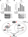
RSK2 mediates antianoikis signals through both ASK1 and CREB transcription targets. (A and B) Knockdown of RSK2 results in increased sensitivity to anoikis induction, and knockdown of ASK1 significantly rescues this phenotype, while simultaneous knockdown of ASK1 and overexpression of PTK6 (A) or knockdown of ING3 (B) results in a further rescue effect. Stable RSK2 knockdown cells were transiently cotransfected with ASK1 siRNA and Flag-PTK6 or ING3 siRNA for 24 h, prior to transfer onto an agar-treated plate and culture for 48 h. Apoptotic cell death was assessed by annexin V staining. (C) Proposed model of RSK2-mediated antianoikis signaling in metastatic cancer cells. RSK2 is a signal integrator in metastatic cells, which phosphorylates and regulates multiple protein factors in both acute and chronic ways to provide antianoikis signals.

Similar articles

Cited by

References

    1. Gupta GP, Massague J. 2006. Cancer metastasis: building a framework. Cell 127:679–695 - PubMed
    1. Fidler IJ. 2003. The pathogenesis of cancer metastasis: the ‘seed and soil’ hypothesis revisited. Nat. Rev. Cancer 3:453–458 - PubMed
    1. Kang S, Elf S, Lythgoe K, Hitosugi T, Taunton J, Zhou W, Xiong L, Wang D, Muller S, Fan S, Sun SY, Marcus AI, Gu TL, Polakiewicz RD, Chen GZ, Khuri FR, Shin DM, Chen J. 2010. p90 ribosomal S6 kinase 2 promotes invasion and metastasis of human head and neck squamous cell carcinoma cells. J. Clin. Invest. 120:1165–1177 - PMC - PubMed
    1. Blenis J. 1993. Signal transduction via the MAP kinases: proceed at your own RSK. Proc. Natl. Acad. Sci. U. S. A. 90:5889–5892 - PMC - PubMed
    1. Frodin M, Gammeltoft S. 1999. Role and regulation of 90 kDa ribosomal S6 kinase (RSK) in signal transduction. Mol. Cell. Endocrinol. 151:65–77 - PubMed

Publication types

MeSH terms

LinkOut - more resources