. 2013 Jul;154(7):1080-91.
doi: 10.1016/j.pain.2013.03.021. Epub 2013 Mar 15.

mTORC1 inhibition induces pain via IRS-1-dependent feedback activation of ERK

Affiliations

mTORC1 inhibition induces pain via IRS-1-dependent feedback activation of ERK

Ohannes K Melemedjian et al. Pain. 2013 Jul.

Abstract

Mammalian target of rapamycin complex 1 (mTORC1) inhibitors are extensively used as immunosuppressants to prevent transplant rejection and in treatment of certain cancers. In patients, chronic treatment with rapamycin or its analogues (rapalogues) has been reported to lead to sensory hypersensitivity and pain conditions via an unknown mechanism. Here, we show that pharmacological or genetic inhibition of mTORC1 activates the extracellular signal-regulated kinase (ERK) pathway in sensory neurons via suppression of S6K1 to insulin receptor substrate 1 negative feedback loop. As a result, increased ERK activity induces sensory neuron sensitization, mechanical hypersensitivity, and spontaneous pain. The clinically available adenosine monophosphate-activated protein kinase activator, metformin, which is an antidiabetic drug, prevents rapamycin-induced ERK activation and the development of mechanical hypersensitivity and spontaneous pain. Taken together, our findings demonstrate that activation of the ERK pathway in sensory neurons as a consequence of mTORC1 inhibition leads to the development of pain. Importantly, this effect is abolished by co-treatment with metformin, thus providing a potential treatment option for rapalogue-evoked pain. Our findings highlight the physiological relevance of feedback signaling through mTORC1 inhibition and have important implications for development of pain therapeutics that target the mTOR pathway.


Figures

Fig. 1
Fig. 1
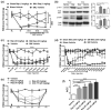
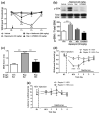
Rapamycin treatment induces allodynia and increases AKT and extracellular signal-regulated kinase (ERK) phosphorylation.(a) Daily intraperitoneal injection of rapamycin in rats with spinal nerve ligation (SNL) (treatment started 14 days post-SNL) leads to a partial reversal of mechanical allodynia but causes allodynia in sham-operated animals. (b) Treatment with rapamycin causes significant increases in ERK (Thr202/Tyr204) and AKT (S473) phosphorylation in the sciatic nerve of sham-operated animals and rapamycin fails to inhibit these kinases in SNL rats. (c) Daily intraperitoneal injection of rapamycin in mice with spared nerve injury (SNI) partially reverses mechanical allodynia on day 7 (#, P < 0.05), however, sham-operated animals develop allodynia within 3 hours after rapamycin injection. (d) Daily injection with metformin (Met, 200 mg/kg) for 7 days in mice with SNI 7 weeks postsurgery reverses neuropathic allodynia for at least 61 days after the cessation of the treatment. (e) Naïve mice develop allodynia when treated with rapamycin in a dose dependent manner and CCI-779 treatment induces allodynia in naïve mice. (f) Area-over-the-curve (AOC) analysis demonstrates dose-related induction of allodynia in response to rapalogue treatment. n = 6 per condition. *P < 0.05; **P < 0.01; ***P < 0.001.
Fig. 2
Fig. 2
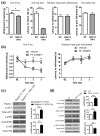
Raptor deletion leads to tactile allodynia. Raptor floxed mice were injected into the hind paw with herpes simplex viral (HSV) vector expressing Cre. (a) The levels of Raptor and rS6 phosphorylation decreased in the dorsal root ganglia (DRG) of Raptor floxed mice (Raptorflox/flox) 2 weeks after the injection compared to the floxed mice injected with HSV without Cre or C57BL/6 mice injected with HSV-Cre. (b) Raptor deletion leads to mechanical allodynia, whereas thermal thresholds are not altered (n = 8 per group). **P < 0.01.
Fig. 3
Fig. 3
Reduction of S6K activity leads to enhanced tactile sensation. (a) Mechanical sensitivity in automated von Frey and tail clip tests is increased in S6K1/2 double-knockout (DKO) mice as compared to wild-type (WT) littermates (n = 8 per group). No change in thermal sensitivity was measured in radiant heat paw withdrawal and hot plate tests. (b) Daily intraperitoneal administration of S6K1 inhibitor PF 4708671 (50 mg/kg) leads to reduction of mechanical threshold (left panel; n = 4 per group), whereas no change in thermal sensation was observed (right panel; n = 6). Phosphorylation of AKT and extracellular signal-regulated kinase (ERK) is increased in sciatic nerves following Raptor deletion (c, n = 6) and in dorsal root ganglia of S6K1/2 DKO mice, while insulin receptor substrate (IRS) S636 phosphorylation is decreased in these mice (d, n = 6). *P < 0.05; **P < 0.01; ***P < 0.001.
Fig. 4
Fig. 4
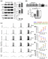
Rapamycin promotes extracellular signal-regulated kinase (ERK)-dependent hyperexcitability of sensory neurons. (a) Treatment of mouse primary sensory neurons in culture with rapamycin for 1 hour results in concentration-related increases in p-ERK and p-AKT (S473). Rapamycin inhibits the phosphorylation rS6, a downstream target of mammalian target of rapamycin complex 1 (mTORC1)-S6K (n = 6). (b) Co-immunoprecipitation analysis reveals that the treatment of primary sensory neurons in culture with rapamycin (100 nM) for 1 hour enhances the association of ERK1 with Nav1.7 (n = 6). ERK2 does not immunoprecipitate with Nav1.7. Rapamycin-enhanced association of ERK1 with Nav1.7 is blocked by U0126 (10 μM) but not affected by U0124 (10 μM) treatment. (c) Patch clamp analysis of mouse sensory neurons in culture treated with rapamycin (n = 13) for 1 hour demonstrates that: rapamycin dose-dependently (d) increases the number of action potentials evoked by ramp currents and (e) reduces the latency to first action potential in response to escalating ramp currents. (f and g) Rapamycin-induced hyperexcitability is blocked by U0126 (n = 10) but unaffected by U0124 (n = 11). Color code of the stars: red (30 nm Rap vs vehicle), orange (100 nM Rap vs vehicle), brown (100 nM Rap + 10 μM U0124 vs vehicle), and green (100 nM Rap + 10 μM U0126 vs 100 nM Rap). *P < 0.05; **P < 0.01; ***P < 0.001.
Fig. 5
Fig. 5
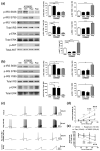
Rapamycin activates extracellular signal-regulated kinase (ERK) by suppressing insulin receptor substrate (IRS)-1 phosphorylation, while adenosine monophosphate-activated protein kinase (AMPK) activator A769662 inhibits the ERK activation by enhancing the phosphorylation of IRS-1. Treatment of dorsal root ganglion (a) or trigeminal ganglia (b) neurons in culture with rapamycin (100 nM) for 1 hour suppresses the phosphorylation of IRS-1 on S636. Treatment of the cultures with A769662 (200 μM) enhances S789 phosphorylation on IRS-1. The net outcome of these phosphorylation events is enhanced Y895 phosphorylation of IRS-1 in response to rapamycin (100 nM) treatment alone. Phosphorylation of Y895 is associated with the activation of ERK (n = 6). (b) AMPK activator A769662 blocks rapamycin-enhanced excitability of sensory neurons. Patch-clamp electrophysiology reveals that the treatment of sensory neurons in culture (n = 10) with rapamycin (100 nM) for 1 hour results in an increase in the number of action potentials evoked by ramp currents and reduces latency to first action potential in response to ramp currents. Co-treatment of sensory neurons in culture (n = 10 per group) with A769662 (200 μM) and rapamycin (100 nM) profoundly attenuates the number of action potential evoked by ramp currents (c) and increases the time to first action potential in response to ramp currents (d). *P < 0.05; **P < 0.01; ***P < 0.001.
Fig. 6
Fig. 6
Adenosine monophosphate-activated protein kinase (AMPK) activators block rapamycin-induced behavioral and biochemical changes. (a) Treatment of naïve mice with rapamycin (20 mg/kg) induces mechanical allodynia that peaks at 3 hours and subsides by 4 hours. Co-treatment with rapamycin and either metformin (200 mg/kg) or A769662 (30 mg/kg) blocks the development of rapamycin-induced allodynia. (b) Sciatic nerves from these animals show a significant increase in p-ERK (extracellular signal-regulated kinase) and its blockade with either metformin or A769662 treatment at 3 hours posttreatment. (c) Rapamycin (20 mg/kg) treatment causes a significant increase in Mouse Grimace Scale (MGS) score at 2–3 hours after systemic administration of drug. This effect is completely blocked by co-administration of metformin (200 mg/kg, Sal = saline). (d) Control (open circles) and Raptor knockout mice (Raptorflox/flox + HSV-Cre; filled circles) were injected daily with metformin (200 mg/kg). Tactile allodynia, exhibited by Raptor-deleted mice, was resolved after 4 days of treatment with metformin. (e) No change in thermal threshold was measured after 4 days of metformin treatment. n = 6 per group. *P < 0.05; **P < 0.01; ***P < 0.001.
Fig. 7
Fig. 7
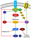
Schematic diagram summarizing the signaling pathways relevant to this study. Activation of receptor tyrosine kinases (RTK) results in the recruitment of the adaptor protein insulin receptor substrate (IRS), which results in the activation of PI3K-AKT- mammalian target of rapamycin (mTOR) and Ras-Raf-Mek-ERK (extracellular signal-regulated kinase) pathways. Serine phosphorylation of IRS on the 636/639 and 789 residues is associated with reduced signaling from IRS, whereas Y895 phosphorylation is associated with enhanced signaling. Blockade of mTOR complex 1 (mTORC1) with rapamycin results in the inhibition of S6K, which leads to reduced IRS S636/639 phosphorylation, enhanced phosphorylation of IRS Y895 residue, and the recruitment of the adaptor protein Grb2, resulting in the activation of the ERK pathway. Activated ERK1 associates and phosphorylates Nav1.7, leading to hyperexcitability of sensory neurons. Activation of adenosine monophosphate-activated protein kinase (AMPK) in response to metformin treatment, leads to enhanced IRS S789 phosphorylation (which is an inhibitory phospho site) and reduced phosphorylation of IRS Y895 (which is a phospho site associated with enhanced IRS activity) residue, which suppresses the recruitment of Grb2, thus reversing rapamycin-induced activation of the ERK pathway.

Similar articles

Cited by

References

    1. Asante CO, Wallace VC, Dickenson AH. Formalin-induced behavioural hypersensitivity and neuronal hyperexcitability are mediated by rapid protein synthesis at the spinal level. Mol Pain. 2009;5:27. - PMC - PubMed
    1. Asante CO, Wallace VC, Dickenson AH. Mammalian target of rapamycin signaling in the spinal cord is required for neuronal plasticity and behavioral hypersensitivity associated with neuropathy in the rat. J Pain. 2010;11:1356–67. - PMC - PubMed
    1. Asiedu MN, Tillu DV, Melemedjian OK, Shy A, Sanoja R, Bodell B, Ghosh S, Porreca F, Price TJ. Spinal protein kinase M zeta underlies the maintenance mechanism of persistent nociceptive sensitization. J Neurosci. 2011;31:6646–53. - PMC - PubMed
    1. Bailey CJ, Turner RC. Metformin. N Engl J Med. 1996;334:574–9. - PubMed
    1. Bourquin AF, Suveges M, Pertin M, Gilliard N, Sardy S, Davison AC, Spahn DR, Decosterd I. Assessment and analysis of mechanical allodynia-like behavior induced by spared nerve injury (SNI) in the mouse. PAIN®. 2006;122:e11–4. - PubMed

Publication types

MeSH terms