Study on injury effect of food additive citric acid on liver tissue in mice
- PMID: 23606053
- PMCID: PMC3918259
- DOI: 10.1007/s10616-013-9567-1
Study on injury effect of food additive citric acid on liver tissue in mice
Erratum in
-
Erratum to: Study on injury effect of food additive citric acid on liver tissue in mice.Cytotechnology. 2016 Oct;68(5):2209. doi: 10.1007/s10616-016-9978-x. Cytotechnology. 2016. PMID: 27142653 Free PMC article. No abstract available.
Abstract
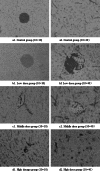
To investigate the damaging effect and action mechanism of the food additive citric acid (CA) on mouse liver, 40 healthy male Kunming mice were randomly divided into control group (0.9 % saline), low CA dose (120 mg/kg), middle dose (240 mg/kg) and high dose groups (480 mg/kg). All experimental mice have received peritoneal injection of the corresponding reagent each week for 3 weeks. After 7 days since the third injection, morphological changes were observed by light microscope; activities of T-SOD, glutathione peroxidase (GSH-Px), caspase-3, and contents of hydrogen peroxide (H2O2) and malonyldialdehyde (MDA) in the liver were evaluated using the corresponding assay kits; DNA fragmentation was assayed using agarose gel electrophoresis. Microscopical detection showed a series of hispathological changes in mouse livers treated with CA, such as indiscriminate liver cell cord, blood clot in central veins, and lymphocyte infiltrating. Biochemical examination suggested the gradually but moderately reduced T-SOD activity and elevated H2O2 level with the increase of CA dose (P > 0.05), and the gradually reduced GSH-Px activity and increased MDA content depending on graded doses with a significant difference (P < 0.05) between the high dose group and the control group. According to cell apoptosis assays, caspase-3 activity were significantly higher in all treatment groups than in the control (P < 0.05) in a dose-dependent manner. Contrasting to the control, characteristic DNA laddering was observed when injected with any of the three graded doses. It can be concluded that certain concentrations of CA cause oxidative damage of the liver by means of the decrease of antioxidative enzyme activities, thus resulting in MDA level elevation and DNA fragmentation inducing active caspase-3.
Figures


Similar articles
-
Effects of the food additive, citric acid, on kidney cells of mice.Biotech Histochem. 2015 Jan;90(1):38-44. doi: 10.3109/10520295.2014.937745. Epub 2014 Sep 8. Biotech Histochem. 2015. PMID: 25196033
-
Effect of Anoectochilus roxburghii flavonoids extract on H2O2 - Induced oxidative stress in LO2 cells and D-gal induced aging mice model.J Ethnopharmacol. 2020 May 23;254:112670. doi: 10.1016/j.jep.2020.112670. Epub 2020 Mar 3. J Ethnopharmacol. 2020. PMID: 32135242
-
Effects of low dose pre-irradiation on hepatic damage and genetic material damage caused by cyclophosphamide.Eur Rev Med Pharmacol Sci. 2014;18(24):3889-97. Eur Rev Med Pharmacol Sci. 2014. PMID: 25555880
-
[Study on genetic toxicity of gaseous benzene to mouse bone marrow cells].Zhonghua Lao Dong Wei Sheng Zhi Ye Bing Za Zhi. 2014 Apr;32(4):246-50. Zhonghua Lao Dong Wei Sheng Zhi Ye Bing Za Zhi. 2014. PMID: 24754935 Chinese.
-
[The protective effect of regulation of paraoxonase 1 gene on liver oxidative stress injury induced by dichlorvos poisoning in mice].Zhonghua Wei Zhong Bing Ji Jiu Yi Xue. 2015 Apr;27(4):285-90. doi: 10.3760/cma.j.issn.2095-4352.2015.04.012. Zhonghua Wei Zhong Bing Ji Jiu Yi Xue. 2015. PMID: 25891459 Chinese.
Cited by
-
Intraoperative parameters and postoperative follow-up of foam-based intraperitoneal chemotherapy (FBIC).Front Pharmacol. 2023 Nov 14;14:1276759. doi: 10.3389/fphar.2023.1276759. eCollection 2023. Front Pharmacol. 2023. PMID: 38035016 Free PMC article.
-
Antioxidative and Cytoprotective Effects of Rosa Roxburghii and Metabolite Changes in Oxidative Stress-Induced HepG2 Cells Following Rosa Roxburghii Intervention.Foods. 2024 Nov 4;13(21):3520. doi: 10.3390/foods13213520. Foods. 2024. PMID: 39517304 Free PMC article.
-
Tartrazine induces structural and functional aberrations and genotoxic effects in vivo.PeerJ. 2017 Feb 23;5:e3041. doi: 10.7717/peerj.3041. eCollection 2017. PeerJ. 2017. PMID: 28243541 Free PMC article.
References
-
- Aktaç T, Kaboğlu A, Ertan F, Ekinci F, Huseyinova G. The short-term effects of single toxic dose of citric acid in mice. J Cell Mol Biol. 2003;2:19–23.
-
- Flora SJ, Mehta A, Satsangi K, Kannan GM, Gupta M. Aluminum-induced oxidative stress in rat brain: response to combined administration of citric acid and HEDTA. Comp Biochem Physiol C: Toxicol Pharmacol. 2003;134:319–328. - PubMed
-
- García-Gil FA, Albendea CD, López-Pingarrón L, Royo-Dachary P, Martínez-Guillén J, Piedrafita E, Martínez-Díez M, Soria J, García JJ. Altered cellular membrane fluidity levels and lipid peroxidation during experimental pancreas transplantation. J Bioenerg Biomembr. 2012;44:571–577. doi: 10.1007/s10863-012-9459-7. - DOI - PubMed
-
- Heo JM, Opapeju FO, Pluske JR, Kim JC, Hampson DJ, Nyachoti CM. Gastrointestinal health and function in weaned pigs: a review of feeding strategies to control post-weaning diarrhoea without using in-feed antimicrobial compounds. J Anim Physiol Anim Nutr (Berl) 2013;97:207–237. doi: 10.1111/j.1439-0396.2012.01284.x. - DOI - PubMed
LinkOut - more resources
Full Text Sources
Other Literature Sources
Research Materials

