

Fractalkine and its receptor mediate extracellular matrix accumulation in diabetic nephropathy in mice
- PMID: 23604552
- PMCID: PMC4737593
- DOI: 10.1007/s00125-013-2907-z
Fractalkine and its receptor mediate extracellular matrix accumulation in diabetic nephropathy in mice
Abstract
Aims/hypothesis: Fractalkine (FKN) is a unique chemokine that works as a chemoattractant and an adhesion molecule. Previous studies have demonstrated that FKN plays a role in ischaemic and protein-overload renal injury via its cognate receptor chemokine (C-X3-C motif) receptor 1 (CX3CR1). However, involvement of the FKN/CX3CR1 system in diabetic nephropathy remains unclear. We examined the role of FKN/CX3CR1 in diabetic mice and mouse mesangial cells (MMCs).
Methods: Streptozotocin (50 mg kg(-1) day(-1)) was intraperitoneally administered for 5 days to male Cx3cr1-knockout (KO) mice and wild-type (WT) mice. MMCs transfected with Fkn (also known as Cx3cl1) or Cx3cr1 siRNA, respectively, were used to elucidate the role of FKN/CX3CR1 in extracellular matrix (ECM) synthesis.
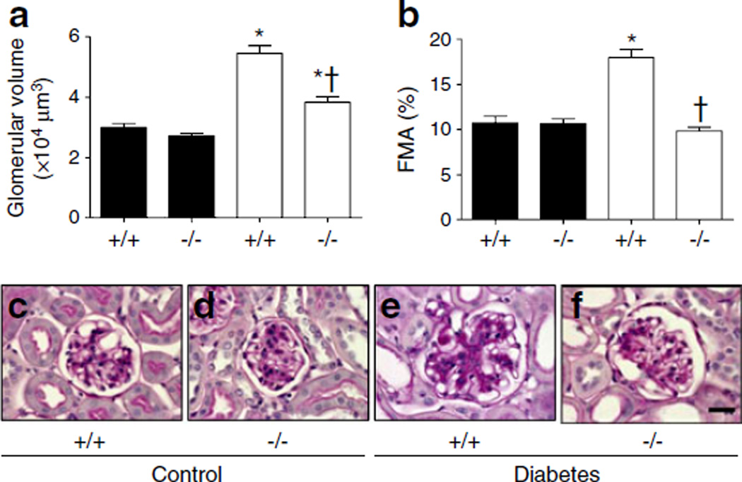
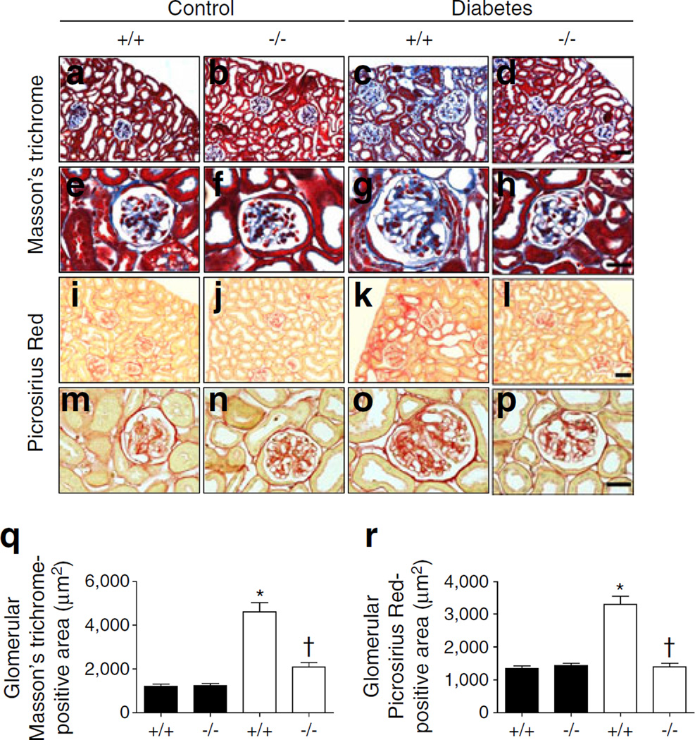
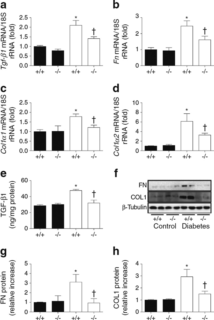
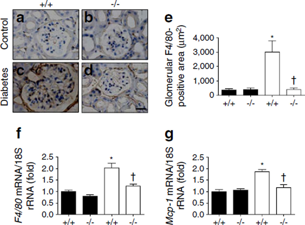
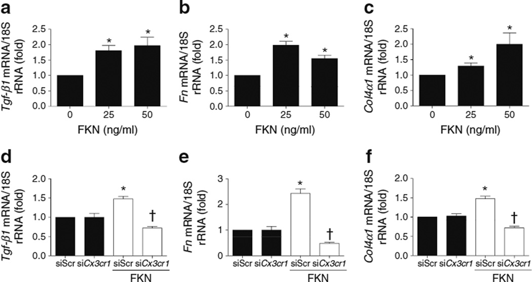
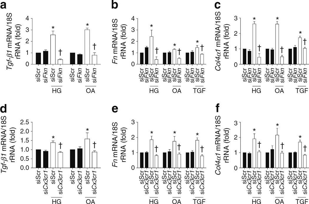
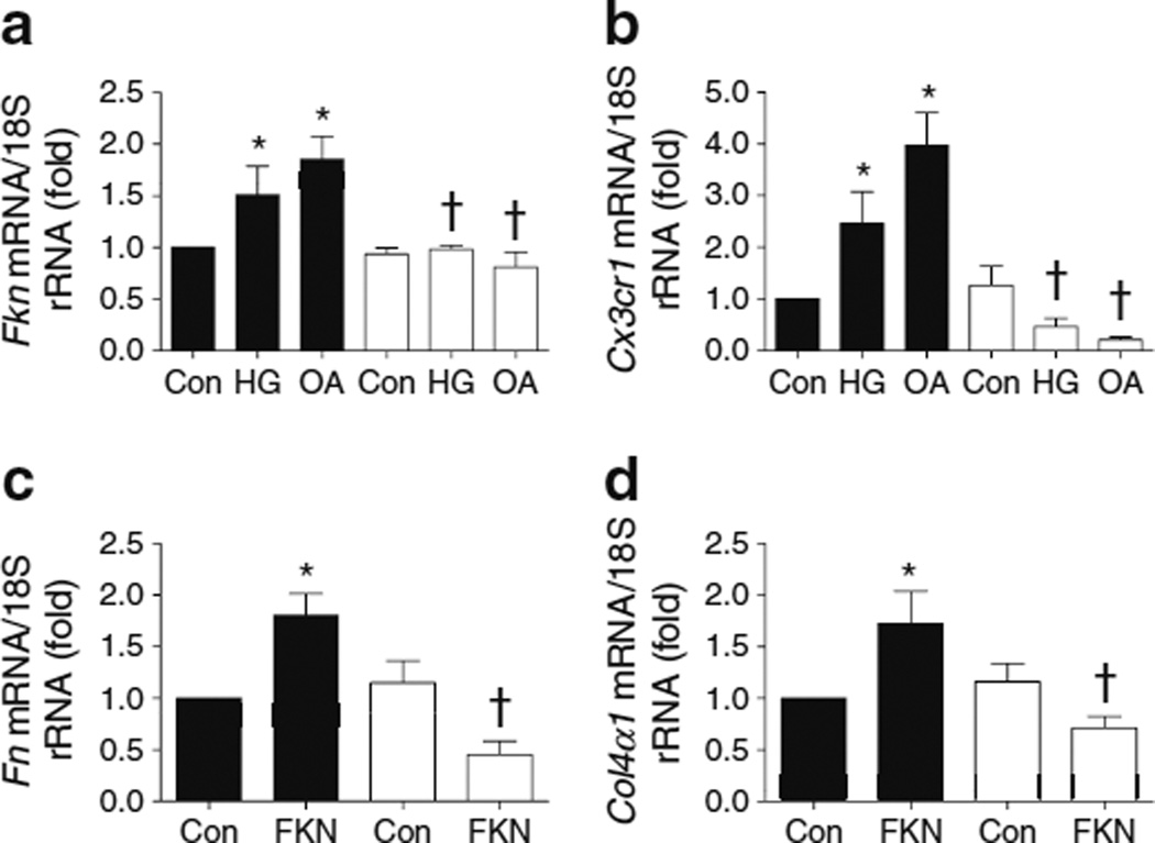
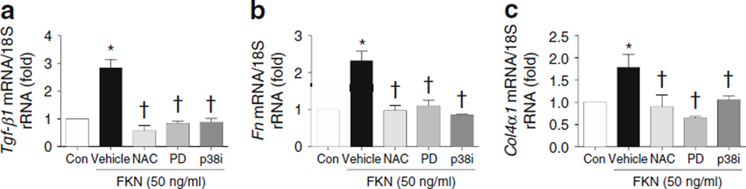
Results: At 12 weeks, diabetic Cx3cr1 KO mice showed no significant changes in plasma glucose, but markers of renal inflammation, fibrosis and ECM, such as the fractional mesangial area, fibronectin and collagen, were significantly lower in diabetic Cx3cr1 KO mice compared with diabetic WT mice. High glucose, oleic acid and TGF-β1 stimulated FKN and CX3CR1 expression, together with the expression of ECM proteins in MMCs, but the effects were significantly attenuated by Fkn or Cx3cr1 siRNA. More importantly, FKN itself increased mesangial ECM through CX3CR1 and subsequent activation of reactive oxygen species and mitogen-activated protein kinases. A neutralising TGF-β antibody inhibited FKN/CX3CR1 in MMCs treated with diabetic stimuli and decreased FKN-induced ECM accumulation.
Conclusions/interpretation: These results demonstrate that FKN/CX3CR1 may play an important role in diabetic renal injury through upregulation of ECM synthesis and could therefore be a therapeutic target for preventing diabetic nephropathy.
Figures

Similar articles
-
Reduced inflammatory and neuropathic pain and decreased spinal microglial response in fractalkine receptor (CX3CR1) knockout mice.J Neurochem. 2010 Aug;114(4):1143-57. doi: 10.1111/j.1471-4159.2010.06837.x. Epub 2010 May 28. J Neurochem. 2010. PMID: 20524966
-
Fractalkine/CX3CR1 axis modulated the development of pancreatic ductal adenocarcinoma via JAK/STAT signaling pathway.Biochem Biophys Res Commun. 2017 Dec 2;493(4):1510-1517. doi: 10.1016/j.bbrc.2017.10.006. Epub 2017 Oct 3. Biochem Biophys Res Commun. 2017. PMID: 28986258
-
Activation of fractalkine/CX3CR1 by vascular endothelial cells induces angiogenesis through VEGF-A/KDR and reverses hindlimb ischaemia.Cardiovasc Res. 2008 May 1;78(2):333-40. doi: 10.1093/cvr/cvm067. Epub 2007 Nov 11. Cardiovasc Res. 2008. PMID: 18006432
-
Fractalkine/CX3CR1 signalling in chronic pain and inflammation.Curr Pharm Biotechnol. 2011 Oct;12(10):1707-14. doi: 10.2174/138920111798357465. Curr Pharm Biotechnol. 2011. PMID: 21466443 Review.
-
CX3CL1 (Fractalkine)-CX3CR1 Axis in Inflammation-Induced Angiogenesis and Tumorigenesis.Int J Mol Sci. 2024 Apr 25;25(9):4679. doi: 10.3390/ijms25094679. Int J Mol Sci. 2024. PMID: 38731899 Free PMC article. Review.
Cited by
-
Regulation and function of CX3CR1 and its ligand CX3CL1 in kidney disease.Cell Tissue Res. 2021 Aug;385(2):335-344. doi: 10.1007/s00441-021-03473-0. Epub 2021 May 19. Cell Tissue Res. 2021. PMID: 34009468 Free PMC article. Review.
-
Tenophages: a novel macrophage-like tendon cell population expressing CX3CL1 and CX3CR1.Dis Model Mech. 2019 Dec 16;12(12):dmm041384. doi: 10.1242/dmm.041384. Dis Model Mech. 2019. PMID: 31744815 Free PMC article.
-
Associations of fractalkine receptor (CX3CR1) and CCR5 gene variants with hypertension, diabetes and atherosclerosis in chronic renal failure patients undergoing hemodialysis.Int Urol Nephrol. 2016 Jul;48(7):1163-70. doi: 10.1007/s11255-016-1293-0. Epub 2016 Apr 27. Int Urol Nephrol. 2016. PMID: 27118566
-
Targeted inhibition of CX3CL1 limits podocytes ferroptosis to ameliorate cisplatin-induced acute kidney injury.Mol Med. 2023 Oct 24;29(1):140. doi: 10.1186/s10020-023-00733-3. Mol Med. 2023. PMID: 37875838 Free PMC article.
-
Identification of a novel immune landscape signature as effective diagnostic markers related to immune cell infiltration in diabetic nephropathy.Front Immunol. 2023 Mar 8;14:1113212. doi: 10.3389/fimmu.2023.1113212. eCollection 2023. Front Immunol. 2023. PMID: 36969169 Free PMC article.
References
-
- Tervaert TW, Mooyaart AL, Amann K, et al. Pathologic classification of diabetic nephropathy. J Am Soc Nephrol. 2010;21:556–563. - PubMed
-
- Whiting DR, Guariguata L, Weil C, Shaw J. IDF diabetes atlas: global estimates of the prevalence of diabetes for 2011 and 2030. Diabetes Res Clin Pract. 2011;94:311–321. - PubMed
-
- Belobradkova J, Filipensky B, Roztocil A, et al. The effect of self-monitoring on perinatal outcome in insulin therapy of diabetic women during pregnancy. Vnitr Lek. 1992;38:1077–1081. - PubMed
-
- Gilbert RE, Cooper ME. The tubulointerstitium in progressive diabetic kidney disease: more than an aftermath of glomerular injury? Kidney Int. 1999;56:1627–1637. - PubMed
-
- Sassy-Prigent C, Heudes D, Mandet C, et al. Early glomerular macrophage recruitment in streptozotocin-induced diabetic rats. Diabetes. 2000;49:466–475. - PubMed
Publication types
MeSH terms
Substances
Grants and funding
LinkOut - more resources
Full Text Sources
Other Literature Sources
Medical
Molecular Biology Databases
Research Materials
Miscellaneous
