. 2013 May 15;32(10):1451-68.
doi: 10.1038/emboj.2013.87. Epub 2013 Apr 19.

The nascent polypeptide-associated complex is a key regulator of proteostasis

Affiliations

The nascent polypeptide-associated complex is a key regulator of proteostasis

Janine Kirstein-Miles et al. EMBO J. .

Abstract

The adaptation of protein synthesis to environmental and physiological challenges is essential for cell viability. Here, we show that translation is tightly linked to the protein-folding environment of the cell through the functional properties of the ribosome bound chaperone NAC (nascent polypeptide-associated complex). Under non-stress conditions, NAC associates with ribosomes to promote translation and protein folding. When proteostasis is imbalanced, NAC relocalizes from a ribosome-associated state to protein aggregates in its role as a chaperone. This results in a functional depletion of NAC from the ribosome that diminishes translational capacity and the flux of nascent proteins. Depletion of NAC from polysomes and re-localisation to protein aggregates is observed during ageing, in response to heat shock and upon expression of the highly aggregation-prone polyglutamine-expansion proteins and Aβ-peptide. These results demonstrate that NAC has a central role as a proteostasis sensor to provide the cell with a regulatory feedback mechanism in which translational activity is also controlled by the folding state of the cellular proteome and the cellular response to stress.


Conflict of interest statement

The authors declare that they have no conflict of interest.

Figures

Figure 1
Figure 1
NAC prevents polyQ aggregation and is an important component of the cellular proteostasis network. (A) Depletion of NAC leads to a higher aggregation propensity of polyQ proteins. The aggregation propensity of the threshold polyQ model Q35-YFP expressed in muscle cells was analysed on day 4 upon knockdown of αNAC, βNAC and α+βNAC. The pictures show representative images of the head region. PolyQ aggregates are indicated by red triangles. The scale bars are 25 μm. (B) Quantification of the number of Q35 aggregates in the whole nematode. Error bars represent mean±s.d. of 50 animals.
Figure 2
Figure 2
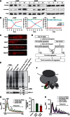
NAC shifts from a soluble ribosome-associated state to the insoluble fraction upon ageing. (A) The protein content of 100 mg (wet weight) synchronized days 3, 5, 7 and 10 old C. elegans cultures were fractionated according to their solubility (see Materials and Methods). The total, (not fractionated), soluble and insoluble fractions of all samples were subjected to SDS–PAGE and subsequent western blot analysis using antibodies against NAC and GFP/YFP. (B) The quantification of the relative signals for αNAC (left), βNAC (middle) and YFP (right) in each of the total (green), soluble (blue) and insoluble (red) fractions reveals that the total amount of NAC stays constant during ageing, whereas the amount in the soluble fraction decreases and concomitantly increases in the insoluble fraction. YFP serves as a control for a protein that remains soluble throughout the lifespan of the animal. (C) NAC localises to foci during ageing. Animals of days 3, 7, 10 and 16 were analysed using immunohistochemistry for NAC localisation. NAC localises to foci with the progression of ageing. The nucleus is highlighted with blue triangles and foci, probably representing protein aggregates, with white triangles. The scale bars are 10 μm. (D) Experimental set-up for the in vitro titration assay. (E) NAC is actively targeted to protein aggregates. The insoluble fraction from day 12-old animals was isolated, washed with detergents and subsequently incubated with the soluble fraction of day 3-old animals. Western blotting reveals that NAC is found in pre-wash insoluble fractions and that it can be removed from aggregates by washing with detergents and rebinds to post-wash aggregates after incubation with lysate. Aggregated proteins were then re-isolated from all three samples and analysed by SDS–PAGE with subsequent Coomassie staining and in parallel by western blot using NAC and RPL-17 antibodies. The signal intensities of sample 1 for both NAC and RPL-17 were each quantified and used to normalise the intensities for samples 2. (F) Cartoon of the ribosome associated chaperone complexes NAC and RAC. The α-subunit of NAC is depicted in green and the β-subunit in red. NAC binds to the ribosome in close proximity to the ribosomal exit site via its β-subunit, whereas both subunits contact the nascent polypeptide chain. We could identify DNJ-11as the Hsp40 component of the RAC complex (shown in grey) by sequence alignments, the C. elegans Hsp70 partner is not known. (G) NAC is required for translation-competent ribosomes. Depicted are the polysome profiles starting with the 80 S peak (see Figure 3A for a complete representative polysome profile of C. elegans) of age-synchronized day 3-old C. elegans cultures upon RNAi of αNAC (green), βNAC (red) and the control (black). The quantification of the relative polysome fractions with respect to the total RNA of the αNAC (green) and βNAC (red) knockdowns compared to the control (black) is depicted on the right. Error bars represent mean±s.d. of three independent experiments. (H) The knockdown of non-ribosomal chaperones does not lead to a decrease of polysome profiles. RNAi mediated knockdown of p97 (cdc-48.1; blue), Hsp110 (C30C11.4; grey) and sHsp (hsp-12.6; purple) does not affect the polysome profile compared to control animals (black) on day 3.
Figure 3
Figure 3
Protein synthesis declines during ageing. (A) Polysome profile of a non-synchronized C. elegans population. The relative abundance of 40 S, 60 S, 80 S ribosome species and the polysome fraction, respectively, is reflected in their absorption at 254 nm (y-axis). The x-axis depicts the sedimentation along a sucrose gradient from 15–45% (w/v). The depicted polysome profile is a representative of three independent analyses. (B) Quantification of the relative amount of 40 S, 60 S, 80 S and polysomes of a non-synchronized population (as shown in (a)) in % with respect to the total RNA. Three independent analyses were used to calculate and draw error bars representing mean±s.d. (C) Polysome profiles of synchronized C. elegans populations of day 2 (red), day 4 (black) and day 10 (blue). The three profiles were aligned on the x-axis according to the sedimentation of their 80 S peak. The dashed line indicates the profile of the polysomes. (D) Quantification of the relative proportion of polysomes (in %) with respect to the total RNA level of non-synchronized and age-synchronized C. elegans cultures from day 2 to day 10. The polysome levels for days 2, 4 and 10 are highlighted in the same colour code as in (C). At least three independent analyses were used for each time point to calculate and draw error bars representing mean±s.d.
Figure 4
Figure 4
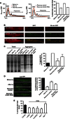
NAC is important for the recovery from heat shock. (A) Heat shock of day 2 old C. elegans at 35°C for 1 h results in a reduction of polysomes (red). The condition before the heat shock (grey) and animals recovered for 24 h (black) serve as controls. The recovery from heat shock (1 h 35°C) is greatly diminished upon knockdown of βNAC (red) during the recovery period of 24 h compared to animals fed with RNAi bacteria expressing the empty vector (black; middle panel). A quantification of the polysome content with respect to the total RNA level is depicted on the right. (B) Heat shock causes reversible foci formation of NAC. Shown here is the localisation of NAC in a muscle cell of C. elegans grown at 20°C (top; left panel) and of C. elegans heat shocked for 30 min at 35°C (middle; left panel) and after 24 h recovery at 20°C (bottom; left panel). The NAC foci formed upon heat shock co-localise with aggregated luciferase-YFP. The images of the middle panel show the separate and overlay images of NAC (red) and Luc-YFP (green) at 20°C. The heat shock conditions are depicted on the right, respectively. The scale bars are 10 μm. (C) The gel shows the total (left lanes) and aggregated protein fractions (right lanes) of animals exposed to heat shock at 35°C for 1 h directly after heat shock and followed by a recovery at 20°C for 24 h upon knockdown of βNAC and of control animals, respectively. The quantification of the aggregated protein fraction of animals before heat shock (black), immediately after heat shock and animals that were fed bacteria-expressing βNAC dsRNA or the empty vector during the recovery period at 20°C for 24 h. The quantification of aggregation is normalised to the aggregation propensity after heat shock. Three independent experiments were used to calculate and draw error bars representing mean±s.d. (D) Aggregation propensity of luciferase-YFP expressing animals before heat shock (top), immediately after heat shock (second from top) and after a recovery at 20°C for 24 h in control animals (third from top) or upon knockdown of βNAC during the recovery period (bottom). The scale bars are 10 μm. An analysis of enzymatic activity of luciferase of the luciferase-YFP expressing C. elegans before heat shock, immediately after heat shock and after a recovery period upon knockdown of βNAC or in animals fed the empty vector during the recovery period are shown on the right. The luciferase activity is normalised to the enzymatic activity before heat shock (black column). Three independent experiments were used to calculate and draw error bars representing mean±s.d. (E) Knockdown of NAC reduces survival after heat shock. Animals grown on RNAi plates since L1 were subjected on day 4 to 6 h of heat-shock at 35°C followed by a recovery period at 20°C for 24 h. Survivors were scored after the 24 h recovery phase in three independent experiments with a total number of 120 animals for each condition. Error bars represent mean±s.d. of the three independent experiments.
Figure 5
Figure 5
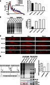
NAC acts as sensor for proteotoxic stress. (A) Analysis of polysome profiles of day 3-old nematodes before heat shock (black) after 10 (blue), 30 (magenta) and 60-min heat shock (red). The quantification of polysome profiles with respect to the total RNA is shown on the right. (B) The gel depicts the total protein levels (left lanes) and the aggregated proteins (right lanes) during the course of heat shock (hs). The time points are indicated on top. The quantification of the aggregated proteins of is shown on the right. The level of aggregation is normalised to the aggregation propensity at the 60-min time point of the heat shock. (C) Immunofluorescence of single muscles cells during the course of heat shock using antibodies against NAC (top row), RPL-4 (middle row) and RPL-17 (bottom row). The time points of heat shock (before, 10 min, 30 min and 60 min) are indicated on top. The foci probably representing insoluble protein are highlighted with white triangles. The scale bars are 10 μm. (D) Schematic presentation of the titration assay. Aggregates from heat-shocked animals were mixed with isolated ribosomes and incubated for 20 min. A parallel of the aggregates and isolated ribosomes were incubated with buffer and would serve as controls. All three samples were subjected first to a low spin to re-isolate protein aggregates and subsequently to a high spin to re-isolate ribosomes. The analysis of all fractions from the titration experiment by SDS–PAGE and subsequent Coomassie staining (top) and in parallel by western blot (bottom) using NAC and RPL-25 antibodies is shown in the middle panel. The αNAC signals are depicted in two different exposure times of the western blot (bottom 2 rows) for better visualisation of the signals. The low spin isolating the aggregated proteins is shown in the left lanes and the high spin isolating the ribosomes is shown in the right lanes. The individual fractions of the titration assay are depicted on top of the gel. A quantification of ribosome-associated NAC after the incubation with protein aggregates relative to the isolated ribosome sample is shown on the right. The quantification of αNAC is normalised to the protein levels of RPL-25 in both samples.
Figure 6
Figure 6
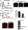
Polysome levels decline upon chronic stress. (A) Chronic stress: expression of 40 consecutive glutamines in the body wall muscle of C. elegans causes a reduction of polysomes. Depicted are the polysome profiles of day 4-old nematodes, which either express Q40-YFP (red), Q35-YFP (blue) or YFP alone (Q0-YFP, black) as control. (B) The images show the aggregation propensity of Q0-YFP (left), Q35-YFP (middle) and Q40-YFP (right) of the head region on day 4. Q40-YFP forms aggregates at day 4, whereas Q0-YFP and Q35-YFP remain soluble at that age (Morley et al, 2002) (see also Figure 1A and Supplementary Figure S1H). The scale bars are 25 μm. (C) Chronic stress: expression of Aβ causes a severe reduction of polysomes and 80 S levels. Depicted are the polysome profiles of Aβ-expressing nematodes (red) and the N2 wild type (control; black) on day 3. The quantification of the polysome content with respect to the total RNA level of is shown on the right. (D) Nematodes expressing the Aβ-peptide 1–42 in the body wall muscle cells were analysed by immunostaining with NAC antibodies. NAC forms foci in cells expressing Aβ most likely representing amyloid deposits (right). The control using wild type C. elegans (N2) is shown on the left. The scale bars are 10 μm. (E) Interaction of NAC and Aβ-peptide. Immobilised NAC antibodies were first incubated with recombinant NAC protein and the complex was subsequently incubated with extracts of an Aβ-expressing strain (CL2006) grown on α+βNAC RNAi. The precipitate was subjected to SDS–PAGE and western blot. The interaction was analysed using the Aβ-specific antibody 4G8. The antibody detected a strong signal for the Aβ-peptide and an additional signal of about 15 kDa probably representing oligomeric species of Aβ (right lane). A parallel, where no NAC protein was bound to the immobilised NAC antibodies, served as control and did not show an interaction with Aβ (left lane).
Figure 7
Figure 7
Reduction of translation initiation enhances proteostasis. (A) The knockdown of translation initiation factors eIF4G (ifg-1) and eIF4E (ife-2) reduces aggregation of Q35-YFP. Images show the head region of day 5-old Q35-YFP nematodes. The control (empty vector) is shown on the left. The scale bars are 10 μm. (B) The graph shows a quantification of the number of Q35 aggregates in the whole nematode on day 5 for the control and the knockdown of eIF4G and eIF4E. Error bars represent mean±standard deviation (s.d.) of 50 animals. (C) Western blot of the Q35-YFP protein levels in the control and upon knockdown of eIF4G and eIF4E and control.
Figure 8
Figure 8
Model for reversible and age/chronic stress-dependent translational control by NAC. Acute proteotoxic challenges (left branch) lead to a temporary translational attenuation due to a sequestration of NAC by misfolded and aggregated proteins. Re-balancing of proteostasis liberates NAC and allows for re-association with ribosomes and translation resumes. Ageing and chronic stress conditions (right branch) lead to a permanent sequestration of NAC and hence to a decrease in protein synthesis.

Comment in

Similar articles

Cited by

References

    1. Albanese V, Reissmann S, Frydman J (2010) A ribosome-anchored chaperone network that facilitates eukaryotic ribosome biogenesis. J Cell Biol 189: 69–81 - PMC - PubMed
    1. Arsenovic PT, Maldonado AT, Colleluori VD, Bloss TA (2012) Depletion of the C. elegans NAC engages the unfolded protein response, resulting in increased chaperone expression and apoptosis. PLoS One 7: e44038. - PMC - PubMed
    1. Beatrix B, Sakai H, Wiedmann M (2000) The alpha and beta subunit of the nascent polypeptide-associated complex have distinct functions. J Biol Chem 275: 37838–37845 - PubMed
    1. Ben-Zvi A, Miller EA, Morimoto RI (2009) Collapse of proteostasis represents an early molecular event in Caenorhabditis elegans ageing. Proc Natl Acad Sci U S A 106: 14914–14919 - PMC - PubMed
    1. Bloss TA, Witze ES, Rothman JH (2003) Suppression of CED-3-independent apoptosis by mitochondrial betaNAC in Caenorhabditis elegans. Nature 424: 1066–1071 - PubMed

Publication types

MeSH terms