. 2013 Sep;70(17):3231-42.
doi: 10.1007/s00018-013-1334-0. Epub 2013 Apr 19.

Two types of muscarinic acetylcholine receptors in Drosophila and other arthropods

Affiliations

Two types of muscarinic acetylcholine receptors in Drosophila and other arthropods

Caitlin Collin et al. Cell Mol Life Sci. 2013 Sep.

Erratum in

  • Cell Mol Life Sci. 2013 Nov;70(21):4197. de Valdivia, Ernesto Gonzalez [corrected to Gonzalez de Valdivia, Ernesto]

Abstract

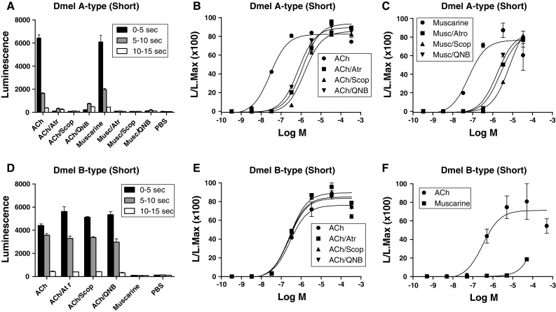
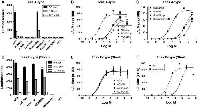
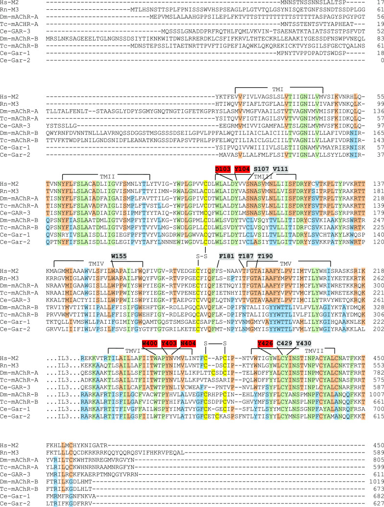
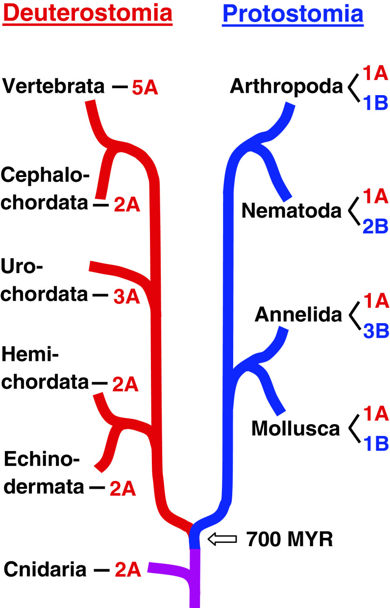
Muscarinic acetylcholine receptors (mAChRs) play a central role in the mammalian nervous system. These receptors are G protein-coupled receptors (GPCRs), which are activated by the agonists acetylcholine and muscarine, and blocked by a variety of antagonists. Mammals have five mAChRs (m1-m5). In this study, we cloned two structurally related GPCRs from the fruit fly Drosophila melanogaster, which, after expression in Chinese hamster ovary cells, proved to be muscarinic acetylcholine receptors. One mAChR (the A-type; encoded by gene CG4356) is activated by acetylcholine (EC50, 5 × 10(-8) M) and muscarine (EC50, 6 × 10(-8) M) and blocked by the classical mAChR antagonists atropine, scopolamine, and 3-quinuclidinyl-benzilate (QNB), while the other (the B-type; encoded by gene CG7918) is also activated by acetylcholine, but has a 1,000-fold lower sensitivity to muscarine, and is not blocked by the antagonists. A- and B-type mAChRs were also cloned and functionally characterized from the red flour beetle Tribolium castaneum. Recently, Haga et al. (Nature 2012, 482: 547-551) published the crystal structure of the human m2 mAChR, revealing 14 amino acid residues forming the binding pocket for QNB. These residues are identical between the human m2 and the D. melanogaster and T. castaneum A-type mAChRs, while many of them are different between the human m2 and the B-type receptors. Using bioinformatics, one orthologue of the A-type and one of the B-type mAChRs could also be found in all other arthropods with a sequenced genome. Protostomes, such as arthropods, and deuterostomes, such as mammals and other vertebrates, belong to two evolutionarily distinct lineages of animal evolution that split about 700 million years ago. We found that animals that originated before this split, such as cnidarians (Hydra), had two A-type mAChRs. From these data we propose a model for the evolution of mAChRs.


Figures

Fig. 1
Fig. 1
Bioluminescence responses of cloned CHO/G-16 cell lines transfected with DNA coding for the D. melanogaster A- and B-type mAChRs. The vertical bars represent SEM (n = 2 or 3), which sometimes are smaller than the symbols used. In these cases, only the symbols are given. Log M written at the abscissae means log concentration given in M. L/L Max written at the ordinates means luminescence divided by maximal luminescence. A Bioluminescence response of CHO/G-16/mAChR A-type (short version) cells after addition of ACh or muscarine alone (final concentration, 3 × 10−7 M), or in the presence of the antagonists atropine (Atr), scopolamine (Scop), or QNB (final concentration, 3 × 10−7 M), or phosphate-buffered saline (PBS). B Dose–response curves of the effects of ACh with or without antagonists. The transfected cells were activated by ACh with an EC50 of 3 × 10−8 M, while in the presence of 3 × 10−7 M of atropine, scopolamine, or QNB, 100-fold more ACh was needed for activation. C Dose–response curves of the effects of muscarine on these cells, with and without antagonists. The transfected cells were activated by muscarine with an EC50 of 6 × 10−8 M, while the antagonists at 3 × 10−7 M, again, shifted the activation of the receptor by 100-fold. D Bioluminescence response of CHO/G-16/muscarinic acetylcholine receptor B-type (short version) cells after addition of ACh or muscarine alone (final concentration, 3 × 10−7 M), or in the presence of antagonists atropine (Atr), scopolamine (Scop), QNB (final concentration 3 × 10−7 M), or phosphate-buffered saline (PBS). In contrast to the A-type receptor, muscarine does not activate, while the antagonists do not block the B-type receptor. E Dose–response curves of the effects of ACh on these cells, with or without antagonists. The transfected cells were activated by ACh with an EC50 of 3 × 10−7 M, while the antagonists (at 3 × 10−7 M) did not block activation of the receptor. F Dose–response curves of the effects of muscarine or ACh on these cells. The transfected cells were only activated by muscarine at about 1,000-fold higher concentrations than ACh
Fig. 2
Fig. 2
Bioluminescence responses of cloned CHO/G-16 cell lines transfected with DNA coding for the T. castaneum A- and B-type mAChRs. The vertical bars represent SEM (n = 2 or 3), which sometimes are smaller than the symbols used. In these cases, only the symbols are given. A Bioluminescence response of CHO/G-16/mAChR A-type cells after addition of ACh or muscarine alone (final concentration, 3 × 10−7 M), or in the presence of the antagonists atropine (Atr), scopolamine (Scop) or QNB (final concentration, 3 × 10−7 M), or phosphate-buffered saline (PBS). B Dose–response curves of the effects of ACh with or without antagonists. The transfected cells were activated by ACh with an EC50 of 3 × 10−8 M, while the antagonists at 3 × 10−7 M shifted the activation of the receptor by 100-fold. C Dose–response curves of the effects of muscarine on these cells, with and without antagonists. The transfected cells were activated by muscarine with an EC50 of 9 × 10−8 M, while the antagonists at 3 × 10−7 M again shifted the activation of the receptor by 100-fold. D Bioluminescence response of CHO/G-16/mAChR B-type (short version) cells after addition of ACh or muscarine alone (final concentration, 3 × 10−7 M), or in the presence of antagonists atropine (Atr), scopolamine (Scop), QNB (final concentration 3 × 10−7 M), or phosphate-buffered saline (PBS). In contrast to the A-type receptor, muscarine does not activate, and the antagonists do not block the receptor. E Dose–response curves of the effects of ACh on these cells, with or without antagonists. The transfected cells were activated by ACh with an EC50 of 3 × 10−8 M, while the antagonists (at 3 × 10−7 M) did not block activation of the receptor. F Dose–response curves of the effects of muscarine or ACh on these cells. The transfected cells were only activated by muscarine at concentrations 100-fold higher than that of ACh
Fig. 3
Fig. 3
qPCR data for the expression of the D. melanogaster A-type mAChR gene CG4356 (A), and B-type mAChR gene CG7918 (B), in different body parts of adult male and female flies. The combined expression (long and short splicing variants) of the A-type and the combined expression (long and short splicing variants) of the B-type receptor were measured. At least 30 body parts were pooled for each mRNA isolation. These pools are the same for each body part column in A and B. The qPCR experiments were run in triplicate. The vertical bars in each column (which are sometimes smaller than the lines of the column) represent SEM. The mRNA concentrations given are relative to column a (a = 1). a female whole body; b female head; c female thorax; d female abdomen; e male whole body; f male head; g male thorax; h male abdomen. Note that both receptor genes are strongly expressed in the head (presumably the brain) of both males and females
Fig. 4
Fig. 4
qPCR data for the expression of the D. melanogaster A-type mAChR gene CG4356 (A) and B-type mAChR gene CG7918 (B) in different developmental stages. The combined expression (long and short splicing variants) of the A-type and the combined expression (long and short splicing variants) of the B-type receptor were measured. At least ten animals were pooled for each developmental stage. These pools are the same for each developmental stage in A and B. The sexes are mixed in columns ae (they could not be discriminated), otherwise they are separated. The mRNA concentrations given are relative to column m (m = 1). Other conditions are as in Fig. 3. a 20–24 h old eggs; b larvae 2 days after egg laying(= first instar); c larvae 3 days after egg laying (= second instar); d larvae 4 days after egg laying (= second instar); e larvae 5 days after egg laying (= third instar); f male third-instar larvae; g female third-instar larvae; h 2-day-old male pupae; i 2-day-old female pupae; j 3-day-old male pupae; k 3-day-old female pupae; l adult male flies; m adult female flies. Note that both receptor genes are strongly expressed in older pupae of both sexes and in adult males
Fig. 5
Fig. 5
Sequence alignments of the human m2 mAChR (Hs-M2), rat m3 (Rn-M3), D. melanogaster A-type (Dm-mAChR-A), T. castaneum A-type (Tc-mAChR-A), C. elegans A-type (Ce-GAR-3), D. melanogaster B-type (Dm-mAChR-B), T. castaneum B-type (Tc-mAChR-B), and the two C. elegans B-type mAChRs (Ce-GAR-1 and -2). The 14 QNB binding residues of the human m2 receptor [19] are indicated by numbers above the m2 sequence and highlighted in both red and grey. The red numbers are the proposed (“modeled”) binding residues for acetylcholine [19]. The transmembrane helices are indicated by TM1-TMVII. Cystine bridges are highlighted in yellow and indicated by “S–S”. The amino acids residues that are identical in all five A-type mAChRs are highlighted in orange; those that are identical in all B-type mAChRs are highlighted in blue. Green indicates residues that are identical in both A- and B-type mAChRs
Fig. 6
Fig. 6
An overview of the emergence of A- and B-type mAChRs in Protostomia (highlighted by blue lines) and Deuterostomia (highlighted by red lines). Cnidarians evolved before the split of Proto- and Deuterostomia, which is indicated by a purple line. The B-type mAChRs appear to be confined to the Protostomia, whereas the A-type receptors occur in both evolutionary lineages and in cnidarians. Our criteria for assigning the receptors as A- or B-type were (1) comparisons of their overall protein sequences, using phylogenetic tree analyses (Fig. S13) and (2) the presence (A-type) or absence (B-type) of residues identical to the m2 QNB-specific binding residues S107, V111, W155, T187, and T190 (See Fig. 5 and “Discussion”). Both criteria should be fulfilled

Similar articles

Cited by

References

    1. Douzéry EJ, Snell EA, Bapteste E, Delsuc F, Philippe H. The timing of eukaryotic evolution: does a relaxed molecular clock reconcile proteins and fossils? Proc Natl Acad Sci USA. 2004;101:15386–15391. doi: 10.1073/pnas.0403984101. - DOI - PMC - PubMed
    1. Yakel JL. Gating of nicotinic Ach receptors: latest insights into ligand binding and function. J Physiol. 2010;588:597–602. doi: 10.1113/jphysiol.2009.182691. - DOI - PMC - PubMed
    1. Millar NS, Harkness PC. Assembly and trafficking of nicotinic acetylcholine receptors (Review) Mol Membr Biol. 2008;25:279–292. doi: 10.1080/09687680802035675. - DOI - PubMed
    1. Jones AK, Sattelle DB. Diversity of insect nicotinic acetylcholine receptor subunits. Adv Exp Med Biol. 2010;683:25–43. doi: 10.1007/978-1-4419-6445-8_3. - DOI - PubMed
    1. Dupuis J, Louis T, Gauthier M, Raymond V. Insights from honeybee (Apis mellifera) and fly (Drosophila melanogaster) nicotinic acetylcholine receptors: from genes to behavioral functions. Neurosci Biobehav Rev. 2012;36:1553–1564. doi: 10.1016/j.neubiorev.2012.04.003. - DOI - PubMed

Publication types