Clinical significance and gene expression study of human hepatic stellate cells in HBV related-hepatocellular carcinoma
- PMID: 23601182
- PMCID: PMC3654985
- DOI: 10.1186/1756-9966-32-22
Clinical significance and gene expression study of human hepatic stellate cells in HBV related-hepatocellular carcinoma
Abstract
Background: Peritumoral activated hepatic stellate cells (HSCs) are versatile myofibroblast-like cells closely related with hepatocellular carcinoma (HCC) progression. So far, comprehensive comparison of gene expression of human HSCs during hepatocarcinogenesis is scanty. Therefore, we identified the phenotypic and genomic characteristics of peritumoral HSCs to explore the valuable information on the prognosis and therapeutic targets of HBV related HCC.
Methods: A tissue microarray containing 224 HBV related HCC patients was used to evaluate the expression of phenotype markers of HSCs including α-SMA, glial fibrillary acidic protein (GFAP), desmin, vinculin and vimentin. HSCs and cancer associated myofibroblasts (CAMFs) were isolated from normal, peritumoral human livers and cancer tissues, respectively. Flow cytometry and gene microarray analysis were performed to evaluate the phenotypic changes and gene expression in HCC, respectively.
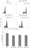
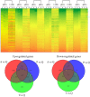
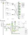
Results: Peritumoral α-SMA positive HSCs showed the prognostic value in time to recurrence (TTR) and overall survival (OS) of HCC patients, especially in early recurrence and AFP-normal HCC patients. Expression of GFAP positive HSCs cell lines LX-2 was significantly decreased after stimulation with tumor conditioned medium. Compared with quiescent HSCs, peritumoral HSCs and intratumoral CAMFs expressed considerable up- and down-regulated genes associated with biological process, cellular component, molecular function and signaling pathways involved in fibrogenesis, inflammation and progress of cancer.
Conclusions: Peritumoral activated HSCs displayed prognostic value in HBV related-HCC, and their genomic characteristics could present rational biomarkers for HCC risk and promising therapeutic targets.
Figures


Similar articles
-
Expression of TREM-1 in hepatic stellate cells and prognostic value in hepatitis B-related hepatocellular carcinoma.Cancer Sci. 2012 Jun;103(6):984-92. doi: 10.1111/j.1349-7006.2012.02273.x. Epub 2012 Apr 19. Cancer Sci. 2012. PMID: 22417086 Free PMC article.
-
miR-106b promotes cancer progression in hepatitis B virus-associated hepatocellular carcinoma.World J Gastroenterol. 2016 Jun 14;22(22):5183-92. doi: 10.3748/wjg.v22.i22.5183. World J Gastroenterol. 2016. PMID: 27298561 Free PMC article.
-
Cluster of differentiation 147 is a key molecule during hepatocellular carcinoma cell-hepatic stellate cell cross-talk in the rat liver.Mol Med Rep. 2015 Jul;12(1):111-8. doi: 10.3892/mmr.2015.3429. Epub 2015 Mar 4. Mol Med Rep. 2015. PMID: 25738354 Free PMC article.
-
Factors predicting occurrence and prognosis of hepatitis-B-virus-related hepatocellular carcinoma.World J Gastroenterol. 2011 Oct 14;17(38):4258-70. doi: 10.3748/wjg.v17.i38.4258. World J Gastroenterol. 2011. PMID: 22090781 Free PMC article. Review.
-
Hepatitis B virus-induced hepatocellular carcinoma.Cancer Lett. 2014 Apr 10;345(2):216-22. doi: 10.1016/j.canlet.2013.08.035. Epub 2013 Aug 25. Cancer Lett. 2014. PMID: 23981576 Review.
Cited by
-
Matrix disequilibrium in Alzheimer's disease and conditions that increase Alzheimer's disease risk.Front Neurosci. 2023 May 26;17:1188065. doi: 10.3389/fnins.2023.1188065. eCollection 2023. Front Neurosci. 2023. PMID: 37304012 Free PMC article. Review.
-
Hepatic stellate cell and monocyte interaction contributes to poor prognosis in hepatocellular carcinoma.Hepatology. 2015 Aug;62(2):481-95. doi: 10.1002/hep.27822. Epub 2015 Apr 28. Hepatology. 2015. PMID: 25833323 Free PMC article.
-
Comparison and validation of the prognostic value of preoperative systemic immune cells in hepatocellular carcinoma after curative hepatectomy.Cancer Med. 2018 Apr;7(4):1170-1182. doi: 10.1002/cam4.1424. Epub 2018 Mar 13. Cancer Med. 2018. PMID: 29533004 Free PMC article.
-
Preoperative neutrophil-to-lymphocyte ratio predicts recurrence of patients with single-nodule small hepatocellular carcinoma following curative resection: a retrospective report.World J Surg Oncol. 2015 Sep 2;13:265. doi: 10.1186/s12957-015-0670-y. World J Surg Oncol. 2015. PMID: 26328917 Free PMC article. Clinical Trial.
-
HBV-DNA Load-Related Peritumoral Inflammation and ALBI Scores Predict HBV Associated Hepatocellular Carcinoma Prognosis after Curative Resection.J Oncol. 2018 Sep 20;2018:9289421. doi: 10.1155/2018/9289421. eCollection 2018. J Oncol. 2018. PMID: 30327670 Free PMC article.
References
Publication types
MeSH terms
Substances
LinkOut - more resources
Full Text Sources
Other Literature Sources
Medical
Molecular Biology Databases
Research Materials
Miscellaneous

