. 2013 Apr;9(4):e1003450.
doi: 10.1371/journal.pgen.1003450. Epub 2013 Apr 4.

Drosophila cyfip regulates synaptic development and endocytosis by suppressing filamentous actin assembly

Affiliations

Drosophila cyfip regulates synaptic development and endocytosis by suppressing filamentous actin assembly

Lu Zhao et al. PLoS Genet. 2013 Apr.

Abstract

The formation of synapses and the proper construction of neural circuits depend on signaling pathways that regulate cytoskeletal structure and dynamics. After the mutual recognition of a growing axon and its target, multiple signaling pathways are activated that regulate cytoskeletal dynamics to determine the morphology and strength of the connection. By analyzing Drosophila mutations in the cytoplasmic FMRP interacting protein Cyfip, we demonstrate that this component of the WAVE complex inhibits the assembly of filamentous actin (F-actin) and thereby regulates key aspects of synaptogenesis. Cyfip regulates the distribution of F-actin filaments in presynaptic neuromuscular junction (NMJ) terminals. At cyfip mutant NMJs, F-actin assembly was accelerated, resulting in shorter NMJs, more numerous satellite boutons, and reduced quantal content. Increased synaptic vesicle size and failure to maintain excitatory junctional potential amplitudes under high-frequency stimulation in cyfip mutants indicated an endocytic defect. cyfip mutants exhibited upregulated bone morphogenetic protein (BMP) signaling, a major growth-promoting pathway known to be attenuated by endocytosis at the Drosophila NMJ. We propose that Cyfip regulates synapse development and endocytosis by inhibiting actin assembly.


Conflict of interest statement

The authors have declared that no competing interests exist.

Figures

Figure 1
Figure 1. cyfip regulates synapse development.
(A–E) Representative NMJ4 synapses from different genotypes co-stained with anti-HRP recognizing the neuronal plasma membrane (green) and an antibody against DLG (red), a postsynaptic scaffold protein. Arrows indicate interbouton spacing; asterisks denote superboutons with multiple satellite boutons attached. Scale bar, 10 µm. (F–H) Statistical results of NMJ length (F), the number of satellite boutons (G), and the number of superboutons (H) in different genotypes. Ubiquitous expression of cyfip controlled by act-Gal4 fully rescued the NMJ phenotypes, whereas pre- and postsynaptic expression of cyfip by elav-Gal4 and C57-Gal4, respectively, partially rescued the satellite bouton phenotype of cyfip85.1 null mutants. n = 20 for each genotype; * p<0.05, *** p<0.01, *** p<0.001; error bars indicate SEM.
Figure 2
Figure 2. Enlarged synaptic vesicles at active zones and more cisternae in the synaptic boutons of cyfip mutants.
(A, B) Electron micrographs of synaptic boutons from wild type (A) and cyfip85.1 mutants (B). Asterisks indicate active zones with T bars. Compared to wild type, cyfip85.1 mutants exhibited significantly more cisternae (arrows in B). Scale bar, 500 nm. (C–E) High magnification view of representative active zones in wild type (C), cyfip85.1 mutants (D), and ubiquitous expression of Cyfip driven by act-Gal4 in cyfip85.1 background (E). Arrowheads in (D) indicate enlarged vesicles near the T-bar. Dashed line defines a 200 nm radius around the active zone for quantitative analysis of SVs. (F, G) Quantification of the number (F) and diameter (G) of SVs within a 200 nm radius of the active zone. n = 446 SVs for wild type, n = 350 SVs for mutants, and n = 445 SVs for the rescue. (H, I) Frequency distributions (H) and cumulative probabilities (I) of SV diameters in the defined area around the active zone.
Figure 3
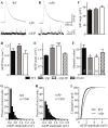
Figure 3. cyfip mutants show normal evoked junctional potential (EJP) amplitudes but larger spontaneous mEJP amplitudes.
(A, B) Representative excitatory junctional potentials (EJPs) and spontaneous miniature EJPs (mEJPs) recorded from wild type and cyfip85.1 mutant NMJs. (C–F) Statistical analysis of mean EJP amplitude (B), mEJP frequency (C), mEJP amplitude (D), and quantal content (E) in different genotypes. The mEJP amplitude is significantly higher whereas the quantal content is lower in cyfip85.1 mutants compared to wild type. The number of animals analyzed is indicated in (F); * p<0.05, *** p<0.001; error bars indicate SEM. (G–I) Frequency distributions (G, H) and cumulative probabilities (I) of mEJP amplitudes recorded from wild type and cyfip85.1 mutants.
Figure 4
Figure 4. cyfip mutants fail to sustain normal neurotransmitter release during high-frequency stimulation.
(A) Statistical analysis of EJP amplitudes under 10 Hz stimulation for 10 min. Comparison of EJPs from wild type (WT), cyfip85.1 (cyfip), cyfip85.1/Df(3R)Exel16174 (cyfip/Df), and cyfip85.1 mutants expressing Cyfip presynaptically driven by elav-Gal4 (rescue) reveals faster rundown of EJP amplitudes in cyfip85.1 and cyfip85.1/Df mutants compared to wild type. (B) Amplitudes of EJPs during tetanic stimulation at 10 Hz for 10 min, and then during low-frequency stimulation at 0.3 Hz for 10 min. cyfip85.1 mutants displayed a significantly slower recovery of EJP amplitudes. n = 10 for each genotype. Error bars indicate SEM. (C) Representative EJP traces recorded from wild type and cyfip85.1 mutants.
Figure 5
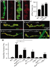
Figure 5. The excess satellite bouton formation in cyfip mutants depends on elevated BMP signaling.
(A, B) Representative synaptic boutons labeled with anti-HRP (red) and anti-pMad (red) in wild type (A) and cyfip85.1 mutants (B). pMad level was increased in cyfip85.1 mutant synapses. Scale bar, 5 µm. (C) Statistical analysis of pMad staining intensity. n≥10 for each genotype. (D–G) Representative NMJ4 synapses double-labeled with anti-HRP (red) and anti-DLG (red) from cyfip85.1 (D), madk00237/+; cyfip85.1 (E), tkv7/+; cyfip85.1 (F), and dadJ1E4/+; cyfip85.1/+ (G). Scale bar, 10 µm. (H) Statistical results for the number of satellite boutons from different genotypes. n≥15 for each genotype. * p<0.05, ** p<0.01, and *** p<0.001. Error bars indicate SEM.
Figure 6
Figure 6. FRAP analysis shows increased F-actin formation at the NMJ terminals of cyfip mutants.
(A, B) Presynaptically expressed GFP-moe driven by elav-Gal4 revealed an uneven distribution of F-actin across different boutons in cyfip85.1 mutants (compare boutons indicated by white and pink arrows in B). Scale bar, 5 µm. (C, D) Decreased total number but increased size of a sub-population at the 75th percentile of GFP-moe patches in cyfip85.1 mutant NMJs. n = 406 for the control; n = 115 for cyfip mutants; n = 211 for cyfip/Df mutants; * p<0.05; ** p<0.01. (E, F) Time-lapse images of GFP-moe expressed in the presynaptic terminals by elav-Gal4 in wild type (E) and cyfip85.1 mutants (F). The rectangular box indicates the region of interest (ROI) for photobleaching. Arrows indicate the position of recovered GFP-moe signals. Scale bar, 2 µm. (G, H) Relative GFP fluorescence intensities within the ROI after photobleaching of GFP-moe (G) and actin-GFP (H) patches. (I) Statistical results of fluorescence recovery rates (s−1) after photobleaching of presynaptically expressed GFP-moe and actin-GFP. n≥17 from 4 animals. Error bars indicate SEM.
Figure 7
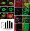
Figure 7. New F-actin assembly is enhanced in cyfip mutants.
(A–F) Synaptic boutons of wild type (A–C) and cyfip85.1 mutants (D–F) under different conditions: vehicle-treated control, treated with jasplakinolide, and after 1 h of jasplakinolide washout. The NMJ boutons were double-labeled with anti-HRP (green) and phalloidin (red) to reveals postsynaptic F-actin. (G) Fluorescence recovery index in the NMJ boutons of different genotypes after jasplakinolide treatment. At least 15 different boutons from 4 animals of each genotype were analyzed. (H–J) Phalloidin labeled (red) cyfip mosaic eye discs of third instar larvae under different conditions: vehicle-treated control (H), treated with jasplakinolide (I), and 90 min recovery after jasplakinolide washout (J). cyfip85.1 mutant clones marked by the absence of GFP and outlined by white dotted lines show enhanced F-actin assembly in a subset of mutant cells (indicated by arrows). (K) F-actin recovery in a wild-type eye disc. Arrowheads indicate MF; horizontal arrow in H points to posterior.
Figure 8
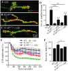
Figure 8. Cyfip functionally antagonizes SCAR in multiple contexts.
(A–C) Representative NMJ terminals of different genotypes. Scale bar, 10 µm. (D) Statistical analysis of the number of satellite boutons in different genotypes. The heterozygous SCAR mutant alleles SCARΔ37 or SCARk13811 rescued the excessive satellite bouton phenotype of cyfip85.1 mutants. (E) Mutating a copy of SCAR rescued the enhanced decline of EJP amplitudes during 10 Hz stimulation in cyfip85.1 mutants. (F) Mutating a copy of SCAR restored the increased F-actin assembly rate in cyfip85.1 mutants to the wild-type level. n≥8 for each genotype; * p<0.05, ** p<0.01, and *** p<0.001; error bars indicate SEM.

Similar articles

Cited by

References

    1. Collins CA, DiAntonio A (2007) Synaptic development: insights from Drosophila. Curr Opin Neurobiol 17: 35–42. - PubMed
    1. Giagtzoglou N, Ly CV, Bellen HJ (2009) Cell adhesion, the backbone of the synapse: “vertebrate” and “invertebrate” perspectives. Cold Spring Harb Perspect Biol 1: a003079. - PMC - PubMed
    1. Packard M, Mathew D, Budnik V (2003) Wnts and TGF beta in synaptogenesis: old friends signalling at new places. Nat Rev Neurosci 4: 113–120. - PMC - PubMed
    1. Ball RW, Warren-Paquin M, Tsurudome K, Liao EH, Elazzouzi F, et al. (2010) Retrograde BMP signaling controls synaptic growth at the NMJ by regulating trio expression in motor neurons. Neuron 66: 536–549. - PubMed
    1. Dillon C, Goda Y (2005) The actin cytoskeleton: integrating form and function at the synapse. Annu Rev Neurosci 28: 25–55. - PubMed

Publication types

MeSH terms

Grants and funding

This work was supported by grants from the National Science Foundation of China (NSFC: 31000487) to DW and from the Strategic Priority Research Program B of the Chinese Academy of Sciences (KSCX2-EW-R-05 and XDB02020400) and the NSFC (30930033 and 30871388) to YQZ. The funders had no role in study design, data collection and analysis, decision to publish, or preparation of the manuscript.

LinkOut - more resources