. 2013 Jun 15;378(2):94-106.
doi: 10.1016/j.ydbio.2013.03.023. Epub 2013 Apr 10.

Cyclin dependent kinase 5 is required for the normal development of oligodendrocytes and myelin formation

Affiliations

Cyclin dependent kinase 5 is required for the normal development of oligodendrocytes and myelin formation

Yan Yang et al. Dev Biol. .

Abstract

The development of oligodendrocytes, the myelinating cells of the vertebrate CNS, is regulated by a cohort of growth factors and transcription factors. Less is known about the signaling pathways that integrate extracellular signals with intracellular transcriptional regulators to control oligodendrocyte development. Cyclin dependent kinase 5 (Cdk5) and its co-activators play critical roles in the regulation of neuronal differentiation, cortical lamination, neuronal cell migration and axon outgrowth. Here we demonstrate a previously unrecognized function of Cdk5 in regulating oligodendrocyte maturation and myelination. During late embryonic development Cdk5 null animals displayed a reduction in the number of MBP+ cells in the spinal cord, but no difference in the number of OPCs. To determine whether the reduction of oligodendrocytes reflected a cell-intrinsic loss of Cdk5, it was selectively deleted from Olig1+ oligodendrocyte lineage cells. In Olig1(Cre/+); Cdk5(fl/fl) conditional mutants, reduced levels of expression of MBP and PLP mRNA were observed throughout the CNS and ultrastructural analyses demonstrated a significant reduction in the proportion of myelinated axons in the optic nerve and spinal cord. Pharmacological inhibition or RNAi knockdown of Cdk5 in vitro resulted in the reduction in oligodendrocyte maturation, but had no effect on OPC cell proliferation. Conversely, over-expression of Cdk5 promoted oligodendrocyte maturation and enhanced process outgrowth. Consistent with this data, Cdk5(-/-) oligodendrocytes developed significantly fewer primary processes and branches than control cells. Together, these findings suggest that Cdk5 function as a signaling integrator to regulate oligodendrocyte maturation and myelination.


Figures

Figure 1
Figure 1
The maturation of spinal cord oligodendrocytes is compromised in the absence of Cdk5 although the proliferation of OPCs is not significantly affected. Fig. 1 A–F At E 18 the relative number of Olig2+ cells is not significantly different in the spinal cords of Cdk5−/− animals and WT littermates. Total cell proliferation was also not significantly altered in Cdk5−/− animals. Fig. 1 G–L The number of MBP+ cells was significantly reduced in the spinal cords of Cdk5−/− animals and their distribution was also more restricted to ventral midline regions. The extent of BrdU incorporation in Olig 2+ and MBP+ cells between WT and Cdk5−/− was not significantly different. Fig 1 M Quantitation of the relative numbers of Olig2+, MBP+ and double labeled cells in WT and Cdk5−/− spinal cord sections. The data is taken from at least 4 sections from 3–5 animals in 3 independent experiments L= lateral spinal cord, V= ventral spinal cord. ** p < 0.05, Scale bar = 25μm.
Figure 2
Figure 2
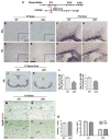
The expression of Mbp and Plp1 mRNA is reduced during postnatal development in Olig1Cre/+; Cdk5fl/fl cKO animals compared to Olig1+/+; Cdk5fl/+ littermate controls. A, An outline of the genetic approach of conditional deletion of Cdk5 in olig1+ cells used in the current study. Exons II–V encoding vital Cdk5 catalytic-domain components were flanked with loxP elements and excised by Olig1-Cre. B–E At P7, in situ hybridization reveals a reduction of Mbp and Plp1 mRNA in the corpus callosum and overlying cortex of Olig1Cre/+; Cdk5fl/fl cKO (C & E) compared to their WT littermates (B & D). F–I the reduced expression mRNA for Mbp and Plp1 is more pronounced at P14 in the corpus callosum of Olig1Cre/+; Cdk5fl/fl cKO (G & I) compared to their WT littermates (F & H). J–L, at P7 spinal cord, the total number of plp1+ cells in both grey matter (GM) and white matter (WM) is significantly reduced in Olig1Cre/+; Cdk5fl/fl cKO revealed by in situ hybridization. No significant difference is seen in the number of pdgf R+ OPCs following a 2 h pulse of BrdU by double labeling of BrdU+ (brown)/pdgf R+ (blue) cells (arrows) in the cortex (Ctx) and in corpus callosum (CC) between WT (M & O) and Olig1Cre/+; Cdk5fl/fl cKO (N & P). Quantitative data is shown in bar graphs Q. The data represent the mean ± SD taken from at least 8 sections from 3 independent animals of each genotype. Scale bar = 25μm.
Figure 3
Figure 3
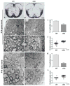
Conditional deletion of Cdk5 in Olig1+ cells results in myelination defects. Reduced myelination in the spinal cord and the optic nerve of Olig1Cre/+; Cdk5fl/fl cKO. A–B In situ hybridization of mRNA expression of Mbp shows reduced level in the spinal cord of Olig1Cre/+; Cdk5fl/fl cKO at P14 compared to their WT littermates. C–J At P14 Olig1Cre/+; Cdk5fl/fl cKO contained significantly reduced number of myelinated axons compared to their WT littermates in both spinal cord (C–F) and the optic nerve (G–J). The relative thickness of the myelin was also reduced in the Olig1Cre/+; Cdk5fl/fl cKO animals and g ratio for spinal cord and optical nerve axons shows a significant reduction in Olig1Cre/+; Cdk5fl/fl cKO (J & L). Quantitative analysis of the proportion of myelinated axons in the spinal cord and optic nerve at P14 reveals a significant reduction in the absence of Cdk5 in oligodendrocyte lineage cells (I & K). P < 0.001. Scale bar = 25μm.
Figure 4
Figure 4
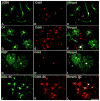
Cdk5 is expressed in cells during multiple distinct stages of development of the oligodendrocyte lineage. A–C In purified primary A2B5+ cell cultures from rat spinal cord, Cdk5 is expressed in the perinuclear cytoplasm and nuclei (red) of virtually every A2B5+ cell (green). D–F Cdk5 (red) is expressed in the majority of O4+ cells (green) as well as in more mature (G–I) MBP+ cells. J–L The expression of Cdk5 in immature cells of the oligodendrocyte lineage is retained in vivo where Cdk5 (red) is expressed in NG2+ cells (green) in the spinal cord of adult mouse. Bar = 25μm.
Figure 5
Figure 5
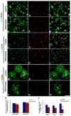
Blocking Cdk5 with roscovitine inhibits oligodendrocyte maturation but does not affect OPC proliferation. A–F The number of NG2+ cells present in A2B5 pan purified cell cultures was only slightly reduced following inhibition of Cdk5 activity for 4 days by treatment with roscovitine and their proliferation as measured by incorporation of BrdU was not significantly different. G–L The number of Olig2+ cells present in A2B5 pan purified cell cultures was slightly reduced following inhibition of Cdk5 activity for 4 days by treatment with roscovitine and their proliferation as measured by incorporation of BrdU was not significantly different. M–R, the number of MBP+ cells that developed in A2B5 pan purified cell cultures was significantly reduced following inhibition of Cdk5 activity for 4 days by treatment with roscovitine. The morphology of MBP+ cells in roscovitine treated cultures was notably less complex that in vehicle treated control cultures. The cells contained fewer processes and lacked characteristic membrane sheets of mature MBP+ cells. Quantitation is shown in histogram (S, T) and represents the mean ± SD taken from at least 3 independent coverslips form 3 different experiments. Bar = 25μm.
Figure 6
Figure 6
The maturation of oligodendrocytes is modulated by knockdown or overexpression of Cdk5 in spinal cord cultures. A In spinal cord cultures, cells transfected with RNAi for Cdk5. (Ab-b″) showed approximately 98% endogenous Cdk5 knockdown compared with cells transfected with empty-vector (EGFP-C3) as a control (Aa-a″). B The morphological complexity of cells transfected with control (Ba-a″) constructs was significantly more pronounced than cells transfected with Cdk5 RNAi (Bb-b″) (green) and co-labeled with O4+ (red) and MBP+ (red) (Bc-d″). Cdk5 RNAi transfected cells had shorten processes and a less complex morphology compared to control cells. Be-e″ Cells that escaped Cdk5 knockdown retained a complex morphology in experimental cultures demonstrating the specificity to the Cdk5 effect. C Over expression of Cdk5 results in increased complexity of cultured O1+ (Cb-b″) and MBP+ oligodendrocytes (Cd-d″). More than 6% of the MBP+ cells had an increased complexity of their processes in cultures driven to overexpress Cdk5-EGFP compared to control. Quantitative data were shown in graphics. Scale bar = 25μm.
Figure 7
Figure 7
Absence of Cdk5 results in a specific reduction in the extent of mature oligodendrocyte processes outgrowth and branching. A, Brain mixed dissociated cell cultures from WT and E18 Cdk5 KO were labeled with O4, O1 and MBP antibodies at 2.5, 7 and 9 days in vitro (DIV) respectively. Although a slight reduction in the proportion O4+ or O1+ oligodendrocytes in Cdk5−/− cultures was seen it was not significant (7A a–d). By contrast, the number of MBP+ cells was significantly reduced in Cdk5−/− cultures (7A e–f). Quantification shows in (7A g). A slight reduction in the number of processes and the extent of branching in O4+ of Cdk5−/− cells but a robust reduction of the number of processes and the extent of branching in MBP+ cells that developed in Cdk5−/− cultures compared to WT (Figure 7Ah). ***p<0.001.. B, The perturbed development of oligodendrocytes in the absence of Cdk5 is not rescued by addition of exogenous PDGF in spinal cord explant cultures. a, b Addition of exogenous PDGF (10ng/ul) in spinal cord explant culture of E18 Cdk5−/− did not enhance the number or morphology of O4+ cells in Cdk5−/− cultures compared to Cdk5+/+. c, d Addition of exogenous PDGF did not significantly alter the morphology or the number of MBP+ cell that developed in explant cultures of E18 Cdk5−/− animals. Quantitative analysis of the average footprint for individual MBP+ cells from WT or Cdk5−/− animals treated with PDGF treatment showed a substantially smaller footprint from Cdk5−/− cells (Fig. 7Be). Scale bar = 25μm in A and B.
Figure 7
Figure 7
Absence of Cdk5 results in a specific reduction in the extent of mature oligodendrocyte processes outgrowth and branching. A, Brain mixed dissociated cell cultures from WT and E18 Cdk5 KO were labeled with O4, O1 and MBP antibodies at 2.5, 7 and 9 days in vitro (DIV) respectively. Although a slight reduction in the proportion O4+ or O1+ oligodendrocytes in Cdk5−/− cultures was seen it was not significant (7A a–d). By contrast, the number of MBP+ cells was significantly reduced in Cdk5−/− cultures (7A e–f). Quantification shows in (7A g). A slight reduction in the number of processes and the extent of branching in O4+ of Cdk5−/− cells but a robust reduction of the number of processes and the extent of branching in MBP+ cells that developed in Cdk5−/− cultures compared to WT (Figure 7Ah). ***p<0.001.. B, The perturbed development of oligodendrocytes in the absence of Cdk5 is not rescued by addition of exogenous PDGF in spinal cord explant cultures. a, b Addition of exogenous PDGF (10ng/ul) in spinal cord explant culture of E18 Cdk5−/− did not enhance the number or morphology of O4+ cells in Cdk5−/− cultures compared to Cdk5+/+. c, d Addition of exogenous PDGF did not significantly alter the morphology or the number of MBP+ cell that developed in explant cultures of E18 Cdk5−/− animals. Quantitative analysis of the average footprint for individual MBP+ cells from WT or Cdk5−/− animals treated with PDGF treatment showed a substantially smaller footprint from Cdk5−/− cells (Fig. 7Be). Scale bar = 25μm in A and B.

Similar articles

Cited by

References

    1. Alexander K, Yang HS, Hinds PW. Cellular senescence requires CDK5 repression of Rac1 activity. Mol Cell Biol. 2004;24:2808–2819. - PMC - PubMed
    1. Barres BA, Hart IK, Coles HS, Burne JF, Voyvodic JT, Richardson WD, Raff MC. Cell death and control of cell survival in the oligodendrocyte lineage. Cell. 1992;70:31–46. - PubMed
    1. Benavides DR, Quinn JJ, Zhong P, Hawasli AH, DiLeone RJ, Kansy JW, Olausson P, Yan Z, Taylor JR, Bibb JA. Cdk5 modulates cocaine reward, motivation, and striatal neuron excitability. J Neurosci. 2007;27:12967–12976. - PMC - PubMed
    1. Boccaccio GL, Carminatti H, Colman DR. Subcellular fractionation and association with the cytoskeleton of messengers encoding myelin proteins. Journal of neuroscience research. 1999;58:480–491. - PubMed
    1. Boccaccio GL, Colman DR. Myelin basic protein mRNA localization and polypeptide targeting. Journal of neuroscience research. 1995;42:277–286. - PubMed

Publication types

MeSH terms