Monitoring plaque inflammation in atherosclerotic rabbits with an iron oxide (P904) and (18)F-FDG using a combined PET/MR scanner
- PMID: 23582588
- PMCID: PMC4128694
- DOI: 10.1016/j.atherosclerosis.2013.03.019
Monitoring plaque inflammation in atherosclerotic rabbits with an iron oxide (P904) and (18)F-FDG using a combined PET/MR scanner
Abstract
Purpose: The aim of this study was to compare the ability of (18)F-FDG PET and iron contrast-enhanced MRI with a novel USPIO (P904) to assess change in plaque inflammation induced by atorvastatin and dietary change in a rabbit model of atherosclerosis using a combined PET/MR scanner.
Materials and methods: Atherosclerotic rabbits underwent USPIO-enhanced MRI and (18)F-FDG PET in PET/MR hybrid system at baseline and were then randomly divided into a progression group (high cholesterol diet) and a regression group (chow diet and atorvastatin). Each group was scanned again 6 months after baseline imaging. R2* (i.e. 1/T2*) values were calculated pre/post P904 injection. (18)F-FDG PET data were analyzed by averaging the mean Standard Uptake Value (SUVmean) over the abdominal aorta. The in vivo imaging was then correlated with matched histological sections stained for macrophages.
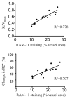
Results: (18)F-FDG PET showed strong FDG uptake in the abdominal aorta and P904 injection revealed an increase in R2* values in the aortic wall at baseline. At 6 months, SUVmean values measured in the regression group showed a significant decrease from baseline (p = 0.015). In comparison, progression group values remained constant (p = 0.681). R2* values showed a similar decreasing trend in the regression group suggesting less USPIO uptake in the aortic wall. Correlations between SUVmean or Change in R2* value and macrophages density (RAM-11 staining) were good (R(2) = 0.778 and 0.707 respectively).
Conclusion: This experimental study confirms the possibility to combine two functional imaging modalities to assess changes in the inflammation of atherosclerotic plaques. (18)F-FDG-PET seems to be more sensitive than USPIO P904 to detect early changes in plaque inflammation.
Copyright © 2013 Elsevier Ireland Ltd. All rights reserved.
Conflict of interest statement
E.L., S.B., P.R. and C.C. are employees of Guerbet; Z.A.F. received partial funding from Guerbet.
Figures


Similar articles
-
Combined PET/DCE-MRI in a Rabbit Model of Atherosclerosis: Integrated Quantification of Plaque Inflammation, Permeability, and Burden During Treatment With a Leukotriene A4 Hydrolase Inhibitor.JACC Cardiovasc Imaging. 2018 Feb;11(2 Pt 2):291-301. doi: 10.1016/j.jcmg.2017.11.030. JACC Cardiovasc Imaging. 2018. PMID: 29413439
-
FDG-PET can distinguish inflamed from non-inflamed plaque in an animal model of atherosclerosis.Int J Cardiovasc Imaging. 2010 Jan;26(1):41-8. doi: 10.1007/s10554-009-9506-6. Epub 2009 Sep 22. Int J Cardiovasc Imaging. 2010. PMID: 19784796
-
Pioglitazone modulates vascular inflammation in atherosclerotic rabbits noninvasive assessment with FDG-PET-CT and dynamic contrast-enhanced MR imaging.JACC Cardiovasc Imaging. 2011 Oct;4(10):1100-9. doi: 10.1016/j.jcmg.2011.04.020. JACC Cardiovasc Imaging. 2011. PMID: 21999870 Free PMC article.
-
Inflammation as a Predictor of Abdominal Aortic Aneurysm Growth and Rupture: A Systematic Review of Imaging Biomarkers.Eur J Vasc Endovasc Surg. 2016 Sep;52(3):333-42. doi: 10.1016/j.ejvs.2016.05.002. Epub 2016 Jun 6. Eur J Vasc Endovasc Surg. 2016. PMID: 27283346 Review.
-
Variability and uncertainty of 18F-FDG PET imaging protocols for assessing inflammation in atherosclerosis: suggestions for improvement.J Nucl Med. 2015 Apr;56(4):552-9. doi: 10.2967/jnumed.114.142596. Epub 2015 Feb 26. J Nucl Med. 2015. PMID: 25722452 Review.
Cited by
-
Assessment of atherosclerosis in large vessel walls: A comprehensive review of FDG-PET/CT image acquisition protocols and methods for uptake quantification.J Nucl Cardiol. 2015 Jun;22(3):468-79. doi: 10.1007/s12350-015-0069-8. Epub 2015 Apr 1. J Nucl Cardiol. 2015. PMID: 25827619 Review.
-
MRI study of atherosclerotic plaque progression using ultrasmall superparamagnetic iron oxide in Watanabe heritable hyperlipidemic rabbits.Br J Radiol. 2015 Sep;88(1053):20150167. doi: 10.1259/bjr.20150167. Epub 2015 Jun 17. Br J Radiol. 2015. PMID: 26083261 Free PMC article.
-
Feasibility of simultaneous PET/MR in diet-induced atherosclerotic minipig: a pilot study for translational imaging.Am J Nucl Med Mol Imaging. 2014 Aug 15;4(5):448-58. eCollection 2014. Am J Nucl Med Mol Imaging. 2014. PMID: 25143863 Free PMC article.
-
Simultaneous 18-FDG PET and MR imaging in lower extremity arterial disease.Front Cardiovasc Med. 2024 Feb 9;11:1352696. doi: 10.3389/fcvm.2024.1352696. eCollection 2024. Front Cardiovasc Med. 2024. PMID: 38404725 Free PMC article.
-
The Role of MR Enterography in Assessing Crohn's Disease Activity and Treatment Response.Gastroenterol Res Pract. 2016;2016:8168695. doi: 10.1155/2016/8168695. Epub 2015 Dec 27. Gastroenterol Res Pract. 2016. PMID: 26819611 Free PMC article. Review.
References
-
- Ross R. Atherosclerosis is an inflammatory disease. Am Heart J. 1999;138:S419–20. - PubMed
-
- Crisby M, Nordin-Fredriksson G, Shah PK, Yano J, Zhu J, Nilsson J. Pravastatin treatment increases collagen content and decreases lipid content, inflammation, metalloproteinases, and cell death in human carotid plaques: implications for plaque stabilization. Circulation. 2001;103:926–33. - PubMed
-
- Sukhova GK, Williams JK, Libby P. Statins reduce inflammation in atheroma of nonhuman primates independent of effects on serum cholesterol. Arterioscler Thromb Vasc Biol. 2002;22:1452–8. - PubMed
-
- Rudd JH, Warburton EA, Fryer TD, et al. Imaging atherosclerotic plaque inflammation with [18F]-fluorodeoxyglucose positron emission tomography. Circulation. 2002;105:2708–11. - PubMed
-
- Tang TY, Muller KH, Graves MJ, et al. Iron oxide particles for atheroma imaging. Arterioscler Thromb Vasc Biol. 2009;29:1001–8. - PubMed
Publication types
MeSH terms
Substances
Grants and funding
LinkOut - more resources
Full Text Sources
Other Literature Sources
Medical

