. 2013 Apr 30;110(18):7452-7.
doi: 10.1073/pnas.1302164110. Epub 2013 Apr 8.

Genome-wide siRNA screen identifies the retromer as a cellular entry factor for human papillomavirus

Affiliations

Genome-wide siRNA screen identifies the retromer as a cellular entry factor for human papillomavirus

Alex Lipovsky et al. Proc Natl Acad Sci U S A. .

Abstract

Despite major advances in our understanding of many aspects of human papillomavirus (HPV) biology, HPV entry is poorly understood. To identify cellular genes required for HPV entry, we conducted a genome-wide screen for siRNAs that inhibited infection of HeLa cells by HPV16 pseudovirus. Many retrograde transport factors were required for efficient infection, including multiple subunits of the retromer, which initiates retrograde transport from the endosome to the trans-Golgi network (TGN). The retromer has not been previously implicated in virus entry. Furthermore, HPV16 capsid proteins arrive in the TGN/Golgi in a retromer-dependent fashion during entry, and incoming HPV proteins form a stable complex with retromer subunits. We propose that HPV16 directly engages the retromer at the early or late endosome and traffics to the TGN/Golgi via the retrograde pathway during cell entry. These results provide important insights into HPV entry, identify numerous potential antiviral targets, and suggest that the role of the retromer in infection by other viruses should be assessed.


Conflict of interest statement

The authors declare no conflict of interest.

Figures

Fig. 1.
Fig. 1.
Genome-wide RNA interference screen for human genes required for HPV16 infection. (A) Scatter plot showing percent inhibition of HPV16-GFP infection for two different replicates of the genome-wide screen. Each spot represents a single siRNA pool. (B) Pie chart of 216 hits tested for HPV16-GFP inhibition in the confirmation screen, showing the number (and percentage) of active siRNAs for each hit. (C) HeLa-S3 cells were transfected with a control RISC-free siRNA (con) or siRNAs against APH1A (a γ-secretase subunit), ATP6AP2 (a V-ATPase subunit), or VPS29 (a retromer subunit). (Left) Knock-down efficiency of the targeted mRNA was assayed by qRT-PCR 48 h after transfection, and mRNA levels in knock-down cells were normalized to the cognate mRNA in control cells. (Right) Knock-down cells were infected with HPV16-GFP, and GFP expression was assayed 2 d later by flow cytometry. The fraction of GFP positive cells was normalized to the fraction of GFP positive cells in cells transfected with control siRNA. The results of an experiment performed in triplicate are shown; similar results were obtained in multiple independent experiments. (D) DAVID software was used to classify the 1,000 highest-ranked primary hits according to cellular compartment, and fold-enrichment of each category in the hit list compared with the complete siRNA library was calculated. Only gene categories enriched by more than twofold are shown. qRT-PCR, quantitative reverse transcriptase-PCR.
Fig. 2.
Fig. 2.
Retrograde transport is required for HPV infection. (A) Rab knock-down specifically inhibits HPV16-GFP infection. Effect of siRNAs on infection efficiency was assayed as described in the legend to Fig. 1C, Right. Black bars, HPV16-GFP infection; gray bars, Ad5-GFP infection. Results are the average (±SD) of multiple independent experiments. Statistical significance relative to cells transfected with control siRNA was determined by a paired two-tailed t test: *P < 0.05; **P < 0.01. (B) HeLa cells were infected with HPV16-GFP (black bars) or Ad5-GFP (gray bars) and treated with the indicated concentrations of Retro-2 at the time of infection. GFP expression was assayed by flow cytometry 40 h postinfection, and the fraction of GFP-positive cells was normalized to the fraction of GFP-positive cells in untreated sample. Results are displayed as described in A. (C) HeLa cells were transfected with RISC-free siRNA (Left and Center) or VPS26 siRNA (Right). Forty-eight hours later, cells were infected with HPV16-GFP at an MOI of 100. Cells in the Center panel were treated with 100 μM Retro-2 at the time of infection. Twenty hours postinfection, cells were immunostained with the polyclonal L1 antiserum and visualized by immunofluorescence microscopy. A single confocal slice is shown in each panel.
Fig. 3.
Fig. 3.
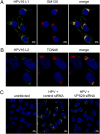
HPV localizes to a Golgi-like compartment. (A) HeLa cells were infected with HPV16-GFP for 16 h at an MOI of 50 and immunostained with polyclonal antiserum against L1 (green) and monoclonal antibody against GM130 (red). Areas of strongest overlap are pseudocolored white in the Right panel using an ImageJ colocalization plugin. The same confocal slice is shown in all three panels. (B) Samples were processed as in A except cells were stained with anti-L2 (RG1) and TGN46. (C) HeLa cells were transfected with a RISC-free siRNA (Left and Center) or VPS29 siRNA (Right). Forty-eight hours later, cells were mock-infected (Left) or infected with HPV16-GFP at an MOI of 100 (Center and Right). Sixteen hours postinfection, the cells were reacted with antibodies against L1 and GM130 and processed for PLA. A single confocal slice is shown in each panel.
Fig. 4.
Fig. 4.
The retromer is required for HPV infection. (A) HeLa-S3 cells were transfected with a RISC-free siRNA (control), or siRNAs against VPS26, VPS29, or VPS35. Knock-down cells were infected 48 h later with HPV16-GFP (black bars) or Ad5-GFP (gray bars). Infection efficiency was assayed as described in the legend to Fig. 1C, Right. Results are displayed as in Fig. 2A. (B) The ability of HPV16-GFP to infect human cervical keratinocytes was analyzed and displayed as in A. (C) HeLa cells were transfected with a RISC-free siRNA (control), or siRNAs against VPS26, VPS29, or VPS35. Knock-down cells were infected 48 h later with HPV16-GFP (black bars), HPV18-GFP (dark gray bars), or HPV5-GFP (light gray bars). Infection efficiency was assayed as described in the legend to Fig. 1C, Right. Results are displayed as in Fig. 2A.
Fig. 5.
Fig. 5.
The retromer forms a complex with incoming HPV16 capsids. (A) HeLa-sen2 cells were transfected with RISC-free siRNA (Left three panels) or VPS26 siRNA (Right). After 48 h, cells were infected with HPV16-GFP.L2-HA at an MOI of 200. Twelve hours postinfection, cells were immunostained with antibodies recognizing HA (red) or VPS35 (green). Areas of strongest overlap are pseudocolored white in the two Right panels by using an ImageJ colocalization plug-in. The same confocal slice is shown in the Left three panels. A single confocal slice is shown in the Right panel. (B) HeLa-sen2 cells were infected with HPV16-GFP L2-HA at an MOI of 200. Twelve hours postinfection, cells were immunostained with antibodies recognizing HA (green, HPV16 L2) or EEA1 (red). Areas of strongest overlap are pseudocolored white in the Right panel as in A. The same confocal slice is shown in all three panels. (C) 293T cells were transfected with genes encoding myc-tagged retromer subunits VPS26, VPS29, and VPS35 (Ret) or with an empty vector (Vec). As indicated, cells were then mock-infected (−) or infected with HPV16-GFP.L2-HA at an MOI of 50 for 8 h (+). Cells were then harvested in Nonidet P-40 buffer and precipitated with a myc (M) or an isotype- and species-matched control (C) antibody. Samples were analyzed by SDS/PAGE and immunoblotting with an HA antibody. Arrows indicate HA-tagged L2. Upper shows immunoprecipitated material; Lower shows 5% of total extract.

Comment in

Similar articles

Cited by

References

    1. Inoue T, Moore P, Tsai B. How viruses and toxins disassemble to enter host cells. Annu Rev Microbiol. 2011;65:287–305. - PubMed
    1. Chaturvedi AK, et al. Human papillomavirus and rising oropharyngeal cancer incidence in the United States. J Clin Oncol. 2011;29(32):4294–4301. - PMC - PubMed
    1. Parkin DM, Bray F. Chapter 2: The burden of HPV-related cancers. Vaccine. 2006;24(Suppl 3):S11–S25. - PubMed
    1. Modis Y, Trus BL, Harrison SC. Atomic model of the papillomavirus capsid. EMBO J. 2002;21(18):4754–4762. - PMC - PubMed
    1. Giroglou T, Florin L, Schäfer F, Streeck RE, Sapp M. Human papillomavirus infection requires cell surface heparan sulfate. J Virol. 2001;75(3):1565–1570. - PMC - PubMed

Publication types