. 2013 Apr 3;33(14):6191-202.
doi: 10.1523/JNEUROSCI.4051-12.2013.

Na+ channel-dependent recruitment of Navβ4 to axon initial segments and nodes of Ranvier

Affiliations

Na+ channel-dependent recruitment of Navβ4 to axon initial segments and nodes of Ranvier

Shelly A Buffington et al. J Neurosci. .

Abstract

The axon initial segment (AIS) and nodes of Ranvier are the sites of action potential initiation and regeneration in axons. Although the basic molecular architectures of AIS and nodes, characterized by dense clusters of Na(+) and K(+) channels, are similar, firing patterns vary among cell types. Neuronal firing patterns are established by the collective activity of voltage-gated ion channels and can be modulated through interaction with auxiliary subunits. Here, we report the neuronal expression pattern and subcellular localization of Navβ4, the modulatory Na(+) channel subunit thought to underlie resurgent Na(+) current. Immunostaining of rat tissues revealed that Navβ4 is strongly enriched at the AIS of a select set of neuron types, including many characterized by high-frequency firing, and at nodes of Ranvier in the PNS and some nodes in the CNS. By introducing full-length and mutant GFP-tagged Navβ4 into cultured neurons, we determined that the AIS and nodal localization of Navβ4 depends on its direct interaction with Na(+) channel α subunits through an extracellular disulfide bond. Based on these results, we propose that differences in the specific composition of the Na(+) channel complexes enriched at the AIS and nodes contribute to the diverse physiologies observed among cell types.


Figures
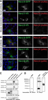
Figure 1.
Figure 1.
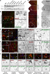
α-Navβ4 antibodies do not cross-react with Navβ1, Navβ2, or other AIS proteins. A, B, COS cells expressing Navβ4–GFP, Navβ1–His/V5, or Navβ2–His/V5 immunostained for either GFP or V5 (green), as appropriate, and Navβ4 (red). A, Mouse monoclonal α-Navβ4. B, Rabbit polyclonal α-Navβ4. Nuclei were labeled with Hoechst stain (blue in the merged image). Scale bars, 20 μm. C, Western blotting of COS cell protein lysates immunoblotted using α-Navβ4 antibodies and antibodies to detect either the GFP- or His-tagged Navβ subunits. D, Immunoblots of rat brain membrane (RBM) protein homogenates or Navβ4–GFP-transfected COS cells, as indicated, probed for Navβ4, which has an approximate molecular weight of 38 kDa. A nonspecific band in the rat brain membrane homogenate recognized by the rabbit polyclonal antibody against Navβ4 is denoted with an asterisk. M, Mouse; R, rat.
Figure 2.
Figure 2.
Immunolocalization of Navβ4 reveals AIS enrichment in multiple CNS cell types. A, Western blot of brain-region-specific homogenates probed with the mouse (M) monoclonal α-Navβ4 antibody. Immunoblotting for actin served as a loading control. RBM, Rat brain membrane. B–F, Immunostaining of multiple brain regions for Navβ4 (mouse monoclonal; red) and CB (B, green) or βIV spectrin (C–F, green). Hoechst nuclear staining is shown in blue in the merged images. B, Immunostaining of cerebellar sections for Navβ4 (red) and the Purkinje cell marker CB (green). C, Immunostaining of somatosensory cortex showing layer-specific enrichment of Navβ4 expression. Boxes D and E are expanded in the subsequent panels. F, Representative images of Navβ4-expressing neurons in the indicated brain regions. AIS enrichment is demonstrated by colocalization with βIV spectrin. Scale bars: B, F, 20 μm; C–E, 40 μm.
Figure 3.
Figure 3.
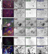
Navβ4 is enriched at interneuron AIS. A–C, Triple immunostaining of Navβ4 (red) (mouse monoclonal antibody) with the calcium binding proteins CB and PV in green or blue, as indicated. Arrowheads highlight Navβ4+ AIS. Scale bars: A, B, 40 μm; C, 80 μm. A, Immunostaining of somatosensory cortex layers V and II/III, as indicated. The Navβ4+ layer V pyramidal neuron AIS is highlighted by the arrow in the top row. The bottom row displays the relatively low expression of Navβ4 among both the excitatory pyramidal neurons and CB and/or PV+ interneurons of layer II/III cortex. B, Immunostaining of Navβ4+ interneurons in the hippocampus. Arrowhead points to the Navβ4+ AIS of the CB/PV+ hippocampal interneuron featured in the top row. The arrowhead in the bottom row highlights the Navβ4+ AIS of the CB-expressing, PV hippocampal neuron featured in the bottom row. C, Immunostaining of the TB showing Navβ4 expression and AIS-enrichment in the CB/PV-expressing cells, indicated by the arrowheads. Scale bars, 80 μm.
Figure 4.
Figure 4.
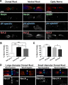
Navβ4 is enriched at a subset of CNS and PNS nodes of Ranvier. A, Immunostaining of rat dorsal and ventral spinal cord roots and optic nerve using antibodies against Navβ4 (rabbit polyclonal; green), nodal βIV spectrin (blue), and juxtaparanodal Kv1.2 channels (red). B, Quantification of the percentage of nodes with detectable Navβ4 immunoreactivity enrichment in the indicated nerve types (n = 100, N = 3). C, Quantification of Navβ4+ nodes in large- vs small-diameter ventral (VR) and dorsal (DR) root axons (n = 100, N = 4). *p < 0.05, ***p < 0.001. Error bars represent SEM. D, Images show representative examples of Navβ4+ and Navβ4 large- and small-diameter dorsal root nodes coimmunostained for βIV spectrin (blue) and Kv1.2 (red), shown in the merged images. Scale bars, 10 μm.
Figure 5.
Figure 5.
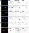
AnkG and Nav1 α subunit expression are required for Navβ4 AIS targeting. A, Hippocampal neurons transfected with GFP-tagged Navβ4 and immunostained for GFP (green), ankG (red), and MAP2 (blue). B, Cultured hippocampal neurons immunolabeled for Navβ4 (green), ankG (red), and MAP2 (blue). C, D, Immunostaining of cultured hippocampal neurons cotransfected with the indicated shRNA and FL Navβ4–GFP constructs. Neurons were labeled with α-βIV spectrin or α-neurofascin-186 (blue), α-GFP (green), and α-ankG, or α-pan Nav channels (red). Transfected cells are marked with an *. Scale bars, 20 μm. C, Immunostaining of cultured neurons transfected with shRNA targeting the AIS-organizing protein ankG or Na+ channel α subunits, as indicated. Arrowhead indicates βIV spectrin+/Navα/Navβ4 AIS in the bottom row, and arrows point toward neighboring control neuron AIS in all panels. D, Knockdown of the AIS proteins βIV spectrin and neurofascin-186. Arrowheads highlight the location of Navβ4+ AIS in all panels.
Figure 6.
Figure 6.
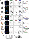
Cysteine 28 is required for Navβ4 AIS localization. A, Neurons transfected with FL, ΔC, or ΔN Navβ4 constructs and immunostained for GFP (green), ankG (red), and MAP2 (blue). Longitudinal line scans through the AIS show fluorescence intensity (F.I.) of the Navβ4–GFP (green), ankG (red), and MAP2 (blue) signals. B–E, Immunostaining of cultured hippocampal neurons transfected with Navβ4-C28A (B) or the indicated Navβ4 single point mutants (C) (C23A, C101A, and G52A). Neurons were immunolabeled using antibodies against GFP (green), ankG (red), and MAP2 (blue). Scale bar, 20 μm. D, Quantitative analysis of Navβ4 point mutant targeting to the AIS in cultured hippocampal neurons. E, Western blotting for Navβ4 in whole-brain membrane protein homogenates denatured in reducing (R) or non-reducing (NR) sample buffer. TM, Transmembrane.
Figure 7.
Figure 7.
Cysteine 28 is required for Navβ4 targeting to nodes of Ranvier. DRG neurons transfected with Navβ4 expression constructs encoding FL-, ΔC- (A), ΔN-, and C28A–Navβ4–GFP (B) constructs cocultured with myelinating Schwann cells. Myelinated axon segments were detected by immunostaining for MBP (red), whereas ankG immunoreactivity (blue) defined heminodes and forming nodes of Ranvier. The FL or mutant GFP-tagged Navβ4 proteins were detected by GFP immunoreactivity (green). Scale bar, 10 μm. C, Quantification of FL-, ΔC-, ΔN-, and C28A–Navβ4+ heminodes in the myelinating cocultures.

Similar articles

Cited by

References

    1. Aman TK, Grieco-Calub TM, Chen C, Rusconi R, Slat EA, Isom LL, Raman IM. Regulation of persistent Na current by interactions between beta subunits of voltage-gated Na channels. J Neurosci. 2009;29:2027–2042. - PMC - PubMed
    1. Bant JS, Raman IM. Control of transient, resurgent, and persistent current by open-channel block by Na channel beta4 in cultured cerebellar granule neurons. Proc Natl Acad Sci U S A. 2010;107:12357–12362. - PMC - PubMed
    1. Barbieri R, Baroni D, Moran O. Identification of an intra-molecular disulfide bond in the sodium channel β1-subunit. Biochem Biophys Res Commun. 2012;420:364–367. - PubMed
    1. Bean BP. The action potential in mammalian central neurons. Nat Rev Neurosci. 2007;8:451–465. - PubMed
    1. Brackenbury WJ, Davis TH, Chen C, Slat EA, Detrow MJ, Dickendesher TL, Ranscht B, Isom LL. Voltage-gated Na+ channel beta1 subunit-mediated neurite outgrowth requires Fyn kinase and contributes to postnatal CNS development in vivo. J Neurosci. 2008;28:3246–3256. - PMC - PubMed

Publication types

MeSH terms

LinkOut - more resources