. 2013;8(3):e59755.
doi: 10.1371/journal.pone.0059755. Epub 2013 Mar 27.

Plasmodium berghei MAPK1 displays differential and dynamic subcellular localizations during liver stage development

Affiliations

Plasmodium berghei MAPK1 displays differential and dynamic subcellular localizations during liver stage development

Jannika Katharina Wierk et al. PLoS One. 2013.

Abstract

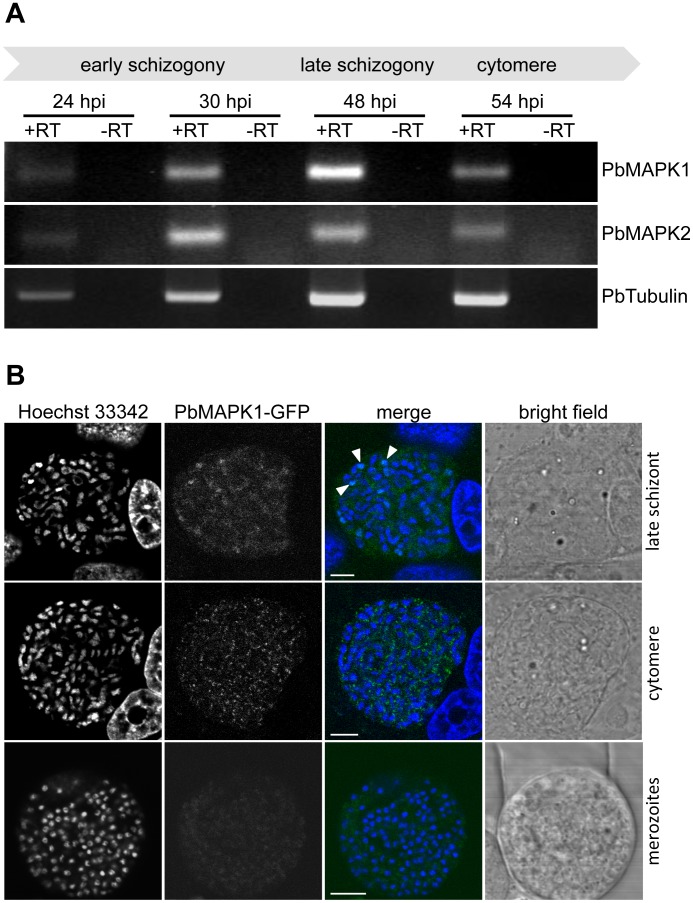
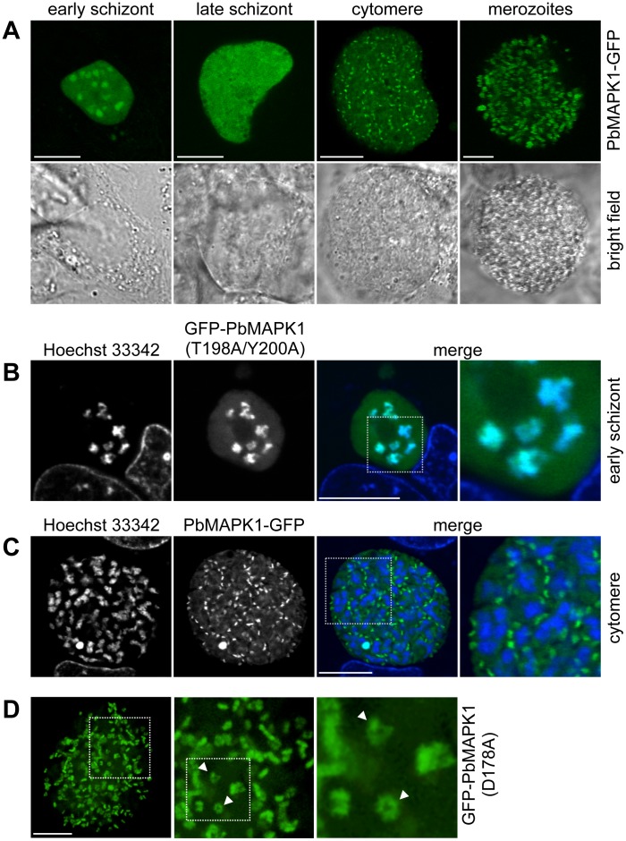
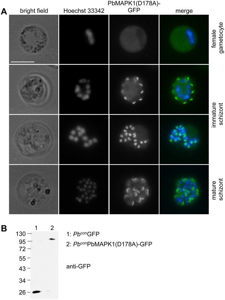
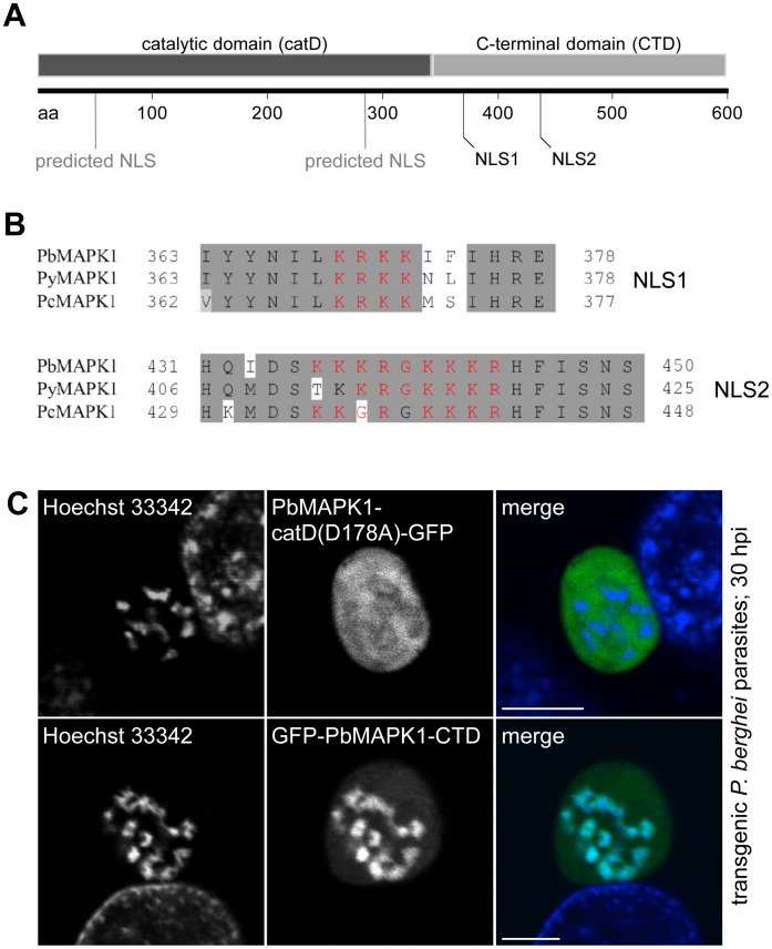
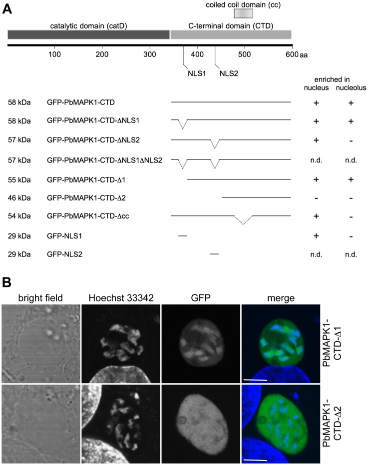
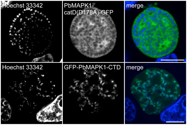
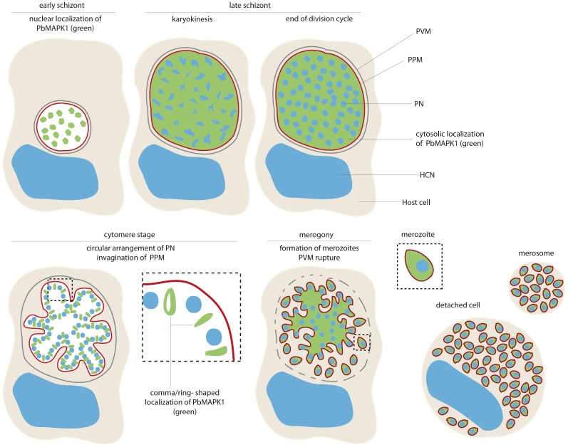
Mitogen-activated protein kinases (MAPKs) regulate key signaling events in eukaryotic cells. In the genomes of protozoan Plasmodium parasites, the causative agents of malaria, two genes encoding kinases with significant homology to other eukaryotic MAPKs have been identified (mapk1, mapk2). In this work, we show that both genes are transcribed during Plasmodium berghei liver stage development, and analyze expression and subcellular localization of the PbMAPK1 protein in liver stage parasites. Live cell imaging of transgenic parasites expressing GFP-tagged PbMAPK1 revealed a nuclear localization of PbMAPK1 in the early schizont stage mediated by nuclear localization signals in the C-terminal domain. In contrast, a distinct localization of PbMAPK1 in comma/ring-shaped structures in proximity to the parasite's nuclei and the invaginating parasite membrane was observed during the cytomere stage of parasite development as well as in immature blood stage schizonts. The PbMAPK1 localization was found to be independent of integrity of a motif putatively involved in ATP binding, integrity of the putative activation motif and the presence of a predicted coiled-coil domain in the C-terminal domain. Although PbMAPK1 knock out parasites showed normal liver stage development, the kinase may still fulfill a dual function in both schizogony and merogony of liver stage parasites regulated by its dynamic and stage-dependent subcellular localization.


Conflict of interest statement

Competing Interests: The authors have declared that no competing interests exist.

Figures

Figure 1
Figure 1. Expression and localization of P. berghei MAPK1 during liver stage development.
(A) Transcripts encoding PbMAPK1 and PbMAPK2 are detectable in P. berghei liver stage parasites. Total RNA was prepared from P. berghei-infected HepG2 cells 24 hpi, 30 hpi, 48 hpi and 54 hpi. RT-PCR analysis was performed using primer pairs specific for pbmapk1, pbmapk2, and the constitutively expressed pbtubulin. To rule out false positive results originating from gDNA contamination, samples lacking reverse transcriptase (-RT) were processed in parallel. (B) Subcellular localization of the endogenous PbMAPK1 protein during liver stage development. HepG2 cells were infected with Pb endPbMAPK1-GFP parasites in which the endogenous mapk1 locus has been gfp-tagged by homologous recombination. Expression and localization of the PbMAPK1-GFP fusion protein was assayed at different time points of liver stage development (late schizont, cytomere, merozoites) by confocal live cell imaging. Parasite and host cell nuclei were visualized using Hoechst 33342. Arrowheads indicate partial co-localization of PbMAPK1 with parasite nuclei in the late schizont. Scale bars: 5 µm.
Figure 2
Figure 2. PbMAPK1 displays differential and dynamic subcellular localization during liver stage development.
(A) Time course of PbMAPK1 localization (live cell imaging). HepG2 cells were infected with Pb LSPbMAPK1-GFP parasites. Live cell imaging was performed at different developmental stages (early schizont ≈30 hpi, late schizont ≈48 hpi, cytomere ≈54 hpi, merozoites ≈60 hpi). (B), (C) Co-staining with nuclei at early schizont and cytomere stages (live cell imaging). HepG2 cells were infected with P. berghei parasites expressing GFP-tagged PbMAPK1. 30 hpi (B, early schizont, Pb conGFP-PbMAPK1(T198A/Y200A)) and 54 hpi (C, cytomere, Pb LSPbMAPK1-GFP), cells were stained with Hoechst 33342 to visualize host cell and parasite nuclei. Areas containing details additionally displayed at a higher magnification are highlighted in the merged pictures. (D) Spinning disc microscopy (live cell imaging). HepG2 cells infected with Pb conGFP-PbMAPK1(D178A) parasites were analyzed by spinning disc live microscopy at 54 hpi (cytomere stage). Highlighted area is also shown at higher magnification; arrowheads indicate ring shaped structures. Scale bars: 10 µm.
Figure 3
Figure 3. PbMAPK1 displays a distinct localization in P. berghei blood stage schizonts.
(A) Pb conPbMAPK1(D178A)-GFP parasites originating from infected mouse blood were analyzed by live epifluorescence microscopy. Parasite nuclei were visualized using Hoechst 33342. Scale bar: 10 µm. (B) Western blot analysis of Pb conGFP and Pb conPbMAPK1(D178A)-GFP parasites. Mixed blood stage parasite protein extracts were prepared from P. berghei infected mouse blood, separated by SDS-PAGE and blotted onto nitrocellulose. Detection was performed using mouse anti-GFP/anti-mouse HRP. Molecular weight of marker proteins: kDa; expected molecular weights: GFP: 26 kDa, PbMAPK1(D178A)-GFP: 97 kDa.
Figure 4
Figure 4. Nuclear localization of PbMAPK1 is mediated by functional NLSs in the C-terminal domain.
(A) Schematic representation of PbMAPK1 domain structure and position of predicted nuclear localization signals in the catalytic domain (aa 41–57; aa 285–289) and the C-terminal domain (aa 369–372; aa 436–444); aa: amino acids. (B) Alignment of putative nuclear localization signals NLS1 and NLS2 (red) in the PbMAPK1 C-terminal domains of rodent malaria parasites P. berghei (Pb), P. yoelii (Py) and P. chabaudi (Pc). (C) HepG2 cells were infected with either Pb conPbMAPK1-catD(D178A)-GFP or Pb conGFP-PbMAPK1-CTD parasites. 30 hpi host cell and parasite nuclei were stained using Hoechst 33342 and live cell imaging was performed. Scale bar: 5 µm.
Figure 5
Figure 5. Characterization of PbMAPK1 NLSs in HepG2 cells and P. berghei liver stage parasites.
(A) Schematic representation of PbMAPK1-CTD mutants tested in HepG2 cells and summary of results (for confocal images see Figure S4). HepG2 cells were transiently transfected with pEGFP-C2-based plasmids encoding the indicated GFP-PbMAPK1-CTD deletion constructs. 24 hours post transfection, nuclei were stained using Hoechst 33342 and subcellular localization of the fusion proteins was analyzed by live cell imaging; aa: amino acids; n.d.: not determined (due to very low expression levels of the respective fusion proteins). (B) HepG2 cells were infected with either Pb conGFP-PbMAPK1-CTD-Δ1 or Pb conGFP-PbMAPK1-CTD-Δ2 parasites. 32 hpi host cell and parasite nuclei were stained using Hoechst 33342 and live cell imaging was performed. Scale bar: 5 µm.
Figure 6
Figure 6. The developmentally regulated re-localization of PbMAPK1 from the nucleus to extra-nuclear comma/ring-shaped structures is dependent on both the catalytic and the C-terminal domain.
HepG2 cells were infected with either Pb conPbMAPK1-catD(D178A)-GFP or Pb conGFP-PbMAPK1-CTD parasites. 54 hpi host cell and parasite nuclei were stained using Hoechst 33342 and live cell imaging was performed. Scale bar: 10 µm.
Figure 7
Figure 7. Schematic representation: stage-dependent subcellular localization of GFP-tagged MAPK1 in P. berghei liver stage parasites.
While PbMAPK1 is observed inside the parasite nuclei at early schizont stage, at cytomere stage a distinct, comma/ring-shaped localization pattern occurs. PVM: parasitophorous vacuole membrane; PPM: parasite plasma membrane; PN: parasite nucleus; HCN: host cell nucleus.

Similar articles

Cited by

References

    1. Sturm A, Amino R, van de Sand C, Regen T, Retzlaff S, et al. (2006) Manipulation of host hepatocytes by the malaria parasite for delivery into liver sinusoids. Science 313: 1287–1290. - PubMed
    1. Baer K, Klotz C, Kappe SH, Schnieder T, Frevert U (2007) Release of hepatic Plasmodium yoelii merozoites into the pulmonary microvasculature. PLoS Pathog 3: e171. - PMC - PubMed
    1. Gerald N, Mahajan B, Kumar S (2011) Mitosis in the human malaria parasite Plasmodium falciparum. Eukaryot Cell 10: 474–482. - PMC - PubMed
    1. Stanway RR, Mueller N, Zobiak B, Graewe S, Froehlke U, et al. (2011) Organelle segregation into Plasmodium liver stage merozoites. Cell Microbiol 13: 1768–1782. - PubMed
    1. Tewari R, Straschil U, Bateman A, Bohme U, Cherevach I, et al. (2010) The systematic functional analysis of Plasmodium protein kinases identifies essential regulators of mosquito transmission. Cell Host Microbe 8: 377–387. - PMC - PubMed

MeSH terms

Substances

Grants and funding

The work was supported by the MALSIG (Signalling in life cycle stages of malaria parasites) and the EVIMalaR (European virtual institute of malaria research) EU consortia. The funders had no role in study design, data collection and analysis, decision to publish, or preparation of the manuscript.