. 2013 May 10;288(19):13808-20.
doi: 10.1074/jbc.M113.460956. Epub 2013 Mar 26.

A separate pool of cardiac phospholemman that does not regulate or associate with the sodium pump: multimers of phospholemman in ventricular muscle

Affiliations

A separate pool of cardiac phospholemman that does not regulate or associate with the sodium pump: multimers of phospholemman in ventricular muscle

Krzysztof J Wypijewski et al. J Biol Chem. .

Abstract

Background: Phospholemman regulates the plasmalemmal sodium pump in excitable tissues.

Results: In cardiac muscle, a subpopulation of phospholemman with a unique phosphorylation signature associates with other phospholemman molecules but not with the pump.

Conclusion: Phospholemman oligomers exist in cardiac muscle.

Significance: Much like phospholamban regulation of SERCA, phospholemman exists as both a sodium pump inhibiting monomer and an unassociated oligomer. Phospholemman (PLM), the principal quantitative sarcolemmal substrate for protein kinases A and C in the heart, regulates the cardiac sodium pump. Much like phospholamban, which regulates the related ATPase SERCA, PLM is reported to oligomerize. We investigated subpopulations of PLM in adult rat ventricular myocytes based on phosphorylation status. Co-immunoprecipitation identified two pools of PLM: one not associated with the sodium pump phosphorylated at Ser(63) and one associated with the pump, both phosphorylated at Ser(68) and unphosphorylated. Phosphorylation of PLM at Ser(63) following activation of PKC did not abrogate association of PLM with the pump, so its failure to associate with the pump was not due to phosphorylation at this site. All pools of PLM co-localized to cell surface caveolin-enriched microdomains with sodium pump α subunits, despite the lack of caveolin-binding motif in PLM. Mass spectrometry analysis of phosphospecific immunoprecipitation reactions revealed no unique protein interactions for Ser(63)-phosphorylated PLM, and cross-linking reagents also failed to identify any partner proteins for this pool. In lysates from hearts of heterozygous transgenic animals expressing wild type and unphosphorylatable PLM, Ser(63)-phosphorylated PLM co-immunoprecipitated unphosphorylatable PLM, confirming the existence of PLM multimers. Dephosphorylation of the PLM multimer does not change sodium pump activity. Hence like phospholamban, PLM exists as a pump-inhibiting monomer and an unassociated oligomer. The distribution of different PLM phosphorylation states to different pools may be explained by their differential proximity to protein phosphatases rather than a direct effect of phosphorylation on PLM association with the pump.

Keywords: Caveolae; FXYD Proteins; Heart; Na,K-ATPase; PP2A; Phospholemman; Protein Palmitoylation; Protein Phosphatase; Protein Phosphorylation; Serine-Threonine Protein Phosphatase.


Figures

FIGURE 1.
FIGURE 1.
PLM phosphorylated at Ser63 co-immunoprecipitates poorly with the Na pump α subunit. A, PLM was immunoprecipitated (IP) from ARVMs using antibodies specific for Ser63 (S63), Ser68 (S68), and Thr69 (T69) phosphorylated and unphosphorylated (UPh) forms. In the representative immunoblots shown, although the PLM-Ser63 antibody precipitates a substantial amount of PLM, co-precipitation of sodium pump α1 subunit is relatively poor, particularly compared with the adjacent Ser68 immunoprecipitation, which precipitates the same amount of PLM but a considerably larger amount of sodium pump α1 subunit. B, quantitative analysis of Western blot data presented in A. For comparison purposes, the quantity of sodium pump α1 subunit co-precipitated by each antibody was normalized to the quantity of PLM precipitated in the same reaction. To allow comparison of multiple experiments, this ratio was arbitrarily defined as equal to 1 for the immunoprecipitation reactions using antibodies specific for the unphosphorylated form of PLM. *, p < 0.05 (n = 7). C, co-immunoprecipitation reactions from homogenates of whole brain, skeletal muscle (quadriceps), and ARVMs using antibodies specific for PLM phosphorylated at Ser63 and Ser68. Na pump α subunits from both brain and skeletal muscle exhibit the same preferential enrichment of the pump α subunit in PLM-Ser68 immunoprecipitation reactions only.
FIGURE 2.
FIGURE 2.
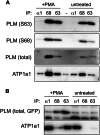
PLM-Ser63 antibody performance in co-immunoprecipitation experiments. A, immunoprecipitation (IP) reactions from untreated and PMA-treated (300 nm, 10 min) ARVMs. In untreated cells, sodium pump α1 subunit co-precipitated poorly with PLM-Ser63. Increasing the quantity of Ser63-phosphorylated PLM by activating PKC increased the quantity of sodium pump α1 subunit co-precipitated. B, immunoprecipitation reactions from FT-293 cells stably expressing PLM-YFP. In the absence of PMA, PLM is largely unphosphorylated in these cells and therefore not immunoprecipitated by Ser63 (S63) and Ser68 (S68) antibodies. Following PMA treatment, sodium pump α1 subunit is co-precipitated considerably more efficiently with Ser63-phosphorylated than Ser68-phosphorylated PLM.
FIGURE 3.
FIGURE 3.
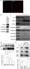
Subcellular localization of Ser63-phosphorylated PLM. A, immunofluorescent localization of Ser63 (S63) and Ser68 (S68) phosphorylated PLM in ARVMs. Both forms are distributed grossly identically in myocyte surface membranes in t-tubules, surface sarcolemma, and intercalated disks. B, cell surface fractions were prepared from ARVMs using cell-impermeable biotinylation reagents and analyzed alongside the unfractionated cell lysate from which they were derived. Ser63- and Ser68-phosphorylated forms are equally enriched in cell surface membranes compared with unfractionated starting material. C, sucrose gradient fractionation of ARVMs to purify a caveolar/lipid raft component. All phosphorylation states and unphosphorylated (UPh) PLM are localized in buoyant membranes from sucrose gradients (fractions 4 and 5), which are also enriched in the caveolar marker protein caveolin 3 (cav3) but not the bulk sarcolemma marker clathrin. T69, Thr69. D, phosphospecific immunoprecipitations (IP) of PLM indicate very poor co-purification of caveolin 3 with Ser63-phosphorylated PLM despite its localization to the caveolar membrane compartment. Quantitative analysis of the relative co-purification of caveolin 3 with PLM in phosphospecific co-immunoprecipitation reactions is shown (for an explanation of the calculation see legend to Fig. 1). SM, immunoprecipitation starting material. *, p < 0.05 (n = 7). E, palmitoylated proteins were purified by acyl rac in the presence (+) and absence (−) of hydroxylamine. UF lanes, unfractionated starting material; UB lanes, proteins not purified by acyl rac; B lanes, proteins purified by acyl rac. Ser63-phosphorylated PLM is purified to a considerably greater extent relative to the unfractionated starting material than Ser68-phosphorylated PLM, indicating that it is considerably more palmitoylated. *, p < 0.05 (n = 5).
FIGURE 4.
FIGURE 4.
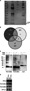
Protein interactions of Ser63-phosphorylated PLM. A, co-immunoprecipitation reactions using antibodies specific for unphosphorylated (C2), Ser63-phosphorylated (S63), and Ser68-phosphorylated (S68) PLM. Despite purifying similar quantities of PLM, the number and abundance of co-purifying proteins are substantially lower in the Ser63 immunoprecipitation reaction compared with the Ser68 immunoprecipitation reaction. B, co-immunoprecipitation reactions shown in A were analyzed by LC-MS/MS to identify all proteins. Venn diagram showing overlap in the proteins identified from each reaction. Protein identifications are shown in Table 1 and supplemental Table S1. UPh, unphosphorylated. C, control and PMA-treated ARVMs (300 nm, 10 min) were treated with the homobifunctional cross-linker DSP (1 mm, 60 min at 4 °C) and immunoblotted as indicated. No unique interactions for Ser63-phosphorylated PLM were revealed. cav3, caveolin 3. D, PP2A catalytic subunit was immunoprecipitated from ARVMs, and co-purifying sodium pump α1 and PLM phosphorylation states were immunoblotted. Ser63-phosphorylated PLM does not co-purify with PP2A. Duplicate immunoprecipitations are shown. SM, immunoprecipitation starting material; IP, immunoprecipitation.
FIGURE 5.
FIGURE 5.
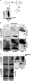
Partial formaldehyde fixation to identify multiprotein complexes. A, reaction scheme for formaldehyde monomer (left) in the presence of water to form formaldehyde polymer (right), which is hydrolyzed back to the monomer on heating/boiling. The primary amine reactive formaldehyde is therefore capable of cross-linking primary amines any multiple of 2 Å apart. A representative caveolin 3 immunoblot shows the result of partially fixing ARVMs with formaldehyde. The partially fixed caveolin 3 (cav3) multimer is resolved up to a nonamer, which may be quantitatively retrieved as a monomer upon hydrolysis of the formaldehyde polymer. SM, immunoprecipitation starting material. B, ARVMs were treated with the concentration of formaldehyde indicated above each lane, and immunoblotted as shown. Formaldehyde fixation reveals no interactions for Ser63-phosphorylated PLM (S63). Ser68-phosphorylated PLM (S68) is observed at 100 kDa (likely PLM interacting with the sodium pump α subunit) and 150 kDa (likely PLM-α-β complexes). Short and long exposures of the same immunoblots are shown. C, ARVMs were treated with PMA (300 nm, 10 min), fixed with 2% formaldehyde in the continued presence of PMA, and immunoblotted as shown without heating prior to electrophoresis. Ser63-phosphorylated PLM may be detected at 150 kDa following formaldehyde fixation in the presence of PMA (arrowhead). A species migrating at 50 kDa is immunoreactive with the Ser68-phosphorylated PLM antibody, consistent with the formation of a PLM tetramer (arrowhead).
FIGURE 6.
FIGURE 6.
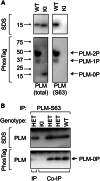
Phospholemman multimers in cardiac muscle. A, characterization of PLM from WT and KI animals on standard and Phos-Tag SDS-PAGE. The mobility of molecular mass standards is indicated alongside the immunoblots. Unphosphorylated (0P, immunoreactive with total PLM antibody only) and singly (1P) and doubly (2P) phosphorylated states (immunoreactive with Ser63 (S63) phosphospecific antibody) are resolved on Phos-Tag PAGE. 3SA PLM migrates entirely as unphosphorylated in Phos-Tag gels. B, Ser63-phosphorylated PLM was immunoprecipitated from WT, heterozygous KI (HET), and homozygous KI hearts under co-immunoprecipitation (Co-IP) and immunoprecipitation (IP) conditions. Purification of unphosphorylated PLM (0P) in co-immunoprecipitation reactions confirms the existence of interactions between WT and 3SA/unphosphorylated PLM.
FIGURE 7.
FIGURE 7.
A pool of PLM that does not regulate the Na pump. A, kinase activation/inhibition causes phosphorylation/dephosphorylation of PLM in ARVMs. veh, vehicle; bis, 1 μm bisindolylmaleimide; PMA, 300 nm PMA; Fors, 10 μm forskolin; PE, 100 μm phenylephrine; Iso, 100 nm isoproterenol, all applied for 10 min at 35 °C (for clarity of presentation, only relevant treatment groups are quantitated). S63, Ser63; S68, Ser68; T69, Thr69. *, p < 0.05 (n = 8). B, kinase activation/inhibition in ARVMs leading to phosphorylation/dephosphorylation of PLM does not alter the quantity of PLM co-immunoprecipitated with the sodium pump α1 subunit. Although bis treatment causes PLM dephosphorylation in unfractionated lysates, the phosphorylation status of pump-associated PLM is not altered. *, p < 0.05 (n = 7). C, whole cell Na pump currents in ARVMs. Although bis treatment causes PLM dephosphorylation in unfractionated lysates, pump currents are not altered because the phosphorylation status of pump-associated PLM is not changed (six cells from three animals for vehicle and seven cells from five animals for bis). IP, immunoprecipitation.
FIGURE 8.
FIGURE 8.
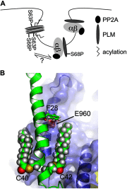
Modeling. A, cartoon depicting the relationships between PLM, Na pump, and PP2A established in this investigation. B, palmitates were added to the NMR structure of PLM in positions Cys40 and Cys42 (Protein Data Bank code 2JO1, shown in green) modeled with pump α subunit (Protein Data Bank code 3B8E, shown in blue) according to Ref. . Palmitate at Cys42 of PLM may impinge on PLM F28 (side chain shown in green)/α subunit Glu960 (side chain shown in red) and therefore alter the association of PLM with the pump.

Similar articles

Cited by

References

    1. Sweadner K. J., Rael E. (2000) The FXYD gene family of small ion transport regulators or channels. cDNA sequence, protein signature sequence, and expression. Genomics 68, 41–56 - PubMed
    1. Geering K. (2006) FXYD proteins. New regulators of Na-K-ATPase. Am. J. Physiol. Renal Physiol. 290, F241–F250 - PubMed
    1. Crambert G., Fuzesi M., Garty H., Karlish S., Geering K. (2002) Phospholemman (FXYD1) associates with Na,K-ATPase and regulates its transport properties. Proc. Natl. Acad. Sci. U.S.A. 99, 11476–11481 - PMC - PubMed
    1. Fuller W., Eaton P., Bell J. R., Shattock M. J. (2004) Ischemia-induced phosphorylation of phospholemman directly activates rat cardiac Na/K-ATPase. FASEB J. 18, 197–199 - PubMed
    1. Fuller W., Howie J., McLatchie L. M., Weber R. J., Hastie C. J., Burness K., Pavlovic D., Shattock M. J. (2009) FXYD1 phosphorylation in vitro and in adult rat cardiac myocytes. Threonine 69 is a novel substrate for protein kinase C. Am. J. Physiol. Cell Physiol. 296, C1346–C1355 - PMC - PubMed

Publication types

MeSH terms

LinkOut - more resources