Clinical Trial
. 2013 Jun 7;288(23):16738-16746.
doi: 10.1074/jbc.M112.431528. Epub 2013 Mar 24.

Discoidin domain receptor 1 protein is a novel modulator of megakaryocyte-collagen interactions

Affiliations
Clinical Trial

Discoidin domain receptor 1 protein is a novel modulator of megakaryocyte-collagen interactions

Vittorio Abbonante et al. J Biol Chem. .

Abstract

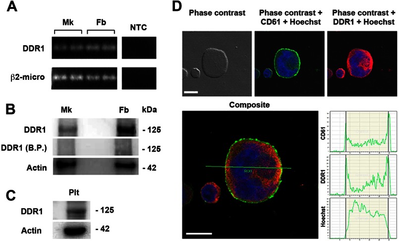
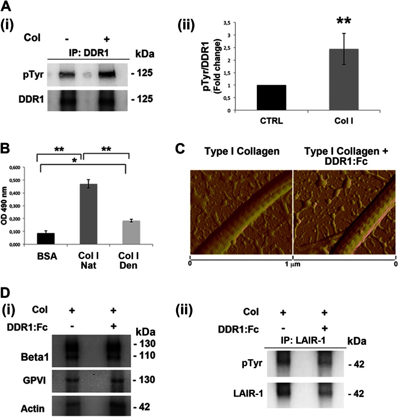
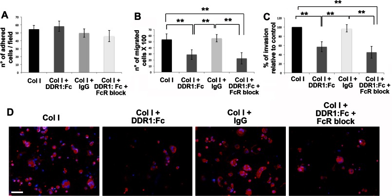
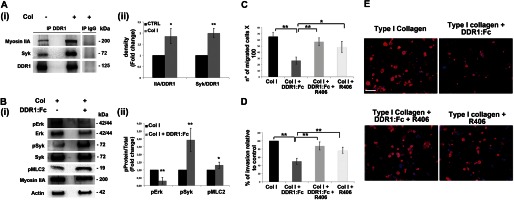
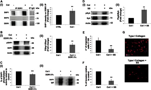
Growing evidence demonstrates that extracellular matrices regulate many aspects of megakaryocyte (MK) development; however, among the different extracellular matrix receptors, integrin α2β1 and glycoprotein VI are the only collagen receptors studied in platelets and MKs. In this study, we demonstrate the expression of the novel collagen receptor discoidin domain receptor 1 (DDR1) by human MKs at both mRNA and protein levels and provide evidence of DDR1 involvement in the regulation of MK motility on type I collagen through a mechanism based on the activity of SHP1 phosphatase and spleen tyrosine kinase (Syk). Specifically, we demonstrated that inhibition of DDR1 binding to type I collagen, preserving the engagement of the other collagen receptors, glycoprotein VI, α2β1, and LAIR-1, determines a decrease in MK migration due to the reduction in SHP1 phosphatase activity and consequent increase in the phosphorylation level of its main substrate Syk. Consistently, inhibition of Syk activity restored MK migration on type I collagen. In conclusion, we report the expression and function of a novel collagen receptor on human MKs, and we point out that an increasing level of complexity is necessary to better understand MK-collagen interactions in the bone marrow environment.

Keywords: Bone Marrow; Discoidin Domain Receptor 1; Extracellular Matrix; Hematopoiesis; Megakaryocytes; Phosphatase; Receptors.


Figures

FIGURE 1.
FIGURE 1.
Human MKs express and synthesize DDR1 tyrosine kinase. A, total cellular RNA was extracted from MKs and fibroblasts (Fb) as positive control. β2-microglobulin was used as housekeeping gene. NTC indicates “no template” controls in the reverse transcriptase and PCR steps. RT-PCR products were loaded in duplicates for each cell type. B, MK and fibroblast lysates were subjected to Western blot analysis using an anti-DDR1 antibody. The anti-DDR1 blocking peptide (B.P.) was used to confirm the specificity of the antibody. Actin was probed to show equal loading. C, DDR1 expression was demonstrated in peripheral blood platelet lysate (Plt) by Western blot. Shown here are representative Western blots out of three independent experiments. D, MKs were cytospun on polylysine-coated glass coverslips, fixed, and stained with an anti-DDR1 antibody (red) and an anti-CD61 antibody (green). The graphs report the intensity of the fluorescence signal along the x axis for each fluorochrome on the optical section. (Immunofluorescence staining, DM IRBE inverted microscope, magnification 40×.) Scale bars are 25 μm. Nuclei were counterstained with Hoechst 33288 (blue).
FIGURE 2.
FIGURE 2.
Type I collagen-dependent DDR1 activation. A, panel i, lysates of MKs, plated on tissue culture plastic or on type I collagen (Col) for 16 h, were immunoprecipitated (IP) with an anti-DDR1 antibody and subjected to Western blotting. Membranes were stained with a monoclonal antibody against phosphotyrosine and with anti-DDR1 antibody. Panel ii, densitometry analysis of the Western blots of 125-KDa phospho-Tyr (pTyr) band on DDR1 immunoprecipitates. B, binding assay to evaluate the DDR1-Fc binding affinity on native type I collagen (Nat Col I) with respect to denatured type I collagen (Den Col I). BSA was used as negative control. Optical density (OD) was measured at 490 nm. C, atomic force microscopy images of dehydrated collagen-coated coverslips in the presence or absence of DDR1-Fc showing similar banded fibrils. D, panel i, MKs were plated, for 2 h, on type I collagen, in the presence or absence of DDR1-Fc. Lysates were subjected to Western blot analysis of active β1 integrin (HUTS-4) and dimeric GPVI. Actin was probed to show equal loading. Panel ii, lysates of megakaryocyte treated as above described were immunoprecipitated with anti-LAIR-1 antibody and subjected to Western blot. Membranes were stained with antibodies anti-phospho-Tyr (pTyr) and anti-LAIR-1. Shown here are representative Western blots out of three independent experiments. Data are presented as means ± S.D. (n = 5 and 4 independent experiments). *, p < 0.05. **, p < 0.01.
FIGURE 3.
FIGURE 3.
DDR1-dependent regulation of MK migration. A, MKs were plated on type I collagen (Col I)-coated coverslips, in the presence of the DDR1-Fc blocking molecule or an IgG as control. Where indicated, MKs were pretreated with FcR blocking solution. After 16 h, adherent MKs were fixed, stained with anti-CD61 antibody, and then counted by fluorescence microscopy. B, MKs were left to migrate in a Transwell plate, upon coating the Transwell filter with type I collagen mixed with the DDR1-Fc blocking molecule or an IgG as control. Where indicated, MKs were pretreated with FcR blocking solution. After 16 h, MKs that had passed in the lower chamber were collected and counted by phase contrast microscopy. C, MKs adhering on the lower side of the Transwell filter were fixed and stained with anti-CD61 antibody and then counted by fluorescence microscopy. D, representative images of MK invasion of type I collagen. Cells adhering to the lower side of the Transwell coated filter, were fixed and stained with anti-CD61 antibody (red) antibody. (Immunofluorescence staining, Olympus BX51 microscope, magnifications 20×.) Scale bars are 100 μm. Nuclei were stained with Hoechst 33288 (blue). Reported results are the means ± S.D. (n = 6 independent experiments). **, p < 0.01.
FIGURE 4.
FIGURE 4.
Syk kinase is involved in DDR1-mediated MK migration on type I collagen. A, panel i, DDR1 was immunoprecipitated (IP) in cell lysates of MKs plated for 16 h on type I collagen (Col) or on tissue culture plastic. A control sample was immunoprecipitated with an unrelated antibody (IgG). Membranes were probed with anti-Syk and anti-myosin IIA antibodies to show DDR1-interacting protein and reprobed with anti-DDR1 antibody to show equal loading. Panel ii, densitometry analysis of the Western blots of Myosin IIA and Syk co-immunoprecipitated with DDR1. B, total cellular lysates of MKs plated for 16 h on type I collagen, inhibiting or not inhibiting DDR1 activation, were subjected to Western blot analysis. Membranes were probed with the indicated antibody, with p indicating the phosphorylated form. Actin was probed to show equal loading. Panel ii, densitometry analysis of the Western blots of phospho-ERK (pERK), phospho-Syk (pSyk), and phospho-MLC2 (pMLC2). C, Transwell migration assay of mature MKs through type I collagen, in the presence of the DDR1-Fc blocking molecule and of Syk specific inhibitor compound R406 (5 μm) either mixed or used singularly. After 16 h, MKs that had passed in the lower chamber were counted by phase contrast microscopy. D, MKs adhering to the lower side of the Transwell filter were fixed and stained with anti-CD61 antibody and then counted by fluorescence microscopy. E, representative images of MK invasion of type I collagen. Cells adhering to the lower side of the Transwell coated filter were fixed and stained with anti-CD61 antibody (red). (Immunofluorescence staining, Olympus BX51 microscope, magnifications 20×.) Scale bars are 100 μm. Nuclei were stained with Hoechst 33288 (blue). Data are presented as means ± S.D.(n = 5, 5, and 5 independent experiment). *, p < 0.05. **, p < 0.01.
FIGURE 5.
FIGURE 5.
SHP1 modulates Syk activation upon type I collagen-DDR1 binding in MKs. A, panel i, DDR1 immunoprecipitates (IP) from MKs plated for 16 h on type I collagen (Col) or on plastic were analyzed by Western blot. A control sample was immunoprecipitated with an unrelated antibody (IgG). Membranes were probed with anti-SHP1 and anti-SHP2 antibodies to show the co-immunoprecipitation and with anti-DDR1 antibody to show equal loading. Panel ii densitometry analysis of the Western blots of SHP1 protein co-immunoprecipitated with DDR1. B, panel i, Western blot analysis of cell lysates of MKs plated for 16 h on type I collagen mixed with or without DDR1-Fc blocking molecule. Membranes were probed with the indicated antibodies with p indicating the phosphorylated form. Actin was probed to show equal loading. Panel ii, densitometry analysis of the Western blots of pSHP1. CTRL, control. C, panel i, SHP1 phosphatase activity measured in a phosphatase assay on SHP1 immunoprecipitates from MK lysates, using p-nitrophenyl phosphate as substrate. MKs were plated for 16 h on type I collagen mixed with or without DDR1-Fc blocking molecule. Panel ii, Western blot analysis of SHP1 immunoprecipitates used for SHP1 phosphatase activity assay. SHP1 phosphatase activity was related to the same concentration of SHP1. Shown here is a representative Western blot out of four independent experiments. D, panel i, MKs were treated with the SHP1 specific inhibitor sodium stibogluconate (SS) (13.4 μm) and plated for 3 h on type I collagen. Cell lysates were subjected to Western blot analysis. Membranes were probed with anti-phospho Syk (Tyr-525/526) antibody and anti-Syk and anti-actin antibodies to show equal loading. Panel ii, densitometry analysis of the Western blots of phospho-Syk (pSyk). E, Transwell migration assay of mature MKs through type I collagen, in the presence of the SHP1 specific inhibitor SS (13.4 μm). After 16 h, MKs that had passed in the lower chamber were counted by phase contrast microscopy. F, MKs adhering to the lower side of the Transwell filter were fixed and stained with anti-CD61 antibody and then counted by fluorescence microscopy. G, representative images of MK invasion of type I collagen. Cells adhering to the lower side of the Transwell coated filter were fixed and stained with anti-CD61 antibody (red) antibody. (Immunofluorescence staining, Olympus BX51 microscope, magnifications 20×.) Scale bars are 100 μm. Nuclei were stained with Hoechst 33288 (blue). Data are presented as means ± S.D. (n = 5, 4, 4, 4, 3, and 3 independent experiments). *, p < 0.05 **, p < 0.01.

Similar articles

Cited by

References

    1. Avecilla S. T., Hattori K., Heissig B., Tejada R., Liao F., Shido K., Jin D. K., Dias S., Zhang F., Hartman T. E., Hackett N. R., Crystal R. G., Witte L., Hicklin D. J., Bohlen P., Eaton D., Lyden D., de Sauvage F., Rafii S. (2004) Chemokine-mediated interaction of hematopoietic progenitors with the bone marrow vascular niche is required for thrombopoiesis. Nat. Med. 10, 64–71 - PubMed
    1. Patel S. R., Hartwig J. H., Italiano J. E., Jr. (2005) The biogenesis of platelets from megakaryocyte proplatelets. J. Clin. Invest. 115, 3348–3354 - PMC - PubMed
    1. Junt T., Schulze H., Chen Z., Massberg S., Goerge T., Krueger A., Wagner D. D., Graf T., Italiano J. E., Jr., Shivdasani R. A., von Andrian U. H. (2007) Dynamic visualization of thrombopoiesis within bone marrow. Science 317, 1767–1770 - PubMed
    1. Nilsson S. K., Debatis M. E., Dooner M. S., Madri J. A., Quesenberry P. J., Becker P. S. (1998) Immunofluorescence characterization of key extracellular matrix proteins in murine bone marrow in situ. J. Histochem. Cytochem. 46, 371–377 - PubMed
    1. Takaku T., Malide D., Chen J, Calado R. T., Kajigaya S., Young N. S. (2010) Hematopoiesis in 3 dimensions: human and murine bone marrow architecture visualized by confocal microscopy. Blood 116, e41–55 - PMC - PubMed

Publication types

MeSH terms

LinkOut - more resources